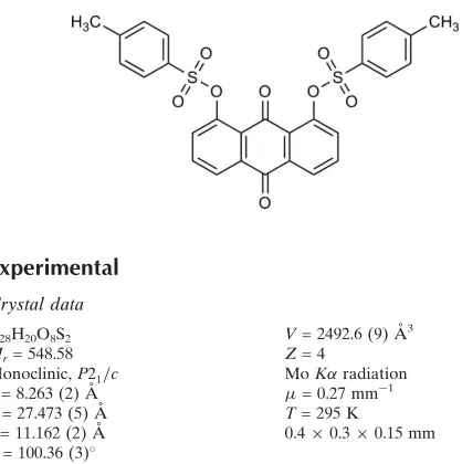
1,8-Bis(tosyloxy)-9,10-anthraquinone
Paweł Niedziałkowski, Damian Trzybin´ski, Artur Sikorski* and Tadeusz Ossowski
Faculty of Chemistry, University of Gdan´sk, J. Sobieskiego 18, 80-952 Gdan´sk, Poland
Correspondence e-mail: [email protected]
Received 23 November 2009; accepted 26 November 2009
Key indicators: single-crystal X-ray study;T= 295 K; mean(C–C) = 0.004 A˚; Rfactor = 0.058;wRfactor = 0.163; data-to-parameter ratio = 12.7.
In the crystal structure of the title compound, C28H20O8S2,
adjacent anthracene skeletons are parallel or inclined at an angle of 20.6 (1). In the molecular structure, the mean plane of the anthracene skeleton makes dihedral angles of 49.6 (1) and 76.8 (1) with the tosyl rings, and the two terminal benzene rings are oriented at an angle of 74.5 (1)with respect to each other. The crystal structure is stabilized by inter-molecular C—H O and C—O interactions.
Related literature
For general background to anthraquinones, see: Cheng & Zee-Cheng (1983); Dzierzbicka et al. (2006); Gattoet al. (1996); Hunger (2003); Krapchoet al.(1991); Nakanishiet al.(2005); Zielske (1987); Zon et al.(2003). For related structures, see: Sereda & Akhvlediani (2003); Slouf (2002); Zain & Ng (2005). For molecular interactions, see: Bianchiet al.(2004); Santos-Contreraset al. (2007); Spek (2009); Steiner (1999). For the synthesis, see: Ossowskiet al.(2000).
Experimental
Crystal data
C28H20O8S2
Mr= 548.58 Monoclinic,P21=c
a= 8.263 (2) A˚ b= 27.473 (5) A˚ c= 11.162 (2) A˚
V= 2492.6 (9) A˚3
Z= 4
MoKradiation
= 0.27 mm1
T= 295 K
0.40.30.15 mm
Data collection
Oxford Diffraction Gemini R ULTRA Ruby CCD diffractometer
18048 measured reflections
4371 independent reflections 3374 reflections withI> 2(I) Rint= 0.050
Refinement
R[F2> 2(F2)] = 0.058 wR(F2) = 0.163
S= 1.15 4371 reflections
345 parameters
H-atom parameters constrained max= 0.46 e A˚
3 min=0.32 e A˚
3
Table 1
Hydrogen-bond geometry (A˚ ,).
D—H A D—H H A D A D—H A
C24—H24 O18i
0.93 2.54 3.332 (4) 143 C33—H33 O30ii
0.93 2.56 3.241 (4) 130 C36—H36 O31iii
0.93 2.58 3.458 (4) 156
Symmetry codes: (i)xþ1;yþ1;zþ2; (ii)x;yþ3 2;z
1
[image:1.610.51.260.538.748.2]2; (iii)x1;y;z.
Table 2
C–O interactions (A˚ ,)..
C O J O J C J C–O J
C10 O27 Cg1iv
3.688 (3) 3.481 (3) 70.71 (17) C10 O27 Cg2v
3.452 (3) 3.528 (3) 83.41 (18)
Symmetry codes: (iv)x,y+ 1,z+ 1; (v)x+ 1,y+ 1,z+ 1.Cg1 andCg2 are the centroids of the C1—C4/C11/C12 and C5—C8/C13/C14 rings respectively.
Data collection: CrysAlis CCD (Oxford Diffraction, 2008); cell refinement: CrysAlis RED (Oxford Diffraction, 2008); data reduc-tion:CrysAlis RED; program(s) used to solve structure:SHELXS97 (Sheldrick, 2008); program(s) used to refine structure:SHELXL97 (Sheldrick, 2008); molecular graphics: ORTEP-3 (Farrugia, 1997); software used to prepare material for publication:SHELXL97and PLATON(Spek, 2009).
This work was supported by the Polish State Committee for Scientific Research (grant Nos. R02 0010 06 and DS 8210–4-0177–9).
Supplementary data and figures for this paper are available from the IUCr electronic archives (Reference: XU2694).
References
Bianchi, R., Forni, A. & Pilati, T. (2004).Acta Cryst.B60, 559–568. Cheng, C. C. & Zee-Cheng, R. K. Y. (1983).Prog. Med. Chem.20, 83–118. Dzierzbicka, K., Sowin´ski, P. & Kołodziejczyk, A. M. (2006).J. Pept. Sci.12,
670–678.
Farrugia, L. J. (1997).J. Appl. Cryst.30, 565.
Gatto, B., Zagotto, G., Sissi, C., Cera, C., Uriarte, E., Palu, G., Capranico, G. & Palumbo, M. (1996).J. Med. Chem.39, 3114–3122.
Hunger, K. (2003). Industrial Dyes Chemistry, Properties, Applications. Weinheim: Wiley-VCH Verlag GmbH & Co. KGaA.
Krapcho, A. P., Getahun, Z., Avery, K. L., Vargas, K. J., Hacker, M. P., Spinelli, S., Pezzoni, G. & Manzotti, C. (1991).J. Med. Chem.34, 2373–2380. Nakanishi, F., Nagasawa, Y., Kabaya, Y., Sekimoto, H. & Shimomura, K.
(2005).Plant Phys. Biochem.43, 921–928.
Ossowski, T., Kira, J., Rogowska, D., Warmke, H. & Młodzianowski, J. (2000). Acta Crystallographica Section E
Structure Reports Online
Oxford Diffraction. (2008). CrysAlis CCD and CrysAlis RED. Oxford Diffraction Ltd, Yarnton, England.
Santos-Contreras, R. J., Martı´nez-Martı´nez, F. J., Garcı´a-Ba´ez, E. V., Padilla-Martı´nez, I. I., Peraza, A. L. & Ho¨pfl, H. (2007).Acta Cryst.C63, o239–o242. Sereda, G. A. & Akhvlediani, D. G. (2003).Tetrahedron Lett.44, 9125–9126. Sheldrick, G. M. (2008).Acta Cryst.A64, 112–122.
Slouf, M. (2002).J. Mol. Struct.611, 139–146.
Spek, A. L. (2009).Acta Cryst.D65, 148–155. Steiner, T. (1999).Chem. Commun.pp. 313–314.
Zain, S. M. & Ng, S. W. (2005).Acta Cryst.E61, o2921–o2923. Zielske, A. G. (1987).J. Org. Chem.52, 1305–1309.
supporting information
Acta Cryst. (2010). E66, o33–o34 [doi:10.1107/S1600536809051009]
1,8-Bis(tosyloxy)-9,10-anthraquinone
Pawe
ł
Niedzia
ł
kowski, Damian Trzybi
ń
ski, Artur Sikorski and Tadeusz Ossowski
S1. Comment
Anthraquinones, its amino and hydroxy derivatives as the largest group of naturally occurring quinines are of great
practical significance in pharmacology, biochemistry and dye chemistry (Hunger, 2003). Anthraquinones are widely
widespread in nature, they are occur in bark, or roots of different plants, and display various pharmacological activities
such as anti-oxidant, anti-microbial, anti-fungal and anti-viral (Nakanishi et al., 2005). The anthraquinone ring system is
often found in antitumor drugs, such as anthracyclines, mitoxantrone, ametantrone and anthrapyrazoles (Cheng &
Zee-Cheng, 1983). Its planarity allows an intercalation between base pairs of DNA in the β conformation, while its redox
properties are linked to the production of radical species in biological systems. The chemical and biological activity of
anthraquinone compounds depends on the different substituents of the planar ring system (Krapcho et al., 1991, Gatto et
al., 1996). Anthraquinones are also interesting compounds for the investigations in analytical and electroanalytical
chemistry due to the fact that they contain several π electrons, the reducible p-quinone system and are electroactive (Zon
et al., 2003). The tosyl group is a very good leaving group, commonly used in organic synthesis in nucleophilic
substitution reaction. This phenomenon is applicable to prepare the various aminoanthraquinone from
(tosy1oxy)anthra-quinone precursors (Zielske, 1987). The 1,8-Bis(tosyloxy)-9,10-anthra(tosy1oxy)anthra-quinone is a very convenient and often used
precursor to obtain the 1,8-diaminoanthraquinones derivatives (Dzierzbicka et al., 2006).
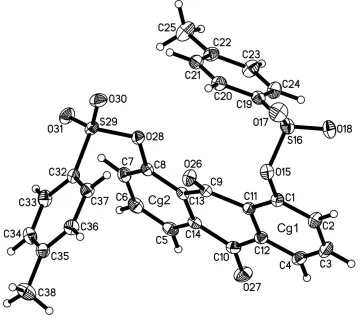
In the molecule of the title compound (Fig. 1) the bond lengths and angles characterizing the geometry of the
anthra-quinone skeleton are typical for this group of compounds (Sereda & Akhvlediani, 2003; Slouf, 2002; Zain & Ng, 2005).
In the packing of molecules of the title compound, the anthracene skeletons, with an average deviations from planarity
of 0.044 (1) Å, are parallel or inclined at an angle of 20.1 (1)°. The mean plane of the anthracene skeleton makes dihedral
angles of 49.6 (1)° and 76.8 (1)°, with the tosyl phenyl rings. Those phenyl rings are oriented at the angle of 74.5 (1)° to
each other.
The crystal structure is stabilized by C–H···O (Table 1, Fig. 2) and C–O···π (Table 1, Fig. 3) intermolecular interactions.
The C–H···O interactions are the hydrogen bond type (Steiner, 1999, Bianchi et al., 2004). All interactions demonstrated
were found by PLATON (Spek, 2009).
S2. Experimental
1,8-Bis(tosyloxy)-9,10-anthraquinone was synthesized according to the method reported in the literature (Ossowski et al.,
2000). To the stirring mixture of 5.0 g (20.8 mmol) 1,8-dihydroxy-9,10-anthraquinone and 5.22 g (27.4 mmol) of p
-toluenesulfonyl chloride in 200 ml dichloromethane was dropwise added over 5 h 15 ml triethylamine in 100 ml
dichloro-methane. The progress of the reaction was monitored by TLC (SiO2, dichloromethane-petroleum ether 1:1 v/v) until the
completion of reaction. The reaction mixture was stirred 6 h at room temperature. The solution was washed with water (3
x 100 ml), the organic phase was dried over MgSO4 and concentrated. The residue was purified by column
(3.64 g, 28%). Single crystals suitable for X-ray diffraction were prepared by slow evaporation of a solution of the title
compound in methanol at room temperature (m.p. = 448–450 K; elemental analysis (% found/calculated: C 61.41/61.30,
H 3.65/3.67, S 11.69/11.69)).
S3. Refinement
H atoms were positioned geometrically, with C—H = 0.93 Å and 0.96 Å for the aromatic and methyl H atoms,
respectively, and constrained to ride on their parent atoms with Uiso(H) = xUeq(C), where x = 1.2 for the aromatic and x =
[image:4.610.126.487.190.513.2]1.5 for the methyl H atoms.
Figure 1
The molecular structure of the title compound showing the atom-labeling scheme. Displacement ellipsoids are drawn at
the 25% probability level, and H atoms are shown as small spheres of arbitrary radius. Cg1 and Cg2 are the centroids of
Figure 2
The arrangement of the molecules in the crystal structure viewed approximately along c axis. The C–H···O interactions
are represented by dashed lines. H atoms not involved in interactions have been omitted. [Symmetry codes: (i) -x + 1, -y
Figure 3
The arrangement of the molecules in the crystal structure viewed approximately along c axis. The C–O···π interactions
are represented by dotted lines. H atoms not involved in interactions have been omitted. [Symmetry codes: (iv) -x, -y + 1,
-z + 1; (v) -x + 1, -y + 1, -z + 1.]
1,8-Bis(tosyloxy)-9,10-anthraquinone
Crystal data
C28H20O8S2 Mr = 548.58
Monoclinic, P21/c Hall symbol: -P 2ybc a = 8.263 (2) Å b = 27.473 (5) Å c = 11.162 (2) Å β = 100.36 (3)° V = 2492.6 (9) Å3 Z = 4
F(000) = 1136 Dx = 1.462 Mg m−3
Mo Kα radiation, λ = 0.71073 Å Cell parameters from 10108 reflections θ = 3.2–29.2°
µ = 0.27 mm−1 T = 295 K Block, yellow 0.4 × 0.3 × 0.15 mm
Data collection
Oxford Diffraction Gemini R ULTRA Ruby CCD
diffractometer
Radiation source: Enhance (Mo) X-ray Source Graphite monochromator
Detector resolution: 10.4002 pixels mm-1 ω scans
18048 measured reflections
4371 independent reflections 3374 reflections with I > 2σ(I) Rint = 0.050
θmax = 25.1°, θmin = 3.2° h = −9→9
k = −28→32 l = −13→11
Refinement
Refinement on F2 Least-squares matrix: full R[F2 > 2σ(F2)] = 0.058 wR(F2) = 0.163 S = 1.15 4371 reflections 345 parameters
0 restraints
Primary atom site location: structure-invariant direct methods
Secondary atom site location: difference Fourier map
H-atom parameters constrained w = 1/[σ2(F
o2) + (0.0997P)2 + 0.3332P] where P = (Fo2 + 2Fc2)/3
(Δ/σ)max = 0.002 Δρmax = 0.46 e Å−3 Δρmin = −0.32 e Å−3
Special details
Geometry. All e.s.d.'s (except the e.s.d. in the dihedral angle between two l.s. planes) are estimated using the full
covariance matrix. The cell e.s.d.'s are taken into account individually in the estimation of e.s.d.'s in distances, angles and torsion angles; correlations between e.s.d.'s in cell parameters are only used when they are defined by crystal symmetry. An approximate (isotropic) treatment of cell e.s.d.'s is used for estimating e.s.d.'s involving l.s. planes.
Fractional atomic coordinates and isotropic or equivalent isotropic displacement parameters (Å2)
x y z Uiso*/Ueq
H25B 0.8771 0.6762 0.8264 0.128* H25C 0.7595 0.6832 0.7007 0.128* O26 0.1427 (3) 0.61106 (7) 0.63596 (19) 0.0530 (5) O27 0.2365 (3) 0.44406 (8) 0.4261 (2) 0.0714 (7) O28 0.3778 (2) 0.65378 (6) 0.53713 (16) 0.0471 (5) S29 0.34049 (9) 0.70699 (2) 0.48060 (7) 0.0481 (2) O30 0.3284 (3) 0.73551 (8) 0.5843 (2) 0.0638 (6) O31 0.4611 (3) 0.71850 (8) 0.4092 (2) 0.0643 (6) C32 0.1493 (3) 0.70184 (10) 0.3851 (3) 0.0444 (6) C33 0.1394 (4) 0.70045 (11) 0.2604 (3) 0.0556 (8) H33 0.2343 0.7015 0.2266 0.067* C34 −0.0135 (4) 0.69754 (12) 0.1866 (3) 0.0586 (8) H34 −0.0206 0.6962 0.1026 0.070* C35 −0.1561 (4) 0.69661 (10) 0.2349 (3) 0.0525 (7) C36 −0.1428 (4) 0.69827 (11) 0.3605 (3) 0.0533 (7) H36 −0.2380 0.6979 0.3941 0.064* C37 0.0083 (3) 0.70044 (10) 0.4368 (3) 0.0488 (7) H37 0.0156 0.7010 0.5209 0.059* C38 −0.3217 (5) 0.69311 (16) 0.1525 (4) 0.0821 (11) H38A −0.3199 0.7114 0.0795 0.123* H38B −0.4047 0.7061 0.1936 0.123* H38C −0.3458 0.6596 0.1320 0.123*
Atomic displacement parameters (Å2)
U11 U22 U33 U12 U13 U23
C23 0.060 (2) 0.0445 (18) 0.092 (3) 0.0124 (14) 0.0231 (17) −0.0039 (16) C24 0.066 (2) 0.0404 (16) 0.070 (2) 0.0106 (14) 0.0173 (16) 0.0004 (14) C25 0.084 (3) 0.064 (2) 0.118 (3) −0.0084 (19) 0.044 (2) −0.004 (2) O26 0.0669 (13) 0.0389 (11) 0.0587 (12) 0.0081 (9) 0.0259 (10) 0.0001 (9) O27 0.0901 (17) 0.0553 (13) 0.0714 (15) −0.0082 (11) 0.0213 (13) −0.0257 (12) O28 0.0543 (11) 0.0439 (11) 0.0429 (11) −0.0036 (8) 0.0085 (8) 0.0006 (8) S29 0.0439 (4) 0.0417 (4) 0.0596 (5) −0.0070 (3) 0.0114 (3) 0.0020 (3) O30 0.0678 (14) 0.0496 (12) 0.0720 (14) −0.0069 (10) 0.0070 (11) −0.0169 (10) O31 0.0490 (12) 0.0625 (14) 0.0855 (16) −0.0099 (10) 0.0230 (11) 0.0141 (12) C32 0.0452 (15) 0.0402 (14) 0.0500 (16) 0.0007 (11) 0.0143 (12) 0.0055 (12) C33 0.0544 (17) 0.0613 (18) 0.0564 (18) 0.0063 (14) 0.0241 (14) 0.0098 (14) C34 0.069 (2) 0.0629 (19) 0.0436 (16) 0.0113 (15) 0.0102 (15) 0.0056 (14) C35 0.0535 (17) 0.0416 (15) 0.0605 (19) 0.0073 (12) 0.0050 (14) 0.0021 (13) C36 0.0437 (16) 0.0584 (18) 0.0600 (19) 0.0032 (13) 0.0156 (14) 0.0024 (14) C37 0.0483 (15) 0.0535 (17) 0.0469 (16) −0.0007 (12) 0.0144 (12) 0.0011 (13) C38 0.066 (2) 0.099 (3) 0.073 (2) 0.010 (2) −0.0094 (18) −0.001 (2)
Geometric parameters (Å, º)
S16—C19 1.745 (3) C38—H38B 0.9600 C19—C24 1.378 (4) C38—H38C 0.9600
O18—S16—O15 108.09 (12) C32—C37—H37 120.7 O17—S16—C19 110.62 (15) C35—C38—H38A 109.5 O18—S16—C19 109.39 (14) C35—C38—H38B 109.5 O15—S16—C19 104.49 (12) H38A—C38—H38B 109.5 C24—C19—C20 120.4 (3) C35—C38—H38C 109.5 C24—C19—S16 120.0 (2) H38A—C38—H38C 109.5 C20—C19—S16 119.6 (2) H38B—C38—H38C 109.5
O27—C10—C14—C5 −2.9 (4) C35—C36—C37—C32 1.1 (4) C12—C10—C14—C5 173.9 (2) C33—C32—C37—C36 −0.8 (4) O27—C10—C14—C13 176.9 (2) S29—C32—C37—C36 177.5 (2)
Hydrogen-bond geometry (Å, º)
D—H···A D—H H···A D···A D—H···A
C24—H24···O18i 0.93 2.54 3.332 (4) 143 C33—H33···O30ii 0.93 2.56 3.241 (4) 130 C36—H36···O31iii 0.93 2.58 3.458 (4) 156