N
,
N
-Dimethylanilinium
2,4,6-trinitro-phenolate
Nagarajan Vembua* and Frank R. Fronczekb
aDepartment of Chemistry, Urumu Dhanalakshmi College, Tiruchirappalli 620 019,
India, andbDepartment of Chemistry, Louisiana State University, Baton Rouge, LA
70803-1804, USA
Correspondence e-mail: [email protected]
Received 5 December 2008; accepted 9 December 2008
Key indicators: single-crystal X-ray study;T= 90 K; mean(C–C) = 0.002 A˚;Rfactor = 0.023;wRfactor = 0.060; data-to-parameter ratio = 10.0.
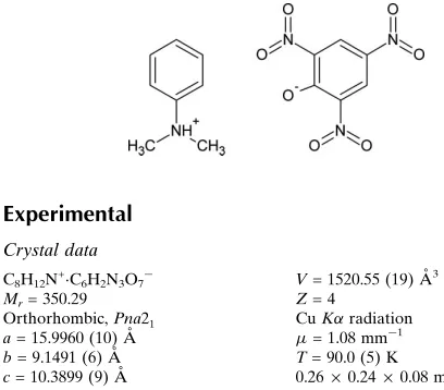
In the title compound, C8H12N +
C6H2N3O7
, there are N— H O and C—H O interactions which generate R2
1
(5),
R2 1
(6) andR1 2
(6) ring motifs. The supramolecular aggregation is completed by the presence of edge-to-face and offset face-to-face–interactions with centroid–centroid distances of 3.673 and 3.697 A˚ , respectively.
Related literature
For a detailed account of the design of organic polar crystals, see: Pecaut & Bagieu-Beucher (1993). For hydrogen bonding in nitrophenol complexes, see: Inet al.(1997); Zadrenkoet al.
(1997); Mizutani et al.(1998). For the supramolecular archi-tecture of molecular complexes of trinitrophenols, see: Botoshanskyet al.(1994); Vembuet al.(2003). For details of the monoclinic polymorph of the title compound, see: Takayanagi et al.(1996). For hydrogen-bonding criteria, see: Desiraju & Steiner (1999); Desiraju (1989); Jeffrey (1997). For graph-set notation, see: Bernsteinet al.(1995); Etter (1990).
Experimental
Crystal data
C8H12N+C6H2N3O7
Mr= 350.29 Orthorhombic,Pna21 a= 15.9960 (10) A˚ b= 9.1491 (6) A˚ c= 10.3899 (9) A˚
V= 1520.55 (19) A˚3
Z= 4
CuKradiation
= 1.08 mm1 T= 90.0 (5) K 0.260.240.08 mm
Data collection
Bruker Kappa APEXII CCD area-detector diffractometer Absorption correction: multi-scan
(SADABS; Sheldrick, 1996) Tmin= 0.767,Tmax= 0.919
16980 measured reflections 2823 independent reflections 2755 reflections withI> 2(I) Rint= 0.033
Refinement
R[F2> 2(F2)] = 0.023
wR(F2) = 0.060 S= 1.03 2823 reflections 283 parameters 1 restraint
H atoms treated by a mixture of independent and constrained refinement
max= 0.14 e A˚3
min=0.16 e A˚3
Absolute structure: Flack (1983), 1309 Friedel pairs
[image:1.610.44.248.562.741.2]Flack parameter: 0.07 (12)
Table 1
Hydrogen-bond geometry (A˚ ,).
D—H A D—H H A D A D—H A
N7—H7 O16 0.892 (17) 1.825 (18) 2.7128 (14) 172.8 (16) N7—H7 O19 0.892 (17) 2.578 (16) 3.0517 (15) 114.0 (12) C2—H2 O16 0.931 (18) 2.341 (17) 3.0464 (16) 132.4 (13) C2—H2 O24 0.931 (18) 2.498 (17) 3.3444 (17) 151.4 (14) C8—H8A O19 1.001 (18) 2.411 (18) 3.1171 (18) 126.9 (13) C9—H9C O19 0.997 (18) 2.592 (17) 3.2311 (17) 121.9 (12) C9—H9B O21i 0.974 (19) 2.571 (19) 3.5074 (17) 161.3 (14) C9—H9B O25ii
0.974 (19) 2.476 (18) 3.0794 (18) 119.9 (13) C4—H4 O21iii
0.924 (19) 2.466 (18) 3.1776 (16) 134.0 (14) C14—H14 O19iv 0.941 (18) 2.564 (18) 3.4988 (16) 172.3 (14) C9—H9C O22v
0.997 (18) 2.502 (18) 3.3283 (16) 140.0 (13)
Symmetry codes: (i)x;yþ1;z; (ii)xþ1 2;yþ
1 2;zþ
1
2; (iii) xþ 1 2;yþ
3 2;z
1 2; (iv)
x1 2;yþ
3 2;z; (v)xþ
1 2;yþ
3 2;z.
Data collection:APEX2(Bruker, 2006); cell refinement:APEX2 andSAINT(Bruker, 2006); data reduction:SAINT; program(s) used to solve structure:SHELXS97(Sheldrick, 2008); program(s) used to refine structure:SHELXL97(Sheldrick, 2008); molecular graphics: PLATON (Spek, 2003); software used to prepare material for publication:SHELXL97.
NV thanks the University Grants Commission (UGC), Government of India, for a minor research project grant [MRP-2219/06(UGC-SERO)].
Supplementary data and figures for this paper are available from the IUCr electronic archives (Reference: SJ2565).
References
Bernstein, J., Davis, R. E., Shimoni, L. & Chang, N. (1995).Angew. Chem. Int. Ed. Engl.34, 1555–1573.
Botoshansky, M., Herbstein, F. H. & Kapon, M. (1994).Acta Cryst.B50, 191– 200.
Bruker (2006).APEX2andSAINT. Bruker AXS Inc., Madison, Wisconsin, USA.
Desiraju, G. R. (1989).Crystal Engineering: The Design of Organic Solids. Amsterdam: Elsevier.
Desiraju, G. R. & Steiner, T. (1999).The Weak Hydrogen Bond in Structural Chemistry and Biology.New York, Oxford University Press.
Etter, M. C. (1990).Acc. Chem. Res.23, 120–126. Flack, H. D. (1983).Acta Cryst.A39, 876–881.
In, Y., Nagata, H., Doi, M., Ishida, T. & Wakahara, A. (1997).Acta Cryst.C53, 367–369.
Jeffrey, G. A. (1997). An Introduction to Hydrogen Bonding. New York, Oxford University Press.
organic compounds
Acta Cryst.(2009). E65, o111–o112 doi:10.1107/S1600536808041743 Vembu and Fronczek
o111
Acta Crystallographica Section E
Structure Reports
Online
(1998).J. Phys. Org. Chem.11, 737–742.
Pecaut, J. & Bagieu-Beucher, M. (1993).Acta Cryst.C49, 834–837. Sheldrick, G. M. (1996).SADABS. University of Gottingen, Germany. Sheldrick, G. M. (2008).Acta Cryst.A64, 112–122.
Spek, A. L. (2003).J. Appl. Cryst.36, 7–13.
Chem. Pharm. Bull.44, 2199–2204.
Vembu, N., Nallu, M., Garrison, J. & Youngs, W. J. (2003).Acta Cryst.E59, o913–o916.
supporting information
sup-1
Acta Cryst. (2009). E65, o111–o112
supporting information
Acta Cryst. (2009). E65, o111–o112 [doi:10.1107/S1600536808041743]
N
,
N
-Dimethylanilinium 2,4,6-trinitrophenolate
Nagarajan Vembu and Frank R. Fronczek
S1. Comment
The design of organic polar crystals for quadratic non-linear optical applications is supported by the observation that the
organic molecules containing π-electron systems asymmetrized by electron donor and acceptor groups are highly
polarizable entities in which problems of transparency and crystal growth may arise from their molecular crystal packing
(Pecaut & Bagieu-Beucher, 1993). It is known that nitrophenols act not only as π-acceptors to form various π-stacking
complexes with other aromatic molecules, but also as acidic ligands to form salts through specific electrostatic or
H-bonding interactions (In et al., 1997). The bonding of electron-donor acceptor complexes strongly depends on the nature
of the partners. The linkage could involve not only electrostatic interactions, but also the formation of molecular
complexes (Zadrenko et al., 1997). It has been reported that proton transferred thermochromic complexes were formed
between phenols and amines in apolar solvents at low temperature if an appropriate H-bonding network between the
phenols and amines was present to stabilize it (Mizutani et al., 1998). Pyridinium picrate has been reported in two
crystalline phases and it appears in both phases as an internally linked H-bonded ion pair. These two phases are referred
to as molecular crystals rather than salts based on their structural arrangements (Botoshansky et al., 1994). A similar
structural arrangement has also been reported for 4-dimethylaminopyridinium picrate (Vembu et al., 2003). The
monoclinic polymorph of the title compound (CSD Reference Code: REYDEE) has been reported previously
(Takayanagi et al., 1996). We have structurally elucidated the orthorhombic polymorph of the title compound as a
forerunner to assessing its optical properties and report its structure here.
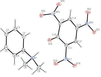
The asymmetric unit of (I) contains one N,N-Dimethylanilinium cation, and one 2,4,6-trinitrophenolate anion. (Fig.1).
The crystal structure of (I) is stabilized by N—H···O and C—H···O interactions. The range of H···O distances (Table 1)
found in (I) agrees with those found for N—H···O (Jeffrey, 1997) and C—H···O hydrogen bonds (Desiraju & Steiner,
1999). The N7—H7···O16 and N7—H7···O19 interactions form a pair of bifurcated donor bonds that link the N,N
-di-methylanilinium cation and 2,4,6-trinitrophenolate anion and also form a motif of graph set R2
1(6) (Bernstein et al., 1995;
Etter, 1990). Another pair of bifurcated donor bonds consists of the C2—H2···O16 and C2—H2···O24 interactions that
also link the cation and the anion and form a R2
1(6) motif. The C8—H8A···O19 and C9—H9C···O19 interactions
constitute a pair of bifurcated bonds forming a R1
2(6) motif that link the cation and the anion. The N7—H7···O19 and C8
—H8A···O19 interactions constitute a pair of bifurcated acceptor bonds that form a R1
2(5) motif. The above two motifs,
R1
2(6) and R12(5), together form a R12(5) motif by the interplay of the trifurcated acceptor bonds formed by N7—
H7···O19, C9—H8A···O19 and C9—H9C···O19 interactions. There are five intermolecular C—H···O interactions (Table
1) that contribute to the supramolecular aggregation of the title compound. The intramolecular N—H···O interactions
mentioned above also contribute to the formation of cooperative H-bonded network (Fig. 2). There is an offset π···π
stacking interaction, Cg1···Cg2 (x, -1+y, z) at 3.697Å with α = 3.19, β = 24.88 and γ = 24.00 and the two perpendicular
distances being 3.377 and 3.354Å. There is also an edge to face π···π stacking interaction, Cg1···Cg2 (1.5-x, -0.5+y,
The interplay of strong N—H···O and weak C—H···O, π···π interactions with different strengths, directional preferences
and distance presents a complex mosaic of interactions. The three dimensional arrangement of the 2,4,6-trinitrophenolate
and N,N-dimethylanilinium moieties in the unit cell, shows that the title compound is an internally linked hydrogen
bonded ion pair and hence can be regarded as a molecular crystal rather than a salt.
S2. Experimental
2,4,6-Trinitrophenol (5.2 mmol) dissolved in aqueous ethanol (25 ml) was added dropwise to N,N-dimethylaniline (5.7
mmol) in aqueous ethanol (25 ml). The above solution was constantly stirred at room temperature for 2 hrs. The
precipitated product was filtered and recrystallized from aqueous ethanol. Yield 75% (3.9 mmol).
S3. Refinement
All H-atoms were located in difference maps and their positions and isotropic displacement parameters freely refined.
The 1309 Friedel pairs (96.2% coverage) were not merged, and the absolute structure was determined by refinement of
[image:4.610.131.486.292.562.2]the Flack (1983) parameter.
Figure 1
The asymmetric unit of (I) with the atoms labelled and displacement ellipsoids depicted at the 50% probability level for
supporting information
sup-3
[image:5.610.127.487.66.457.2]Acta Cryst. (2009). E65, o111–o112
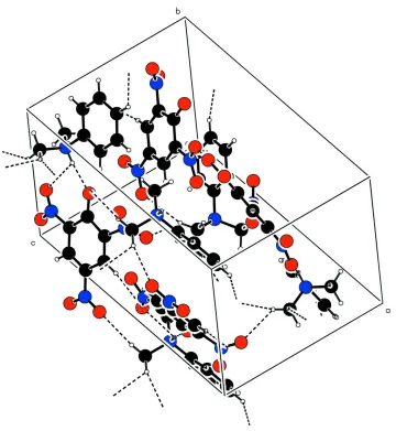
Figure 2
The molecular packing viewed down the b-axis. Dashed lines represent the N—H···O and C—H···O interactions within
the lattice.
N,N-Dimethylanilinium 2,4,6-trinitrophenolate
Crystal data
C8H12N+·C6H2N3O7−
Mr = 350.29
Orthorhombic, Pna21
Hall symbol: P 2c -2n
a = 15.996 (1) Å
b = 9.1491 (6) Å
c = 10.3899 (9) Å
V = 1520.55 (19) Å3
Z = 4
F(000) = 728
Dx = 1.530 Mg m−3
Melting point: 401 K
Cu Kα radiation, λ = 1.54178 Å Cell parameters from 9564 reflections
θ = 5.5–70.2°
µ = 1.08 mm−1
T = 90 K Plate, yellow
Bruker Kappa APEXII CCD area-detector diffractometer
Radiation source: fine-focus sealed tube Graphite monochromator
φ and ω scans
Absorption correction: multi-scan (SADABS; Sheldrick, 1996)
Tmin = 0.767, Tmax = 0.919
16980 measured reflections 2823 independent reflections 2755 reflections with I > 2σ(I)
Rint = 0.033
θmax = 70.2°, θmin = 5.6°
h = −19→19
k = −10→10
l = −12→12
Refinement
Refinement on F2
Least-squares matrix: full
R[F2 > 2σ(F2)] = 0.023
wR(F2) = 0.060
S = 1.03 2823 reflections 283 parameters 1 restraint
Primary atom site location: structure-invariant direct methods
Secondary atom site location: difference Fourier map
Hydrogen site location: inferred from neighbouring sites
H atoms treated by a mixture of independent and constrained refinement
w = 1/[σ2(F
o2) + (0.0386P)2 + 0.2146P]
where P = (Fo2 + 2Fc2)/3
(Δ/σ)max < 0.001
Δρmax = 0.14 e Å−3
Δρmin = −0.16 e Å−3
Absolute structure: Flack (1983), 1309 Friedel pairs
Absolute structure parameter: 0.07 (12)
Special details
Geometry. All e.s.d.'s (except the e.s.d. in the dihedral angle between two l.s. planes) are estimated using the full covariance matrix. The cell e.s.d.'s are taken into account individually in the estimation of e.s.d.'s in distances, angles and torsion angles; correlations between e.s.d.'s in cell parameters are only used when they are defined by crystal symmetry. An approximate (isotropic) treatment of cell e.s.d.'s is used for estimating e.s.d.'s involving l.s. planes.
Refinement. Refinement of F2 against ALL reflections. The weighted R-factor wR and goodness of fit S are based on F2,
conventional R-factors R are based on F, with F set to zero for negative F2. The threshold expression of F2 > σ(F2) is used
only for calculating R-factors(gt) etc. and is not relevant to the choice of reflections for refinement. R-factors based on F2
are statistically about twice as large as those based on F, and R- factors based on ALL data will be even larger.
Fractional atomic coordinates and isotropic or equivalent isotropic displacement parameters (Å2)
x y z Uiso*/Ueq
C1 0.37023 (8) 1.31731 (14) 0.30638 (13) 0.0151 (3)
C2 0.28455 (9) 1.29778 (15) 0.30264 (13) 0.0165 (3)
C3 0.23581 (8) 1.40616 (15) 0.24585 (13) 0.0178 (3)
C4 0.27257 (10) 1.53064 (15) 0.19438 (13) 0.0195 (3)
C5 0.35871 (9) 1.54844 (15) 0.20120 (14) 0.0200 (3)
C6 0.40878 (9) 1.44179 (15) 0.25752 (13) 0.0180 (3)
N7 0.42143 (7) 1.20264 (12) 0.37028 (12) 0.0150 (2)
C8 0.49639 (9) 1.15827 (16) 0.29319 (15) 0.0212 (3)
C9 0.44558 (8) 1.25020 (15) 0.50295 (14) 0.0200 (3)
C10 0.28722 (8) 0.85997 (14) 0.41862 (13) 0.0141 (3)
C11 0.34012 (7) 0.75029 (14) 0.47878 (12) 0.0142 (2)
C12 0.31105 (8) 0.62014 (14) 0.52863 (12) 0.0141 (3)
C13 0.22695 (8) 0.58924 (13) 0.52313 (13) 0.0149 (3)
C14 0.17059 (8) 0.68496 (14) 0.46412 (12) 0.0146 (3)
supporting information
sup-5
Acta Cryst. (2009). E65, o111–o112
O16 0.31146 (5) 0.97691 (10) 0.36915 (10) 0.0183 (2)
N17 0.42940 (7) 0.77360 (12) 0.48867 (11) 0.0160 (2)
O18 0.47489 (6) 0.66722 (10) 0.50973 (10) 0.0207 (2)
O19 0.45735 (6) 0.89878 (10) 0.47807 (11) 0.0247 (2)
N20 0.19719 (7) 0.45202 (12) 0.57365 (11) 0.0167 (2)
O21 0.24919 (6) 0.36384 (10) 0.61486 (10) 0.0194 (2)
O22 0.12141 (6) 0.42767 (12) 0.57227 (11) 0.0267 (2)
N23 0.13932 (7) 0.90790 (12) 0.35088 (11) 0.0162 (2)
O24 0.14260 (6) 1.04028 (10) 0.36928 (12) 0.0240 (2)
O25 0.08701 (6) 0.84815 (11) 0.28202 (9) 0.0207 (2)
H2 0.2611 (10) 1.2148 (19) 0.3398 (17) 0.017 (4)*
H3 0.1778 (12) 1.3963 (19) 0.2403 (18) 0.023 (4)*
H4 0.2380 (11) 1.600 (2) 0.1572 (16) 0.016 (4)*
H5 0.3856 (11) 1.633 (2) 0.1698 (17) 0.023 (4)*
H6 0.4678 (11) 1.4531 (17) 0.2608 (17) 0.017 (4)*
H7 0.3883 (10) 1.1243 (18) 0.3740 (17) 0.018 (4)*
H8A 0.5236 (11) 1.0734 (18) 0.3373 (18) 0.025 (4)*
H8B 0.5363 (12) 1.2445 (19) 0.2860 (19) 0.028 (5)*
H8C 0.4773 (12) 1.125 (2) 0.208 (2) 0.036 (5)*
H9A 0.4780 (10) 1.3372 (18) 0.4959 (17) 0.017 (4)*
H9B 0.3966 (11) 1.2756 (18) 0.5538 (18) 0.022 (4)*
H9C 0.4760 (11) 1.1692 (19) 0.5467 (17) 0.020 (4)*
H12 0.3479 (11) 0.5528 (18) 0.5656 (17) 0.017 (4)*
H14 0.1134 (11) 0.6615 (17) 0.4590 (17) 0.017 (4)*
Atomic displacement parameters (Å2)
U11 U22 U33 U12 U13 U23
C1 0.0177 (6) 0.0136 (6) 0.0140 (6) 0.0022 (5) −0.0007 (5) −0.0020 (5)
C2 0.0197 (6) 0.0135 (6) 0.0161 (6) −0.0011 (5) 0.0012 (5) −0.0010 (5)
C3 0.0172 (6) 0.0170 (6) 0.0193 (7) 0.0013 (5) −0.0022 (5) −0.0039 (5)
C4 0.0281 (7) 0.0146 (6) 0.0158 (6) 0.0041 (6) −0.0028 (5) −0.0002 (5)
C5 0.0286 (7) 0.0137 (7) 0.0175 (7) −0.0029 (5) 0.0024 (6) −0.0003 (5)
C6 0.0198 (7) 0.0166 (7) 0.0175 (7) −0.0021 (5) 0.0024 (5) −0.0017 (5)
N7 0.0142 (5) 0.0132 (5) 0.0174 (5) −0.0006 (4) 0.0011 (4) −0.0003 (5)
C8 0.0193 (6) 0.0192 (7) 0.0251 (7) 0.0028 (5) 0.0058 (6) 0.0008 (6)
C9 0.0214 (6) 0.0200 (7) 0.0187 (7) 0.0008 (6) −0.0046 (6) −0.0016 (6)
C10 0.0146 (6) 0.0140 (6) 0.0137 (6) −0.0002 (5) −0.0013 (5) −0.0026 (5)
C11 0.0132 (6) 0.0156 (6) 0.0138 (6) 0.0004 (5) 0.0006 (5) −0.0023 (5)
C12 0.0161 (6) 0.0136 (6) 0.0127 (6) 0.0030 (5) −0.0003 (5) −0.0009 (5)
C13 0.0170 (6) 0.0120 (6) 0.0155 (6) 0.0002 (5) 0.0001 (5) 0.0005 (5)
C14 0.0123 (6) 0.0162 (6) 0.0152 (6) 0.0002 (5) 0.0008 (5) −0.0023 (5)
C15 0.0149 (6) 0.0140 (6) 0.0145 (6) 0.0031 (5) −0.0007 (5) 0.0000 (5)
O16 0.0177 (4) 0.0152 (5) 0.0219 (5) −0.0020 (3) −0.0016 (4) 0.0040 (4)
N17 0.0142 (5) 0.0177 (5) 0.0162 (5) −0.0005 (4) −0.0008 (4) 0.0008 (5)
O18 0.0143 (4) 0.0205 (5) 0.0274 (5) 0.0040 (4) −0.0012 (4) 0.0025 (4)
O19 0.0181 (5) 0.0180 (5) 0.0378 (6) −0.0050 (4) −0.0061 (4) 0.0062 (5)
O22 0.0146 (5) 0.0267 (5) 0.0389 (6) −0.0060 (4) −0.0035 (4) 0.0098 (5)
N23 0.0135 (5) 0.0193 (6) 0.0156 (5) 0.0017 (4) 0.0009 (4) 0.0034 (5)
O24 0.0221 (5) 0.0140 (5) 0.0360 (6) 0.0036 (4) −0.0007 (5) 0.0041 (4)
O25 0.0162 (4) 0.0271 (5) 0.0188 (5) 0.0020 (4) −0.0047 (4) 0.0004 (4)
Geometric parameters (Å, º)
C1—C2 1.3826 (19) C9—H9C 0.997 (18)
C1—C6 1.3910 (19) C10—O16 1.2488 (17)
C1—N7 1.4874 (16) C10—C15 1.4537 (18)
C2—C3 1.3926 (19) C10—C11 1.4538 (18)
C2—H2 0.931 (18) C11—C12 1.3794 (19)
C3—C4 1.389 (2) C11—N17 1.4475 (16)
C3—H3 0.934 (18) C12—C13 1.3758 (18)
C4—C5 1.389 (2) C12—H12 0.935 (18)
C4—H4 0.924 (19) C13—C14 1.3985 (18)
C5—C6 1.391 (2) C13—N20 1.4417 (17)
C5—H5 0.942 (19) C14—C15 1.3661 (18)
C6—H6 0.951 (18) C14—H14 0.941 (18)
N7—C9 1.4962 (18) C15—N23 1.4652 (16)
N7—C8 1.4980 (18) N17—O19 1.2344 (14)
N7—H7 0.892 (17) N17—O18 1.2348 (14)
C8—H8A 1.001 (18) N20—O22 1.2326 (15)
C8—H8B 1.017 (18) N20—O21 1.2353 (15)
C8—H8C 0.99 (2) N23—O24 1.2273 (15)
C9—H9A 0.953 (17) N23—O25 1.2291 (15)
C9—H9B 0.974 (19)
C2—C1—C6 122.34 (12) H9A—C9—H9B 106.3 (14)
C2—C1—N7 117.85 (11) N7—C9—H9C 109.3 (10)
C6—C1—N7 119.76 (11) H9A—C9—H9C 113.0 (14)
C1—C2—C3 118.35 (12) H9B—C9—H9C 108.9 (14)
C1—C2—H2 119.5 (10) O16—C10—C15 122.53 (12)
C3—C2—H2 122.1 (10) O16—C10—C11 125.98 (11)
C4—C3—C2 120.66 (13) C15—C10—C11 111.39 (11)
C4—C3—H3 118.4 (11) C12—C11—N17 115.67 (11)
C2—C3—H3 120.9 (11) C12—C11—C10 124.13 (11)
C3—C4—C5 119.77 (13) N17—C11—C10 120.21 (11)
C3—C4—H4 117.9 (11) C13—C12—C11 119.44 (12)
C5—C4—H4 122.3 (11) C13—C12—H12 119.8 (10)
C4—C5—C6 120.67 (13) C11—C12—H12 120.7 (10)
C4—C5—H5 122.1 (11) C12—C13—C14 121.33 (12)
C6—C5—H5 117.2 (11) C12—C13—N20 119.13 (11)
C1—C6—C5 118.20 (13) C14—C13—N20 119.47 (11)
C1—C6—H6 121.1 (10) C15—C14—C13 118.50 (11)
C5—C6—H6 120.7 (10) C15—C14—H14 121.0 (10)
supporting information
sup-7
Acta Cryst. (2009). E65, o111–o112
C1—N7—C8 113.16 (11) C14—C15—C10 125.18 (12)
C9—N7—C8 111.39 (11) C14—C15—N23 116.42 (11)
C1—N7—H7 105.0 (10) C10—C15—N23 118.37 (11)
C9—N7—H7 110.3 (12) O19—N17—O18 122.24 (10)
C8—N7—H7 106.3 (11) O19—N17—C11 119.19 (10)
N7—C8—H8A 108.2 (10) O18—N17—C11 118.55 (10)
N7—C8—H8B 109.3 (11) O22—N20—O21 123.25 (11)
H8A—C8—H8B 111.3 (14) O22—N20—C13 118.55 (11)
N7—C8—H8C 108.4 (12) O21—N20—C13 118.20 (10)
H8A—C8—H8C 108.1 (15) O24—N23—O25 123.96 (11)
H8B—C8—H8C 111.3 (16) O24—N23—C15 118.88 (11)
N7—C9—H9A 108.2 (11) O25—N23—C15 117.15 (11)
N7—C9—H9B 111.2 (11)
C6—C1—C2—C3 1.1 (2) C12—C13—C14—C15 −2.10 (19)
N7—C1—C2—C3 178.45 (12) N20—C13—C14—C15 −178.99 (12)
C1—C2—C3—C4 0.0 (2) C13—C14—C15—C10 0.2 (2)
C2—C3—C4—C5 −0.9 (2) C13—C14—C15—N23 178.23 (11)
C3—C4—C5—C6 0.9 (2) O16—C10—C15—C14 177.99 (13)
C2—C1—C6—C5 −1.1 (2) C11—C10—C15—C14 1.40 (19)
N7—C1—C6—C5 −178.45 (12) O16—C10—C15—N23 0.00 (19)
C4—C5—C6—C1 0.1 (2) C11—C10—C15—N23 −176.58 (11)
C2—C1—N7—C9 −100.90 (13) C12—C11—N17—O19 −161.31 (13)
C6—C1—N7—C9 76.55 (15) C10—C11—N17—O19 19.03 (18)
C2—C1—N7—C8 133.51 (13) C12—C11—N17—O18 17.45 (17)
C6—C1—N7—C8 −49.04 (16) C10—C11—N17—O18 −162.20 (12)
O16—C10—C11—C12 −177.78 (13) C12—C13—N20—O22 177.52 (12)
C15—C10—C11—C12 −1.34 (18) C14—C13—N20—O22 −5.53 (19)
O16—C10—C11—N17 1.8 (2) C12—C13—N20—O21 −3.32 (18)
C15—C10—C11—N17 178.28 (11) C14—C13—N20—O21 173.63 (12)
N17—C11—C12—C13 −179.98 (11) C14—C15—N23—O24 138.87 (13)
C10—C11—C12—C13 −0.3 (2) C10—C15—N23—O24 −42.97 (17)
C11—C12—C13—C14 2.17 (19) C14—C15—N23—O25 −40.50 (17)
C11—C12—C13—N20 179.06 (12) C10—C15—N23—O25 137.66 (12)
Hydrogen-bond geometry (Å, º)
D—H···A D—H H···A D···A D—H···A
N7—H7···O16 0.892 (17) 1.825 (18) 2.7128 (14) 172.8 (16)
N7—H7···O19 0.892 (17) 2.578 (16) 3.0517 (15) 114.0 (12)
C2—H2···O16 0.931 (18) 2.341 (17) 3.0464 (16) 132.4 (13)
C2—H2···O24 0.931 (18) 2.498 (17) 3.3444 (17) 151.4 (14)
C8—H8A···O19 1.001 (18) 2.411 (18) 3.1171 (18) 126.9 (13)
C9—H9C···O19 0.997 (18) 2.592 (17) 3.2311 (17) 121.9 (12)
C9—H9B···O21i 0.974 (19) 2.571 (19) 3.5074 (17) 161.3 (14)
C9—H9B···O25ii 0.974 (19) 2.476 (18) 3.0794 (18) 119.9 (13)
C9—H9C···O22v 0.997 (18) 2.502 (18) 3.3283 (16) 140.0 (13)
![Methyl 3 [(6 nitro 4 oxo 3 phenyl 3,4 dihydroquinazolin 2 yl)sulfanyl]propanoate](data:image/gif;base64,R0lGODlhAQABAIAAAP///wAAACH5BAEAAAAALAAAAAABAAEAAAICRAEAOw==)