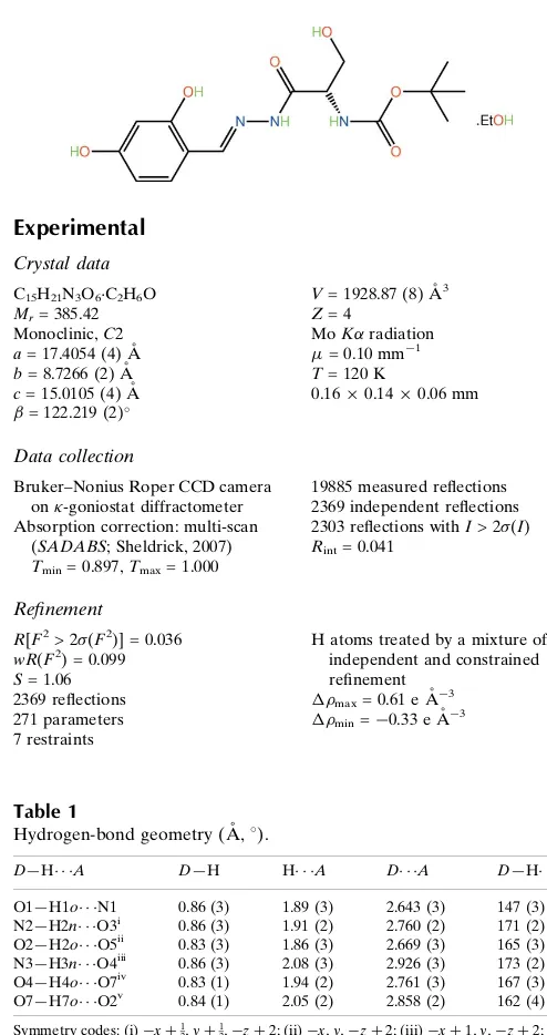
tert
-Butyl
N
-{(1
S
)-1-[(2,4-dihydroxy-
benzylidene)hydrazinecarbonyl]-2-hydroxyethyl}carbamate ethanol
monosolvate
Alessandra C. Pinheiro,aMarcus V. N. de Souza,a Edward R. T. Tiekink,b* Solange M. S. V. Wardellcand James L. Wardelld‡
a
Fundac¸a˜o Oswaldo Cruz, Instituto de Tecnologia em Fa´rmacos – Farmanguinhos, R. Sizenando Nabuco, 100, Manguinhos, 21041-250, Rio de Janeiro, RJ, Brazil, b
Department of Chemistry, University of Malaya, 50603 Kuala Lumpur, Malaysia, c
CHEMSOL, 1 Harcourt Road, Aberdeen AB15 5NY, Scotland, anddCentro de Desenvolvimento Tecnolo´gico em Sau´de (CDTS), Fundac¸a˜o Oswaldo Cruz (FIOCRUZ), Casa Amarela, Campus de Manguinhos, Av. Brasil 4365, 21040-900 Rio de Janeiro, RJ, Brazil
Correspondence e-mail: edward.tiekink@gmail.com
Received 27 January 2011; accepted 30 January 2011
Key indicators: single-crystal X-ray study;T= 120 K; mean(C–C) = 0.004 A˚; disorder in solvent or counterion;Rfactor = 0.036;wRfactor = 0.099; data-to-parameter ratio = 8.7.
The molecule of the title ethanol solvate, C15H21N3O6C2H6O,
adopts a curved shape; the conformation about the imine bond [N N = 1.287 (3) A˚ ] is E. The amide residues occupy positions almost orthogonal to each other [dihedral angle = 85.7 (2)]. In the crystal, a network of O—H O, O—H N
and N—H O hydrogen bonds leads to the formation of supramolecular arrays in the ab plane with the ethanol molecules lying to the periphery on either side. Disorder in the solvent ethanol molecule was evident with two positions being resolved for the C atoms [site occupancy of the major component = 0.612 (10)].
Related literature
For background to the use of l-serine derivatives in
anti-tumour therapy, see: Jiao et al.(2009); Yakura et al.(2007); Takahashiet al.(1988); Sinet al.(1998). For background toN -acylhydrazone derivatives from l-serine for anti-tumour
testing, see: Rollas & Ku¨c¸u¨kgu¨zel (2007); Terziog˘lu & Gu¨rsoy (2003). For related structures, see: Pinheiro et al. (2010); de Souzaet al.(2010).
Experimental
Crystal data
C15H21N3O6C2H6O
Mr= 385.42
Monoclinic,C2 a= 17.4054 (4) A˚ b= 8.7266 (2) A˚ c= 15.0105 (4) A˚ = 122.219 (2)
V= 1928.87 (8) A˚3
Z= 4
MoKradiation = 0.10 mm1 T= 120 K
0.160.140.06 mm
Data collection
Bruker–Nonius Roper CCD camera on-goniostat diffractometer Absorption correction: multi-scan
(SADABS; Sheldrick, 2007) Tmin= 0.897,Tmax= 1.000
19885 measured reflections 2369 independent reflections 2303 reflections withI> 2(I) Rint= 0.041
Refinement
R[F2> 2(F2)] = 0.036
wR(F2) = 0.099 S= 1.06 2369 reflections 271 parameters 7 restraints
H atoms treated by a mixture of independent and constrained refinement
max= 0.61 e A˚
3
min=0.33 e A˚
[image:1.610.308.555.74.541.2]3
Table 1
Hydrogen-bond geometry (A˚ ,).
D—H A D—H H A D A D—H A
O1—H1o N1 0.86 (3) 1.89 (3) 2.643 (3) 147 (3) N2—H2n O3i
0.86 (3) 1.91 (2) 2.760 (2) 171 (2) O2—H2o O5ii
0.83 (3) 1.86 (3) 2.669 (3) 165 (3) N3—H3n O4iii
0.86 (3) 2.08 (3) 2.926 (3) 173 (2) O4—H4o O7iv
0.83 (1) 1.94 (2) 2.761 (3) 167 (3) O7—H7o O2v
0.84 (1) 2.05 (2) 2.858 (2) 162 (4)
Symmetry codes: (i)xþ1 2;yþ
1
2;zþ2; (ii)x;y;zþ2; (iii)xþ1;y;zþ2; (iv)
xþ1 2;yþ
1
2;zþ1; (v)x;y;zþ1.
Data collection: COLLECT (Hooft, 1998); cell refinement:
DENZO(Otwinowski & Minor, 1997) andCOLLECT; data reduc-tion:DENZO andCOLLECT; program(s) used to solve structure:
SHELXS97(Sheldrick, 2008); program(s) used to refine structure:
SHELXL97 (Sheldrick, 2008); molecular graphics: ORTEP-3
(Farrugia, 1997) andDIAMOND(Brandenburg, 2006); software used to prepare material for publication:publCIF(Westrip, 2010).
The use of the EPSRC X-ray crystallographic service at the University of Southampton, England, and the valuable assis-tance of the staff there is gratefully acknowledged. JLW acknowledges support from CAPES (Brazil).
Supplementary data and figures for this paper are available from the IUCr electronic archives (Reference: HB5793).
organic compounds
Acta Cryst.(2011). E67, o581–o582 doi:10.1107/S1600536811003795 Pinheiroet al.
o581
Acta Crystallographica Section E Structure Reports
Online
ISSN 1600-5368
Brandenburg, K. (2006).DIAMOND. Crystal Impact GbR, Bonn, Germany. Farrugia, L. J. (1997).J. Appl. Cryst.30, 565.
Hooft, R. W. W. (1998).COLLECT. Nonius BV, Delft, The Netherlands. Jiao, X., Wang, L., Xiao, Q., Xie, P. & Liang, X. (2009).J. Asian Nat. Prod. Res.
11, 274–280.
Otwinowski, Z. & Minor, W. (1997). Methods in Enzymology, Vol. 276, Macromolecular Crystallography, Part A, edited by C. W. Carter Jr & R. M. Sweet, pp. 307–326. New York: Academic Press.
Pinheiro, A. C., de Souza, M. V. N., Tiekink, E. R. T., Wardell, J. L. & Wardell, S. M. S. V. (2010).Acta Cryst.E66, o1004–o1005.
Rollas, S. & Ku¨c¸u¨kgu¨zel, S. G. (2007).Molecules,12, 1910–1939.
Sheldrick, G. M. (2007).SADABS. Bruker AXS Inc., Madison, Wisconsin, USA.
Sin, N., Meng, L., Auth, H. & Crews, C. M. (1998).Bioorg. Med. Chem.6, 1209–1217.
Souza, M. V. N. de, Pinheiro, A. C., Tiekink, E. R. T., Wardell, S. M. S. V. & Wardell, J. L. (2010).Acta Cryst.E66, o3253–o3254.
Takahashi, A., Nakamura, H., Ikeda, D., Naganawa, H., Kameyama, T., Kurasawa, S., Okami, Y., Takeuchi, T. & Iitaka, Y. (1988).J. Antibiot.41, 1568–1574.
Terziog˘lu, N. & Gu¨rsoy, A. (2003).Eur. J. Med. Chem.38, 633–643. Westrip, S. P. (2010).J. Appl. Cryst.43, 920–925.
supporting information
sup-1
Acta Cryst. (2011). E67, o581–o582
supporting information
Acta Cryst. (2011). E67, o581–o582 [doi:10.1107/S1600536811003795]
tert
-Butyl
N
-{(1
S
)-1-[(2,4-dihydroxybenzylidene)hydrazinecarbonyl]-2-hydroxy-ethyl}carbamate ethanol monosolvate
Alessandra C. Pinheiro, Marcus V. N. de Souza, Edward R. T. Tiekink, Solange M. S. V. Wardell
and James L. Wardell
S1. Comment
Several L-serine derivatives have been found to have potential in anti-tumour therapy, for example, conagenin, a naturally
occurring serine derivative, was shown to improve the anti-tumour efficacy of adriamycin and mitomycin C against
murine leukemias (Jiao et al., 2009; Yakura et al., 2007). Other L-serine derivatives reported as potential new anti-tumour
agents include the antibiotic thrazarine, which sensitizes tumour cells to macrophage-mediated cytolysis (Takahashi et al.,
1988), and eponemycin, an immunomodulator, which plays a crucial role in tumour progression and metastases by
supplying essential nutrients to B16 melanoma cells (Sin et al., 1998). Following on from such reports, we have
synthesized some N-acylhydrazones derivatives from L-serine to use in anti-tumour testing. The choice of N
-acyl-hydrazonyl derivatives was suggested by publications indicating that compounds with such groups can aid anti-tumoural
activities (Rollas et al., 2007; Terzioğlu et al., 2003). In continuation of on-going structural studies of these compounds
(Pinheiro et al., 2010; de Souza et al., 2010), we now report the structure of the ethanol solvate of tert-butyl
(1S)-2-[2-(2,4-dihydroxybenzylidene)hydrazino]-1-(hydroxymethyl)-2-oxoethylcarbamate, (I).
Although the absolute structure of (I), Fig. 1, could not be determined experimentally, the assignment of the S
-configuration at the C9 atom is based on a starting reagent. The overall conformation of the molecule is curved with the
major kink occurring at the C9 atom. The dihydroxybenzene ring is slightly twisted out of the plane of the hydrazine
residue with the C2—C1—C7—N1 torsion angle being -8.2 (3) °. The conformation about the N1—C7 imine bond
[1.287 (3) Å] is E. Each of the carbonyl groups is diagonally opposite the amine group and the dihedral angle formed
between the two amide residues is 85.7 (2) °.
As expected with four hydroxyl and two amine donors, there is significant hydrogen bonding operating in the crystal
structure, Table 1. While the O1-hydroxy group forms an intramolecular O–H···N hydrogen bond with the hydrazine-N1
atom, the remaining interactions are intermolecular in nature. The O2-hydroxy group forms an O—H···O hydrogen bond
with the O5-carbonyl, and the O3-hydroxyl group linked to the chiral centre is connected to the ethanol molecule which
in turn forms a hydrogen bond to the O2-hydroxyl group. The N2-amine is connected to the O3-carbonyl and the
N3-amine forms a hydrogen bond with the O4-hydroxyl. The result of the hydrogen bonding is the formation of layers of
molecules in the ab plane sandwiched by ethanol molecules. The layers stack along the c axis, Fig. 2.
S2. Experimental
To a stirred solution of tert-butyl (1S)-2-hydrazino-1-(hydroxymethyl)-2-oxoethylcarbamate (1.0 mmol) in ethanol (10
ml) at room temperature was added 2,4-dihydroxybenzaldehyde (1.05 mmol). The reaction mixture was stirred for 4 h. at
was grown from EtOH. 1H NMR (500 MHz, DMSO-d6) δ (p.p.m.): 11.50 (1H, s, NHN), 11.30 (1H, s), 9.92 (1H, s), 8.30
(1H, s, N=CH), 7.26 (1H, d, J = 8.4 Hz, H6), 6.80 (1H, d, J = 7.7 Hz, NHCH), 6.35–6.30 (1H, m, H5), 6.29 (1H, s, H3),
4.95 (1H, s, OH), 4.02 (1H, m, CH), 3.70–3.50 (2H, m, CH2OH); 1.39 (9H, s, (CH3)3C). 13C NMR (125 MHz, DMSO-d6)
δ (p.p.m.): 170.8, 160.3, 157.9, 155.2, 141.8, 128.0, 110.4, 107.6, 102.3, 78.0, 61.1, 53.9, 28.2. IR (cm-1; KBr): 3200 (O
—H), 1678 (COCH and COO). EM/ESI: [M—H]: 338.3.
S3. Refinement
The C-bound H atoms were geometrically placed (C–H = 0.95–0.99 Å) and refined as riding with Uiso(H) = 1.2–
1.5Ueq(C). The O– and N-bound H atoms were located from a difference map and refined with the distance restraints O–
H = 0.84 ± 0.01 and N–H = 0.86±0.01 Å, and with Uiso(H) = zUeq(carrier atom); z = 1.5 for O and z = 1.2 for N. Disorder
was resolved in the solvent ethanol molecule in that two distinct positions were discerned for the C atoms. From
fractional anisotropic refinement, the major component had a site occupancy factor = 0.612 (10). In the absence of
significant anomalous scattering effects, 2067 Friedel pairs were averaged in the final refinement. However, the absolute
configuration was assigned on the basis of the chirality of the L-serine starting material. The maximum and minimum
residual electron density peaks of 0.61 and 0.33 e Å-3, respectively, were located 0.42 Å and 0.37 Å from the H6 and
[image:4.610.120.483.333.495.2]H16a atoms, respectively.
Figure 1
The molecular structure of (I) showing displacement ellipsoids at the 50% probability level. The ethanol molecule of
supporting information
sup-3
[image:5.610.129.486.71.256.2]Acta Cryst. (2011). E67, o581–o582
Figure 2
A view in projection down the b axis of the stacking of two-dimensional supramolecular arrays in the ab plane in (I) with
the O—H···O and N—H···O hydrogen bonding shown as orange and blue dashed lines, respectively.
tert-Butyl N-{(1S)-1-[(2,4-dihydroxybenzylidene)hydrazinecarbonyl]- 2-hydroxyethyl}carbamate ethanol
monosolvate
Crystal data
C15H21N3O6·C2H6O Mr = 385.42 Monoclinic, C2 Hall symbol: C 2y
a = 17.4054 (4) Å
b = 8.7266 (2) Å
c = 15.0105 (4) Å
β = 122.219 (2)°
V = 1928.87 (8) Å3 Z = 4
F(000) = 824
Dx = 1.327 Mg m−3
Mo Kα radiation, λ = 0.71073 Å Cell parameters from 4327 reflections
θ = 2.9–27.5°
µ = 0.10 mm−1 T = 120 K Block, colourless 0.16 × 0.14 × 0.06 mm
Data collection
Bruker–Nonius Roper CCD camera on κ -goniostat
diffractometer
Radiation source: Bruker-Nonius FR591 rotating anode CCD
Graphite monochromator
Detector resolution: 9.091 pixels mm-1 φ and ω scans
Absorption correction: multi-scan (SADABS; Sheldrick, 2007)
Tmin = 0.897, Tmax = 1.000
19885 measured reflections 2369 independent reflections 2303 reflections with I > 2σ(I)
Rint = 0.041
θmax = 27.5°, θmin = 3.2° h = −22→22
k = −11→11
l = −19→19
Refinement
Refinement on F2
Least-squares matrix: full
R[F2 > 2σ(F2)] = 0.036 wR(F2) = 0.099 S = 1.06 2369 reflections
271 parameters 7 restraints
Primary atom site location: structure-invariant direct methods
neighbouring sites
H atoms treated by a mixture of independent and constrained refinement
where P = (Fo2 + 2Fc2)/3
(Δ/σ)max = 0.001
Δρmax = 0.61 e Å−3
Δρmin = −0.33 e Å−3
Special details
Geometry. All s.u.'s (except the s.u. in the dihedral angle between two l.s. planes) are estimated using the full covariance matrix. The cell s.u.'s are taken into account individually in the estimation of s.u.'s in distances, angles and torsion angles; correlations between s.u.'s in cell parameters are only used when they are defined by crystal symmetry. An approximate (isotropic) treatment of cell s.u.'s is used for estimating s.u.'s involving l.s. planes.
Refinement. Refinement of F2 against ALL reflections. The weighted R-factor wR and goodness of fit S are based on F2,
conventional R-factors R are based on F, with F set to zero for negative F2. The threshold expression of F2 > 2σ(F2) is
used only for calculating R-factors(gt) etc. and is not relevant to the choice of reflections for refinement. R-factors based on F2 are statistically about twice as large as those based on F, and R- factors based on ALL data will be even larger.
Fractional atomic coordinates and isotropic or equivalent isotropic displacement parameters (Å2)
x y z Uiso*/Ueq Occ. (<1)
O1 −0.01014 (11) 0.6314 (2) 0.76103 (14) 0.0306 (4)
H1o 0.0421 (13) 0.674 (4) 0.796 (2) 0.046*
O2 −0.30367 (9) 0.6766 (2) 0.69839 (12) 0.0259 (3)
H2o −0.3330 (19) 0.728 (3) 0.717 (2) 0.039*
O3 0.27544 (10) 0.67257 (19) 0.95473 (12) 0.0251 (3)
O4 0.39535 (10) 0.8716 (2) 0.89717 (11) 0.0266 (3)
H4o 0.3474 (13) 0.853 (4) 0.8402 (14) 0.040*
O5 0.39849 (11) 0.7949 (2) 1.22519 (13) 0.0333 (4)
O6 0.53955 (10) 0.7219 (2) 1.26444 (11) 0.0267 (4)
N1 0.11580 (11) 0.8217 (2) 0.89800 (13) 0.0219 (4)
N2 0.20132 (11) 0.8878 (2) 0.95418 (14) 0.0221 (4)
H2n 0.2041 (18) 0.9793 (16) 0.976 (2) 0.027*
N3 0.44121 (11) 0.8160 (2) 1.10779 (13) 0.0206 (4)
H3n 0.4884 (12) 0.824 (3) 1.104 (2) 0.025*
C1 −0.04087 (13) 0.8468 (3) 0.83868 (15) 0.0209 (4)
C2 −0.06834 (13) 0.7128 (2) 0.77631 (15) 0.0204 (4)
C3 −0.15725 (13) 0.6607 (3) 0.72779 (15) 0.0214 (4)
H3 −0.1759 0.5722 0.6843 0.026*
C4 −0.21852 (13) 0.7390 (3) 0.74340 (15) 0.0205 (4)
C5 −0.19388 (13) 0.8728 (3) 0.80333 (16) 0.0237 (4)
H5 −0.2368 0.9262 0.8124 0.028*
C6 −0.10573 (14) 0.9263 (3) 0.84935 (16) 0.0230 (4)
H6 −0.0888 1.0186 0.8889 0.028*
C7 0.05209 (14) 0.9025 (3) 0.89384 (16) 0.0218 (4)
H7 0.0660 0.9998 0.9273 0.026*
C8 0.27609 (13) 0.8076 (2) 0.97909 (15) 0.0193 (4)
C9 0.36197 (12) 0.9053 (2) 1.03518 (15) 0.0192 (4)
H9 0.3536 0.9876 1.0756 0.023*
C10 0.37737 (13) 0.9809 (3) 0.95365 (16) 0.0232 (4)
supporting information
sup-5
Acta Cryst. (2011). E67, o581–o582
H10B 0.4292 1.0528 0.9900 0.028*
C11 0.45577 (14) 0.7797 (3) 1.20233 (17) 0.0227 (4)
C12 0.57832 (15) 0.6887 (3) 1.37713 (16) 0.0303 (5)
C13 0.52980 (18) 0.5534 (4) 1.3883 (2) 0.0379 (6)
H13A 0.4669 0.5818 1.3629 0.057*
H13B 0.5608 0.5234 1.4626 0.057*
H13C 0.5302 0.4672 1.3467 0.057*
C14 0.5758 (2) 0.8318 (4) 1.4328 (2) 0.0445 (7)
H14A 0.5988 0.9190 1.4126 0.067*
H14B 0.6139 0.8166 1.5092 0.067*
H14C 0.5132 0.8521 1.4127 0.067*
C15 0.67546 (16) 0.6455 (4) 1.41358 (19) 0.0429 (7)
H15A 0.6751 0.5548 1.3748 0.064*
H15B 0.7093 0.6227 1.4891 0.064*
H15C 0.7045 0.7309 1.4003 0.064*
O7 0.27404 (12) 0.3530 (2) 0.28565 (16) 0.0403 (4)
H7o 0.292 (3) 0.443 (2) 0.304 (3) 0.060*
C16 0.3454 (10) 0.245 (2) 0.3050 (12) 0.0525 (8) 0.612 (10)
H16A 0.3995 0.3029 0.3182 0.063* 0.612 (10)
H16B 0.3241 0.1816 0.2413 0.063* 0.612 (10)
C17 0.3713 (5) 0.1452 (9) 0.3940 (6) 0.0495 (14) 0.612 (10)
H17A 0.3926 0.2073 0.4574 0.074* 0.612 (10)
H17B 0.4200 0.0765 0.4046 0.074* 0.612 (10)
H17C 0.3186 0.0845 0.3801 0.074* 0.612 (10)
C18 0.3420 (13) 0.249 (3) 0.3037 (18) 0.0525 (8) 0.388 (10)
H18A 0.3795 0.2940 0.2789 0.063* 0.388 (10)
H18B 0.3133 0.1547 0.2621 0.063* 0.388 (10)
C19 0.4010 (8) 0.2074 (14) 0.4154 (9) 0.0495 (14) 0.388 (10)
H19A 0.4427 0.2922 0.4538 0.074* 0.388 (10)
H19B 0.4360 0.1155 0.4219 0.074* 0.388 (10)
H19C 0.3637 0.1869 0.4450 0.074* 0.388 (10)
Atomic displacement parameters (Å2)
U11 U22 U33 U12 U13 U23
O1 0.0239 (7) 0.0322 (9) 0.0381 (9) −0.0014 (7) 0.0182 (7) −0.0059 (7) O2 0.0155 (7) 0.0351 (9) 0.0283 (8) −0.0018 (6) 0.0124 (6) −0.0029 (7) O3 0.0231 (7) 0.0197 (7) 0.0301 (7) −0.0008 (6) 0.0126 (6) −0.0001 (6)
O4 0.0205 (7) 0.0384 (9) 0.0223 (7) 0.0012 (7) 0.0123 (6) 0.0005 (7)
O5 0.0274 (8) 0.0464 (10) 0.0352 (8) 0.0105 (8) 0.0227 (7) 0.0136 (8) O6 0.0211 (7) 0.0410 (10) 0.0188 (7) 0.0088 (7) 0.0111 (6) 0.0070 (7) N1 0.0152 (7) 0.0239 (9) 0.0251 (8) −0.0026 (7) 0.0097 (7) −0.0006 (7) N2 0.0169 (8) 0.0203 (9) 0.0271 (8) −0.0025 (7) 0.0104 (7) −0.0017 (7)
N3 0.0154 (7) 0.0261 (9) 0.0205 (8) 0.0034 (7) 0.0097 (6) 0.0031 (7)
C6 0.0218 (9) 0.0245 (10) 0.0247 (9) 0.0009 (8) 0.0137 (8) −0.0004 (8) C7 0.0199 (9) 0.0218 (10) 0.0231 (9) −0.0018 (8) 0.0109 (8) 0.0000 (8) C8 0.0184 (9) 0.0201 (10) 0.0200 (8) 0.0001 (8) 0.0106 (7) 0.0035 (7)
C9 0.0158 (8) 0.0190 (9) 0.0216 (8) 0.0009 (7) 0.0091 (7) 0.0003 (7)
C10 0.0175 (9) 0.0248 (10) 0.0265 (10) −0.0001 (8) 0.0113 (8) 0.0034 (8) C11 0.0200 (9) 0.0255 (10) 0.0230 (9) 0.0020 (8) 0.0116 (8) 0.0028 (8) C12 0.0291 (11) 0.0417 (13) 0.0195 (9) 0.0073 (10) 0.0127 (8) 0.0072 (9) C13 0.0358 (12) 0.0448 (15) 0.0369 (13) 0.0103 (11) 0.0219 (11) 0.0141 (11) C14 0.0570 (17) 0.0470 (16) 0.0246 (11) 0.0099 (14) 0.0184 (12) 0.0012 (11) C15 0.0255 (11) 0.070 (2) 0.0263 (11) 0.0118 (12) 0.0096 (9) 0.0146 (12) O7 0.0275 (8) 0.0350 (10) 0.0458 (10) 0.0023 (8) 0.0110 (8) 0.0098 (9) C16 0.0547 (19) 0.0421 (17) 0.0537 (18) 0.0098 (15) 0.0241 (16) 0.0009 (14)
C17 0.047 (3) 0.045 (4) 0.054 (3) 0.014 (2) 0.025 (3) 0.012 (3)
C18 0.0547 (19) 0.0421 (17) 0.0537 (18) 0.0098 (15) 0.0241 (16) 0.0009 (14)
C19 0.047 (3) 0.045 (4) 0.054 (3) 0.014 (2) 0.025 (3) 0.012 (3)
Geometric parameters (Å, º)
O1—C2 1.353 (3) C9—H9 1.0000
O1—H1o 0.86 (3) C10—H10A 0.9900
O2—C4 1.372 (2) C10—H10B 0.9900
O2—H2o 0.83 (3) C12—C13 1.512 (4)
O3—C8 1.232 (3) C12—C14 1.516 (4)
O4—C10 1.417 (3) C12—C15 1.521 (3)
O4—H4o 0.833 (10) C13—H13A 0.9800
O5—C11 1.222 (3) C13—H13B 0.9800
O6—C11 1.342 (2) C13—H13C 0.9800
O6—C12 1.478 (2) C14—H14A 0.9800
N1—C7 1.287 (3) C14—H14B 0.9800
N1—N2 1.386 (2) C14—H14C 0.9800
N2—C8 1.343 (3) C15—H15A 0.9800
N2—H2n 0.855 (10) C15—H15B 0.9800
N3—C11 1.340 (3) C15—H15C 0.9800
N3—C9 1.447 (2) O7—C18 1.400 (9)
N3—H3n 0.86 (3) O7—C16 1.460 (8)
C1—C6 1.405 (3) O7—H7O 0.842 (10)
C1—C2 1.412 (3) C16—C17 1.450 (12)
C1—C7 1.453 (3) C16—H16A 0.9900
C2—C3 1.389 (3) C16—H16B 0.9900
C3—C4 1.388 (3) C17—H17A 0.9800
C3—H3 0.9500 C17—H17B 0.9800
C4—C5 1.394 (3) C17—H17C 0.9800
C5—C6 1.385 (3) C18—C19 1.47 (2)
C5—H5 0.9500 C18—H18A 0.9900
C6—H6 0.9500 C18—H18B 0.9900
C7—H7 0.9500 C19—H19A 0.9800
supporting information
sup-7
Acta Cryst. (2011). E67, o581–o582
C9—C10 1.535 (3) C19—H19C 0.9800
C2—O1—H1o 109 (3) O5—C11—N3 123.4 (2)
C4—O2—H2o 108 (2) O6—C11—N3 110.28 (17)
C10—O4—H4o 109 (2) O6—C12—C13 109.8 (2)
C11—O6—C12 122.20 (16) O6—C12—C14 109.9 (2)
C7—N1—N2 114.81 (18) C13—C12—C14 113.6 (2)
C8—N2—N1 121.30 (18) O6—C12—C15 101.66 (17)
C8—N2—H2n 122.1 (18) C13—C12—C15 110.2 (2)
N1—N2—H2n 116.4 (18) C14—C12—C15 111.0 (2)
C11—N3—C9 119.41 (16) C12—C13—H13A 109.5
C11—N3—H3n 116.2 (18) C12—C13—H13B 109.5
C9—N3—H3n 118.0 (19) H13A—C13—H13B 109.5
C6—C1—C2 118.39 (18) C12—C13—H13C 109.5
C6—C1—C7 119.2 (2) H13A—C13—H13C 109.5
C2—C1—C7 122.35 (18) H13B—C13—H13C 109.5
O1—C2—C3 117.67 (19) C12—C14—H14A 109.5
O1—C2—C1 121.84 (18) C12—C14—H14B 109.5
C3—C2—C1 120.49 (18) H14A—C14—H14B 109.5
C2—C3—C4 119.5 (2) C12—C14—H14C 109.5
C2—C3—H3 120.3 H14A—C14—H14C 109.5
C4—C3—H3 120.3 H14B—C14—H14C 109.5
O2—C4—C3 116.6 (2) C12—C15—H15A 109.5
O2—C4—C5 121.99 (18) C12—C15—H15B 109.5
C3—C4—C5 121.43 (18) H15A—C15—H15B 109.5
C6—C5—C4 118.80 (19) C12—C15—H15C 109.5
C6—C5—H5 120.6 H15A—C15—H15C 109.5
C4—C5—H5 120.6 H15B—C15—H15C 109.5
C5—C6—C1 121.4 (2) C18—O7—C16 0 (3)
C5—C6—H6 119.3 C18—O7—H7o 114 (3)
C1—C6—H6 119.3 C16—O7—H7o 114 (3)
N1—C7—C1 120.9 (2) O7—C16—C17 112.6 (7)
N1—C7—H7 119.6 O7—C16—H16A 109.1
C1—C7—H7 119.6 C17—C16—H16A 109.1
O3—C8—N2 124.15 (19) O7—C16—H16B 109.1
O3—C8—C9 123.30 (18) C17—C16—H16B 109.1
N2—C8—C9 112.44 (18) H16A—C16—H16B 107.8
N3—C9—C8 112.03 (17) O7—C18—C19 112.6 (15)
N3—C9—C10 109.30 (16) O7—C18—H18A 109.1
C8—C9—C10 109.66 (16) C19—C18—H18A 109.1
N3—C9—H9 108.6 O7—C18—H18B 109.1
C8—C9—H9 108.6 C19—C18—H18B 109.1
C10—C9—H9 108.6 H18A—C18—H18B 107.8
O4—C10—C9 112.07 (18) C18—C19—H19A 109.5
O4—C10—H10A 109.2 C18—C19—H19B 109.5
C9—C10—H10A 109.2 H19A—C19—H19B 109.5
O4—C10—H10B 109.2 C18—C19—H19C 109.5
O5—C11—O6 126.34 (19)
C7—N1—N2—C8 169.49 (18) N1—N2—C8—C9 176.62 (17)
C6—C1—C2—O1 179.01 (19) C11—N3—C9—C8 −78.9 (2)
C7—C1—C2—O1 −2.6 (3) C11—N3—C9—C10 159.32 (19)
C6—C1—C2—C3 −0.6 (3) O3—C8—C9—N3 −33.9 (3)
C7—C1—C2—C3 177.78 (19) N2—C8—C9—N3 149.72 (17)
O1—C2—C3—C4 178.56 (18) O3—C8—C9—C10 87.7 (2)
C1—C2—C3—C4 −1.8 (3) N2—C8—C9—C10 −88.7 (2)
C2—C3—C4—O2 −176.30 (18) N3—C9—C10—O4 58.1 (2)
C2—C3—C4—C5 2.7 (3) C8—C9—C10—O4 −65.0 (2)
O2—C4—C5—C6 177.85 (19) C12—O6—C11—O5 −9.2 (4)
C3—C4—C5—C6 −1.1 (3) C12—O6—C11—N3 172.7 (2)
C4—C5—C6—C1 −1.5 (3) C9—N3—C11—O5 12.9 (4)
C2—C1—C6—C5 2.3 (3) C9—N3—C11—O6 −168.91 (18)
C7—C1—C6—C5 −176.19 (19) C11—O6—C12—C13 69.9 (3)
N2—N1—C7—C1 −179.27 (17) C11—O6—C12—C14 −55.8 (3)
C6—C1—C7—N1 170.21 (19) C11—O6—C12—C15 −173.4 (2)
C2—C1—C7—N1 −8.2 (3) C18—O7—C16—C17 −98 (83)
N1—N2—C8—O3 0.3 (3) C16—O7—C18—C19 52 (82)
Hydrogen-bond geometry (Å, º)
D—H···A D—H H···A D···A D—H···A
O1—H1o···N1 0.86 (3) 1.89 (3) 2.643 (3) 147 (3)
N2—H2n···O3i 0.86 (3) 1.91 (2) 2.760 (2) 171 (2)
O2—H2o···O5ii 0.83 (3) 1.86 (3) 2.669 (3) 165 (3)
N3—H3n···O4iii 0.86 (3) 2.08 (3) 2.926 (3) 173 (2)
O4—H4o···O7iv 0.83 (1) 1.94 (2) 2.761 (3) 167 (3)
O7—H7o···O2v 0.84 (1) 2.05 (2) 2.858 (2) 162 (4)


![Crystal structure of 2 {5 [2 (2 hydroxyphenyl)diazen 1 yl] 1 methylpyrrol 2 yl}phenol methanol monosolvate](data:image/gif;base64,R0lGODlhAQABAIAAAP///wAAACH5BAEAAAAALAAAAAABAAEAAAICRAEAOw==)