The majority of authors who have devoted them-selves to the ma�er of the fertility of dogs emphasise the primary importance of the high-quality analysis of ejaculates in dogs with an unknown level of re-productive functions and dogs with fertility disor-ders, but also the examination of ejaculates prior to their preservation (Deibel et al., 1976; Chong et al., 1983; Dunphy, 1989; Iguer-Ouada and Verstegen, 2001a,b).
A�ention has been devoted to the high-quality evaluation of dog ejaculates in particular in rela-tion to the results of their long-term conserva-tion (Pena et al., 1999; Pena and Linde-Forsberg, 2000a,b; Szasz et al., 2000). Rigau et al. (2001) stress the importance of sperm motility as an indicator of the quality of dog ejaculates. A number of newer studies value above all the contributions made by automatised methods like CASA (computer aided sperm analysis) or SQA (sperm quality analyser). The criteria for assessing the quality level of dog semen are given by Iguer-Ouada and Verstegen (2001a,b). As in other species, the importance of the morphological examination of sperm in dogs
is emphasised and above all its dynamic repre-sentation in a resistance test (Oettle, 1993; Schafer et al., 1996; Nothling et al., 1997; Dahlbom et al., 1997; Koehler et al., 1998; Root Kustritz et al., 1998; Sirivaidypong et al., 1999). Most authors focus this examination on assessing the results of the long-term conservation of dog semen and consider these findings as conclusive as far as its fertilisation ca-pabilities are concerned. The classic morphology of sperm is accompanied in a number of studies by a view regarding the proportion of pathologi-cally changed sperm in ejaculates at a frequency of up to 20%. Our experience, but also the views of Christiansen (1984) or Root Kustritz et al. (1998) shift this value to 30%. It is, however, desirable that primary malformations do not exceed 10%. The present study evaluates the functional indicators of sperm, represented particularly by their motil-ity, membrane integrity and acrosome integrmotil-ity, in connection with morphological normosperm or the proportion of pathological sperm in ejaculates. The morphological assessment of semen remains constantly at the forefront of attention, especially
Functional evaluation of dog ejaculates with priority
given to the aspect of acrosome integrity
Z. V�����, D. S������, A. Z�������, P. P����������
Veterinary Research Institute, Brno, Czech Republic
ABSTRACT: 38 ejaculates of dogs of various breeds from 2 to 6 years old were examined in a short-term 120 min survival test. The examination focused on the functional parameters of sperm (motility, percentage of live sperma-tozoa, acrosomal integrity) and their morphology. The levels of relationship between individual criteria of sperm analysis were demonstrated. Statistically highly significant correlations between motility, ratio of live spermatozoa, percentage of intact acrosomes and percentage of spermatozoa with altered superficial structures were proven. Our findings proved the sensitivity of superficial structures, primarily plasmatic membranes, and the higher resistance of structures with an organelle basis. Strict morphological sperm analysis provides important information about the rise in qualitative alterations of ejaculates caused by changes in superficial structures during the course of a short-term survival test.
Keywords: dog; ejaculate; sperm analysis; membrane and acrosome integrity
from the perspective of determining the functional level of the sexual organs of the male. The use of ‘strict’ morphological analysis and especially the evaluation of changes in the surface structures of sperm during short-term survival tests permits a more precise diagnosis to be made of disorders in the functions of sexual organs caused both by them directly becoming diseased or under the influence of disorders in the male’s internal environment.
MATERIAL AND METHODS
The examination incorporated 38 ejaculates from dogs of different breeds aged 2 to 6 years old, obtained when they are examined in veterinary surgeries.
Collection of ejaculates and methods of evalua-tion:
Semen was obtained by manual manipulation in glass collectors warmed to 37°C.
• The volume of ejaculate was measured in cali-brated containers to the nearest 0.1 ml.
• The motility of sperm and the percentage of mo-tile sperm was determined by microscopic exami-nation of the native ejaculate and by comparing the number of motile and non-motile sperm. The objective determination of the number of motile sperm was carried out by evaluating the trajec-tories of motile sperm recorded using the LUCIA system of image analysis.
The recording of sperm movement was conducted on a slide covered with a cover slip magnified 240× and recorded at 10 sequences per 2.4 sec-onds. 10 recordings were made from each ejacu-late. In devising this method, we used the method of spermiophotogrammetry we previously devel-oped as a starting point (Veznik, 1992).
Recording analysis: The total number of sperm on the surface described was determined. A�er pro-jecting the recording of the movement, motile and non-motile sperm were counted. By determining and labelling the trajectories, those sperm from among the motile sperm with a progressive direct movement were marked out.
• The speed of sperm movement was routinely determined using the propulsivity method ac-cording to Baker et al. (1957).
• Fluorochrome primulin (0.1% solution), propid-ium iodide (500 nM solution) and Sperm Viability Kit (100 nM SYBR 14 and 12 µM propidium io-dide fy Molecular Probes, Inc., Eugene, OR, USA)
were used to determine live and dead sperm. The smears were assessed in the NIKON Labophot 2 fluorescent microscope magnified 1 000×.
• The integrity of the plasmatic membrane was de-termined using the Sperm Viability Kit supravital staining and a morphological resistance test dur-ing a short-term survival test and staindur-ing usdur-ing the method according to Hancock or Farelly. • The hypoosmotic test (HOS-test) according to
Jeyendran et al. (1984, 1992) was used to deter-mine the membrane integrity of the plasmatic membrane, the membrane on the tail and the consequent morphological changes in the tail for documenting live and dead sperm. The semen was incubated for 30 min in a solution of sodium citrate with 1.35% fructose at 150 mosmoles. The results of the HOS test were placed into four categories: negative eosin + curled tail, negative eosin + straight tail, positive eosin + curled tail and positive eosin + straight tail.
• The survival tests were carried out in laboratory temperature conditions (22°C), the ejaculate dilut-ed to a concentration of 100 000 sp/mm3, a
physi-ological solution buffered with a phosphate buffer at a pH of 7.2 was used as a diluting medium. The interval of laboratory analysis was a�er 120 min, or 240 and 360 minutes.
• The short-term survival test. Apart from func-tional indicators, in which the initial value (IV) and the values a�er 120 min (2V) were found, changes in the surface structures of the sperm were also determined at the start-time and a�er 120 min of the survival test (Veznik, 1970; Veznik and Svecova, 1992).
• The acrosome immunofluorescence test for evaluating the integrity of the acrosome using the monoclonal antibody Ds-1 (IgG) against in-traacrosomal proteins in dog sperm (set according to Geussova et al. (1997) Acrosome IF-test, EXBIO Prague).
• Evaluating the binding activity of Pisum sativum agglutinin (FITC-PSA in a concentration of 0.01% in a physiological solution at a pH 6.8) in the area of the internal part of the outer leaf of the acro-some (Sirivaidyapong et al., 2000).
• Morphological examination based on the mul-tiparametric recording system and determining the index of teratosperm (Menkveld and Kruger, 1995). The SASMO programme was used for evaluation (Veznik et al., 2000; 2001).
RESULTS
Comparison of the results of determining the motility of sperm by both methods i.e. using the microscopic count and evaluating the trajectories brought information about their suitability. The average value of sperm motility obtained through the microscopic assessment of 38 ejaculates was 74.04% (SD = 11.46), while by determining the trajectories an average of 73.76% (SD = 10.43) was a�ained. Both methods of evaluating motility were compared and showed a statistically highly signifi-cant linear regression (P = 0.01) with a correlation coefficient of 0.7857.
The determination of live and dead sperm using individual supravital staining methods were set against one another and a correspondence between results was found, indicating the usability of all methods and their interchangeability. Comparison of the values of live sperm with the supravital stain-ing methods used showed correspondence and no statistically significant differences were found using the Scheffe method.
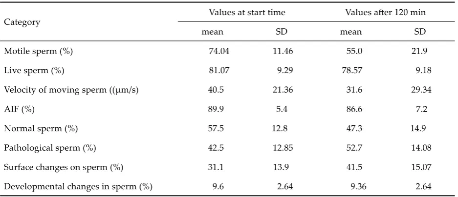
The average values of the spermatoanalytical criteria of semen obtained by examination at the start-time and over the 120 min short-term survival test, indicating the qualitative variety of the com-plex, especially from the high decisive deviations in motility indicators, speed of sperm movement and the high number of pathologically changed sperm, above all surface changes (Table 1).
Changes in the values of individual criteria during the 120 min survival test are a sign of the sensitivity of the indicator and may be ascribed diagnostic weight. The growth of changes on the surface structures of sperm made up 33.4%, which is the largest difference in comparison with the initial values of individual criteria. The fall in values in individual indicators of ejaculates a�ained in order of importance 27.9% in the speed of sperm move-ment, motility fell by 27.7%, normosperm fell by 17.7%, the indicators of membrane integrity fell by 3.5% in both the plasmatic membrane and in the area of the acrosome.
The morphological evaluation of sperm us-ing the system of multiparametric recordus-ing of changes proved the contribution of the abnormali-ties obtained over developmental ones at a ratio of 76.15% to 23.85%. The teratosperm index was set for the sample monitored at 1.27, SD = 0.087. In the enumeration of the frequency of findings made on pathologically changed sperm, secondary changes represent 76% (Figure 1), twel�h position in the overview.
[image:3.595.63.532.109.311.2]The overview of the statistical significance of the relationships between individual criteria for the qualitative evaluation of dog ejaculate (Table 2) indicates a high correlation coefficient and the sta-tistical significance of the percentage of live and motile sperm to the percentage of abnormalities in the surface structures on the sperm. The integrity of the acrosome in relation to pathological sperm Table 1. Average values and SD of indicators of dog semen level (n = 38)
Category Values at start time Values a�er 120 min
mean SD mean SD
Motile sperm (%) 74.04 11.46 55.0 21.9
Live sperm (%) 81.07 9.29 78.57 9.18
Velocity of moving sperm ((µm/s) 40.5 21.36 31.6 29.34
AIF (%) 89.9 5.4 86.6 7.2
Normal sperm (%) 57.5 12.8 47.3 14.9
Pathological sperm (%) 42.5 12.85 52.7 14.08
Surface changes on sperm (%) 31.1 13.9 41.5 15.07
Developmental changes in sperm (%) 9.6 2.64 9.36 2.64
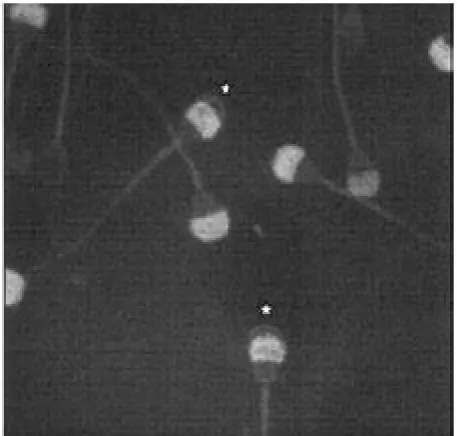
as a whole and to the surface changes in sperm showed a low correlation coefficient without statis-tical significance. Sperm with positive evidence of zymogen in the acrosomal area showed a swelling of the plasmatic membrane in the same part of the sperm (Figure 2).
Using the Acrosome IF-test (AIF) set, the in-tegrity of the acrosome at the initial value of the dynamic test was proven on average in 89.9% of sperm (SD = 5.4). After 120 min this value fell to 86.6% (SD = 7.2). The differences in the averages of positively reacting sperm in the AIF-test and the numbers of live sperm were statistically highly sig-nificant (P = 0.01). The differences between the AIF averages and motile sperm were similarly statisti-cally significant. The results show that the trends in the changes in the values of these criteria point to a correspondence, which is shown by the sta-tistically highly significant correlation coefficients between the motility of sperm, the number of live sperm and the positivity of the AIF-test (Table 2). The acrosomal integrity was also checked using the lectine Pisum sativum. The results obtained were compared with the AIF-test and a statisti-cally highly significant correspondence (P = 0.01) was shown in the correlation coefficient of 0.9370. While the methods of identifying the acrosomal integrity show a morphological persistence on the organelle level, the structural changes of the sur-face membrane are directly linked to the functional level of sperm and their motility.
Evaluating the integrity of the plasmatic mem-brane of the sperm using the effect of hypoosmotic
o o d d d d d d d d d o o d d o
o
o
o d o d o
0 5 10 15 20 25 30 35
1 2 3 4 5 6 7 8 9 10 11 12 13 14 15 16 17 18 19 20 21 22 23
[image:4.595.72.522.96.394.2](%)
Figure 1. Frequency of average pathological findings (n = 38)
1 swelling of acrosome 9 dag defect 17 persistent acroblast 2 bending of tail 10 thickened connecting part 18 ridged head – crista 3 without acrosome 11 abnormal position of tail insertion´ 19 condensation of acrosome 4 curling of tail 12 release of acrosome 20 doubled head
5 changed base of head 13 microcefalia 21 partial absence of mitochondrial spiral 6 residue of cytoplasm 14 macrocefalia 22 vaciolisation of acrosome
7 narrowed elongated head 15 pear-shaped head 23 breaking of tail 8 coiling of tail 16 doubling of tail
o = changes obtained 23.85% findings of developmental chandes d = developmental changes 76.15% findings of changes obtained
[image:4.595.63.292.506.724.2]Table 2. Statistical significance of relationships between individual criteria of qualitative evaluation of canine ejacu-lates
Category Motile sperm
(%)
Live sperm (%)
AIF positive sperm (%)
Pathology of sperm in total
(%)
Surface changes of sperm (%)
Motile sperm xxx xxx xxx xxx
% 0.8090 0.6609 –0.6309 –0.6988
Live sperm xxx xxx xxx xxx
% 0.8090 0.7062 –0.7339 –0.8300
AIF positive sperm xxx xxx 0 0
% 0.6609 0.7062 –0.3728 0.3757
Pathology of sperm in total xxx xxx 0 xxx
% –0.6309 –0.7339 –0.3728 0.5125
Surface changes of sperm xxx xxx 0 xxx
% –0.6988 –0.8300 0.3757 0.5125
AIF = immunofluorescent test of acrosome integrity xxx = highly statistically significant P = 0.01
[image:5.595.62.536.436.535.2]0 = without statistical significance value of correlation coefficient
Table 3. Average values of categories of hypoosmotic sperm test (HOS) (n = 20)
HOS-test
Eosin positive Total of curled tails
Eosin negative
curled tail Live sperm Motile sperm curled tail straight tail
A B C D E F
Mean 21.9% 9.29% 90.7% 72.6% 79.9% 72.9%
SD 18.36 4.66 4.66 12.27 8.75 16.58
C : E – 0.5875, P = 0.05; C : F – 0.6050, P = 0.05; D : E – 0.7820, P = 0.01; D : F – 0.5344, P = 0.05
Table 4. Monitoring of survival rate and sperm motile over a 360-minute perion (n = 8)
Movement of sperm VH 2H 4H 6H
Mean 72.5% 60.0% 45.8% 35.0%
SD 12.16 13.84 18.12 22.73
⊢
0.8827, P = 0.05⊣
Live sperm VH 2H 4H 6H
Mean 78.2% 76.6% 72.9% 70.9%
SD 9.75 10.01 9.85 9.91
0.8397
⊢
without statistical signifikance⊣
[image:5.595.62.540.597.748.2]influence of the environment showed the cor-respondence of the category of eosin in negative sperm with a curled tail to the percentage of live sperm with a highly significant correlation coeffi-cient 0.7820 (P = 0.01) and a correlation coefficient statistically significant for the percentage of motile sperm (0.5344, P = 0.05). The category of eosin in positive sperm with a straight tail and a coiled tail creates a significant prediction value supplementing curled tails in the sum total, which is statistically significant in correlation with both the percentage of live sperm and the percentage of motile sperm (0.5875, P = 0.05 and 0.6050, P = 0.05). The contribu-tion of eosin-positive sperm with a straight tail and the sum of sperm with a pathologically coiled tail according to morphological analysis is entirely in keeping with the number of dead sperm (20.5% to 23.3%) (Table 3).
The predictive importance of the examination us-ing the 120 min survival test is evident from the cor-respondence of the fall in motility of sperm during the 120 min test with their survival rate and motility a�er 6 hours. This correspondence is statistically significant (0.8827, P = 0.05) (Table 4).
DISCUSSION
In this study we have focused on the assess-ment of the diagnostic level of laboratory analy-ses permitting routine use. In connection with the morphological criteria, attention was devoted to the integrity of the plasmatic membrane and the acrosomal complex (Geussova et al., 1997; Pena et al., 1999; Guerin et al., 1999; Kawakami et al., 2000; Sirivaidyapong et al., 2000). Our results showed a relatively important resistance of values point-ing to the integrity of the plasmatic membrane and the integrity of the acrosome. On the other hand, a striking increase was shown in changes of the surface structures, especially the plasmatic membranes in the short-term sperm survival test. These changes were represented above all by a growth of changes typical of changes in the per-meability of the plasmatic membrane in the area of the acrosome.
The assessment of the integrity of the acrosome, whether immunofluorescently using antibodies against intraacrosomal proteins or by connecting PSA lectin, also shows the resistance of this or-ganelle component of sperm in contrast with the membrane covering and functional cell indicators.
The positivity of both reactions, both in the initial value and a�er 120 min, correspondingly shows a fall in intact acrosomes, but in strikingly lower values than are shown by functional shortcomings of the sperm. The quantification of the values of the averages of these indicators shows statistically significant differences, which suggests the higher resistance of organelle membranes. Ashworth et al. (1995) warn that the connection of PSA lectin to the acrosomal matrix of sperm increases the number of dead cells. Pena et al. (1999) discuss this view and ascribe the toxicity for sperm to the concentration of lectin used. In our work we conducted the con-nection of PSA lectin with the acrosomal area both supravitally and post-fixationally. The results of the connection in both cases corresponded a�er 15 min incubation both in number and the character of the reaction. During supravital incubation we showed the significant inhibition of the motility of sperm as early as at 5 minutes.
The integrity of the plasmatic membrane as an important factor for the normal metabolism and function of sperm has been monitored by many authors in different animal species using a reduced osmolarity test. Jeyendran et al. (1992) found in dogs that the changes provoked by the hypoos-motic environment could be used for predicting sperm quality. Our findings also show the usability of this test for interpreting the membrane resist-ance of sperm. The findings showed the statistical significance of comparing the result of the HOS-test with the categories of live and dead sperm and secondary changes on the surface structures of sperm.
The short-term survival test, which rests on the comparison of values obtained at the start-time of the test and a�er 120 min has been used by us since 1970. In bulls this test showed a high diagnostic worth when semen cannot be frozen because of a high increase of structural surface changes in sperm. In the current study we were able to show the importance of this test both in criteria assessing the functional level of sperm and in changes in the morphological structure.
Acknowledgements
REFERENCES
Ashworth P.J.C., Harrison R.A.P., Miller N.G.A., Plummer J.M., Eatson P.F. (1995): Flow cytometric detection of bicarbonate-induced changes in lectin binding in boar and ram sperm populations. Mol. Reprod. Dev., 40, 164–176.
Baker F., Gragle R.G., Salisbury G.W., Van Demark N.L. (1957): Spermatozoa velocities in vitro. A simple method of measurement. Fe�il. Steril., 8, 149–155.
Chong A.P., Walters C.A., Weinrieb S. (1983): The ne-glected laboratory test. J. Androl., 4, 280–282.
Christiansen I.J. (1984): Reproduction in the dos and cat. Bailliere Tindall. London, 99–100.
Dahlbom M., Andersson M., Juga J., Alanko M. (1997): Fertility parameters in male Irish wol�ounds: a two-year follow-up study. J. Small Anim. Pract., 12, 547– 550.
Deibel F.C.Jr., Smith J.E., Crabo B.G., Graham E.F. (1976): Evaluation of six assays of sperm quality by means of their accuracy, precision and sensitivity in separating know induced levels of damage. In: Proc. 8th Int. Congr. Anim. Reprod. an AI, 4, 888–891.
Dunphy B.C., Kay R., Barrat C.L.R., Cooke I.D. (1989): Quality control during the conventional analysis of sperm, an essential exercise. J. Androl., 10, 378–385. Geussova G., Peknicova J., Capkova J., Kalab P., Moos J.,
Philimonenko V.V., Hozak P. (1997): Monoclonal anti-bodies to canine intra-acrosomal sperm proteins rec-ognizing acrosomal status during capacitation and acrosome reaction. Andrologia, 5, 261–268.
Guerin P., Ferrer M., Fontbonne A., Benigni L., Jacquet M., Menezo Y. (1999): In vitro capacitation of dog sper-matozoa as assessed by chlortetracycline staining. Theriogenology, 4, 617–628.
Iguer-Ouada M., Verstagen J.P. (2001a): Validation of the sperm quality analyser (SQA) for dog sperm analysis. Theriogenology, 55, 1143–1158.
Iguer-Ouada M., Verstagen J.P. (2001b): Evaluation of the Hamilton Thorn computer – based automated system for dog semen analysis. Theriogenology, 55, 733–749. Jeyendran R.S., Van Ded Ven H.H., Perez-Pelaez M.,
Crabo B.G., Zaneveld L.J.D. (1984): Development of an assay to asses the functional integrity of the human sperm membrane and its relationship to other semen characteristics. J. Reprod. Fertil., 70, 219–228.
Jeyendran R.S., Van der Ven H.H., Zaneveld L.J.D. (1992): The hypoosmotic swelling test; an update. Arch. An-drol., 29, 105–116.
Kawakami E, Hori T, Tsutsui T. (2000): Azoospermia of dogs with apoptotic germ cells and Leydig cells. J. Vet. Med. Sci., 5, 529–531.
Koehler J.K., Platz C.C. Jr, Waddell W., Jones M.H., Behrns S. (1998): Semen parameters and electron microscope observations of spermatozoa of the red wolf, Canis rufus. J. Reprod. Fertil., 1, 95–101.
Matouskova O., Cigler M., Chalupa J., Hruska K. (1992): STAT Plus – Manual (in Czech). 1st ed. Veterinary Research Institute, Brno. 168 pp.
Menkveld R., Kruger T.F. (1995): Advantages of strict (Tygerberg) criteria for evaluation of sperm morphol-ogy. Int. J. Androl., 2, 36–42.
Nothling J.O., Gerstenberg C., Volkmann D.H. (1997): Semen quality a�er thawing: correlation with fertility and fresh semen quality in dogs. J. Reprod. Fertil., 51, 109–116.
Oe�le E.E. (1993): Sperm morphology and fertility in the dog. J. Reprod. Fertil. 47,Suppl., 257–260.
Pena A., Linde-Forsberg C. (2000a): Effects of spermato-zoal concentration and post-thaw dilution rate on survival a�er thawing of dog spermatozoa. Theriog-enology, 54, 703–718.
Pena A., Linde-Forsberg C. (2000b): Effects of Equex, one- or two-step dilution, and two freezing and thaw-ing rates on post-thaw survival of dog spermatozoa. Theriogenology, 54, 859–875.
Pena A., Johannisson A., Linde-Forsberg C. (1999): Post-thaw evaluation of dog spermatozoa using new triple fluorescent staining and flow cytometry. Theriogenol-ogy, 52, 965–980.
Rigau T., Farré M., Ballester J., Mogas T., Pena A., Rod-rigez-Gil J.E. (2001): Effects of glucose and fructose on motility pa�erns of dog spermatozoa from fresh ejacu-lates. Theriogenology, 56, 801–815.
Root Kustritz M.V., Olson P.N., Johnston S.D., Root T.K. (1998): The effects of stains and investigators on assess-ment of morphology of canine spermatozoa. J. Am. Anim. Hosp. Assoc., 4, 348–352.
Schafer S., Holzman A., Arbeiter K. (1996): Modification of the quality indicators of the ejaculate of beagle dogs due to frequent collections over time. Tierarztl. Prax., 24, 385–390.
Sirivaidyapong S., Bevers M.M., Colenbrander B. (1999): Acrosome reaction in dog sperm is induced by a mem-brane-localized progesterone receptor. J. Androl., 4, 537–544.
Sirivaidyapong S., Cheng F.P., Marks A., Voorhout W.F., Bevers M.M., Colenbrander B. (2000): Effect of sperm diluents on the acrosome reaction in canine sperm. Theriogenology, 3, 789–802.
method to predict the cryopreservability of dog semen. Mol. Reprod. Dev., 55, 289–298.
Veznik Z. (1970): Využití morfologických ukazatelů ke kvalitativnímu hodnocení ejakulátů. Vet. Med. Praha, 15, 571–578.
Veznik Z., Svecova D. (1992): Soubor metod k hodnocení pohlavních funkcí býků, ÚVO Pardubice, 184 s. Veznik Z., Svecova D., Zajicova A. et al. (2000): Hodnocení
semene pro asistovanou reprodukci a výběr plemeníků.
Striktní analýza spermatické morfologie SASMO. VÚVeL, Brno, 140 s.
Veznik Z., Matouskova O., Svecova D., Zajicova A. (2001): The use of the computer technology for the evaluation of the strict morphological sperm analysis. Vet. Med. – Czech, 46, 35–40.
Received: 03–01–02 Accepted a�er corrections: 03–07–02
Corresponding Author