Methyl isonicotinate 1-oxide
Hui Li* and Wen-Ni Zheng
College of Chemistry and Chemical, Engineering, Southeast University, Nanjing 210096, People’s Republic of China
Correspondence e-mail: [email protected]
Received 28 January 2010; accepted 5 February 2010
Key indicators: single-crystal X-ray study;T= 298 K; mean(C–C) = 0.003 A˚;
Rfactor = 0.060;wRfactor = 0.180; data-to-parameter ratio = 16.4.
In the title compound, C7H7NO3, the benzene ring and the methyl ester group are nearly coplanar, forming a dihedral of 3.09 (9). The crystal structure is stabilized by intermolecular C—H O hydrogen bonds, forming layers parallel to (101).
Related literature
For the application of carboxylate derivatives in microelec-tronics and as memory storage devices, see: Fu et al. (2007, 2008); Fu & Xiong (2008).
Experimental
Crystal data
C7H7NO3 Mr= 153.14 Monoclinic,P21=c a= 7.2429 (14) A˚
b= 10.347 (2) A˚ c= 9.898 (2) A˚
= 105.09 (3)
V= 716.2 (3) A˚3
Z= 4
MoKradiation
= 0.11 mm1
T= 298 K
0.300.250.20 mm
Data collection
Rigaku Mercury2 diffractometer Absorption correction: multi-scan
(CrystalClear; Rigaku, 2005) Tmin= 0.96,Tmax= 1.00
7070 measured reflections 1640 independent reflections 972 reflections withI> 2(I) Rint= 0.053
Refinement
R[F2> 2(F2)] = 0.060 wR(F2) = 0.180 S= 1.02 1640 reflections
100 parameters
H-atom parameters constrained
max= 0.18 e A˚
3
min=0.16 e A˚3
Table 1
Hydrogen-bond geometry (A˚ ,).
D—H A D—H H A D A D—H A
C2—H2A O2i
0.93 2.44 3.204 (3) 139
C4—H4A O3ii 0.93 2.42 3.263 (3) 150
Symmetry codes: (i)x;y1 2;z
1
2; (ii)xþ1;yþ 1 2;zþ
1 2.
Data collection: CrystalClear (Rigaku, 2005); cell refinement:
CrystalClear; data reduction:CrystalClear; program(s) used to solve structure:SHELXS97(Sheldrick, 2008); program(s) used to refine structure: SHELXL97 (Sheldrick, 2008); molecular graphics:
SHELXTL(Sheldrick, 2008); software used to prepare material for publication:SHELXTL.
This work was supported by the Innovative Dissertation Fund of Southeast University.
Supplementary data and figures for this paper are available from the IUCr electronic archives (Reference: RZ2415).
References
Fu, D.-W., Song, Y.-M., Wang, G.-X., Ye, Q., Xiong, R.-G., Akutagawa, T., Nakamura, T., Chan, P. W. H., Huang, S.-P. & -, D. (2007).J. Am. Chem. Soc. 129, 5346–5347.
Fu, D.-W. & Xiong, R.-G. (2008).Dalton Trans.pp. 3946–3948.
Fu, D.-W., Zhang, W. & Xiong, R.-G. (2008).Cryst. Growth Des.8, 3461–3464. Rigaku (2005).CrystalClear. Rigaku Corporation, Tokyo, Japan.
Sheldrick, G. M. (2008).Acta Cryst.A64, 112–122.
Acta Crystallographica Section E
Structure Reports Online
supporting information
Acta Cryst. (2010). E66, o584 [doi:10.1107/S1600536810004629]
Methyl isonicotinate 1-oxide
Hui Li and Wen-Ni Zheng
S1. Comment
Carboxylate derivatives attracted more attention as pharmaceutical and phase transition dielectric materials for their
application in micro-electronics and as memory storage devices (Fu et al., 2007; Fu & Xiong 2008; Fu et al., 2008). With
the purpose of obtaining phase transition crystals of carboxylate compounds, the interaction of methyl isonicotinate with
hydrogen peroxide has been studied and we have elaborated a series of new materials including these organic molecules.
In this paper, we describe the crystal structure of the title compound, Methyl isonicotinate 1-oxide.
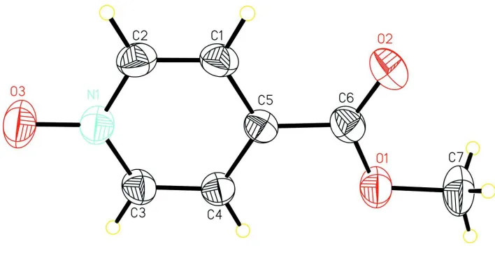
In the title compound (Fig. 1), the benzene ring and the methyl ester group are nearly coplanar, the dihedral angle they
form being 3.09 (9)°). The N1—O3 bond length of the nitrile group (1.292 (2)Å) is within the normal range. The crystal
structure is stabilized by intermolecular C—H···O hydrogen bonds (Table 1) linking the molecules to form layers parallel
to the (101) plane.
S2. Experimental
Methyl isonicotinate 1-oxide (3 mmol, 0.46 g) was dissolved in methanol. The solvent was slowly evaporated in air
affording colourless block-shaped crystals of the title compound suitable for X-ray analysis. Permittivity measurements
show that there is no phase transition within the temperature range (from 100 K to 400 K), and the permittivity is 6.5 at 1
MHz at room temperature.
S3. Refinement
All H atoms attached to C atoms were positioned geometrically and treated as riding, with C—H = 0.93 Å (aromatic),
0.96 Å (methyl) and Uiso(H) = 1.2Ueq(C) and 1.5Ueq(C) for methyl H atoms. A rotating-group model was used for the
Figure 1
A view of the title compound with the atomic numbering scheme. Displacement ellipsoids were drawn at the 30%
probability level.
methyl isonicotinate 1-oxide
Crystal data
C7H7NO3 Mr = 153.14
Monoclinic, P21/c Hall symbol: -P 2ybc a = 7.2429 (14) Å b = 10.347 (2) Å c = 9.898 (2) Å β = 105.09 (3)° V = 716.2 (3) Å3 Z = 4
F(000) = 320 Dx = 1.420 Mg m−3
Mo Kα radiation, λ = 0.71073 Å Cell parameters from 1640 reflections θ = 3.5–27.5°
µ = 0.11 mm−1 T = 298 K Block, colourless 0.30 × 0.25 × 0.20 mm
Data collection
Rigaku Mercury2 diffractometer
Radiation source: fine-focus sealed tube Graphite monochromator
Detector resolution: 13.6612 pixels mm-1 CCD profile fitting scans
Absorption correction: multi-scan (CrystalClear; Rigaku, 2005) Tmin = 0.96, Tmax = 1.00
7070 measured reflections 1640 independent reflections 972 reflections with I > 2σ(I) Rint = 0.053
θmax = 27.5°, θmin = 3.5° h = −9→9
k = −13→13 l = −12→12
Refinement
Refinement on F2 Least-squares matrix: full R[F2 > 2σ(F2)] = 0.060 wR(F2) = 0.180 S = 1.02 1640 reflections 100 parameters
Secondary atom site location: difference Fourier map
Hydrogen site location: inferred from neighbouring sites
H-atom parameters constrained w = 1/[σ2(Fo2) + (0.0868P)2 + 0.0635P]
Special details
Geometry. All esds (except the esd in the dihedral angle between two l.s. planes) are estimated using the full covariance matrix. The cell esds are taken into account individually in the estimation of esds in distances, angles and torsion angles; correlations between esds in cell parameters are only used when they are defined by crystal symmetry. An approximate (isotropic) treatment of cell esds is used for estimating esds involving l.s. planes.
Refinement. Refinement of F2 against ALL reflections. The weighted R-factor wR and goodness of fit S are based on F2, conventional R-factors R are based on F, with F set to zero for negative F2. The threshold expression of F2 > σ(F2) is used only for calculating R-factors(gt) etc. and is not relevant to the choice of reflections for refinement. R-factors based on F2 are statistically about twice as large as those based on F, and R- factors based on ALL data will be even larger.
Fractional atomic coordinates and isotropic or equivalent isotropic displacement parameters (Å2)
x y z Uiso*/Ueq
N1 0.3267 (3) 0.76927 (18) 0.0785 (2) 0.0666 (5)
C5 0.2388 (3) 1.02039 (19) −0.0124 (2) 0.0542 (5)
C6 0.1852 (3) 1.1517 (2) −0.0654 (2) 0.0642 (6)
C4 0.3492 (3) 0.99507 (19) 0.1224 (2) 0.0582 (6)
H4A 0.3944 1.0628 0.1838 0.070*
O3 0.3658 (3) 0.65171 (15) 0.1201 (2) 0.0955 (6)
O1 0.2579 (2) 1.24307 (15) 0.02709 (18) 0.0782 (6)
O2 0.0834 (3) 1.17357 (17) −0.18032 (17) 0.0934 (7)
C3 0.3912 (3) 0.8701 (2) 0.1644 (2) 0.0650 (6)
H3A 0.4662 0.8542 0.2545 0.078*
C2 0.2201 (3) 0.7926 (2) −0.0537 (2) 0.0686 (6)
H2A 0.1772 0.7237 −0.1140 0.082*
C1 0.1753 (3) 0.9152 (2) −0.0993 (2) 0.0642 (6)
H1A 0.1010 0.9290 −0.1900 0.077*
C7 0.2062 (4) 1.3747 (3) −0.0153 (4) 0.1006 (9)
H7A 0.2665 1.4325 0.0590 0.151*
H7B 0.0699 1.3843 −0.0356 0.151*
H7C 0.2479 1.3946 −0.0974 0.151*
Atomic displacement parameters (Å2)
U11 U22 U33 U12 U13 U23
Geometric parameters (Å, º)
N1—O3 1.292 (2) C4—H4A 0.9300
N1—C3 1.350 (3) O1—C7 1.445 (3)
N1—C2 1.357 (3) C3—H3A 0.9300
C5—C1 1.389 (3) C2—C1 1.357 (3)
C5—C4 1.390 (3) C2—H2A 0.9300
C5—C6 1.472 (3) C1—H1A 0.9300
C6—O2 1.205 (3) C7—H7A 0.9600
C6—O1 1.326 (3) C7—H7B 0.9600
C4—C3 1.368 (3) C7—H7C 0.9600
O3—N1—C3 121.0 (2) N1—C3—H3A 119.1
O3—N1—C2 119.8 (2) C4—C3—H3A 119.1
C3—N1—C2 119.1 (2) N1—C2—C1 120.9 (2)
C1—C5—C4 117.49 (19) N1—C2—H2A 119.5
C1—C5—C6 119.2 (2) C1—C2—H2A 119.5
C4—C5—C6 123.3 (2) C2—C1—C5 121.0 (2)
O2—C6—O1 123.6 (2) C2—C1—H1A 119.5
O2—C6—C5 123.3 (2) C5—C1—H1A 119.5
O1—C6—C5 113.05 (19) O1—C7—H7A 109.5
C3—C4—C5 119.77 (19) O1—C7—H7B 109.5
C3—C4—H4A 120.1 H7A—C7—H7B 109.5
C5—C4—H4A 120.1 O1—C7—H7C 109.5
C6—O1—C7 116.4 (2) H7A—C7—H7C 109.5
N1—C3—C4 121.7 (2) H7B—C7—H7C 109.5
C1—C5—C6—O2 2.1 (3) O3—N1—C3—C4 −179.25 (19)
C4—C5—C6—O2 −177.0 (2) C2—N1—C3—C4 1.1 (3)
C1—C5—C6—O1 −178.87 (17) C5—C4—C3—N1 −0.6 (3)
C4—C5—C6—O1 2.0 (3) O3—N1—C2—C1 179.22 (18)
C1—C5—C4—C3 0.1 (3) C3—N1—C2—C1 −1.2 (3)
C6—C5—C4—C3 179.26 (17) N1—C2—C1—C5 0.7 (3)
O2—C6—O1—C7 1.1 (3) C4—C5—C1—C2 −0.1 (3)
C5—C6—O1—C7 −177.87 (18) C6—C5—C1—C2 −179.32 (18)
Hydrogen-bond geometry (Å, º)
D—H···A D—H H···A D···A D—H···A
C2—H2A···O2i 0.93 2.44 3.204 (3) 139
C4—H4A···O3ii 0.93 2.42 3.263 (3) 150