Original Article
MiR-381 functions as a tumor suppressor
in gastric cancer by targeting ROCK2
Yilin Xie1 , Jing Qi2, Congbo Zhu2, Dingmin Zhao2, Guoqing Liao2
1Department of Endoscopy Center, The First Affiliated Hospital of Xiamen University, Xiamen, P. R. China; 2 Depart-ment of Gastrointestinal Surgery, Xiangya Hospital of Central South University, Changsha, Hunan Province, P. R. China
Received February 25, 2017; Accepted May 9, 2017; Epub January 1, 2019; Published January 15, 2019
Abstract: MiR-381 has been reported to be deregulated in many different types of human cancers. However, the clinical significance and function of miR-381 in gastric cancer (GC) remains unclear. Based on real-time quantita-tive PCR (RTq-PCR) analysis, we found that miR-381 was frequently lost or downregulated in GC tissues and cell lines, and decreased miR-381 was correlated with GC tumor size (P=0.012), TNM stage (P=0.007), invasion depth (P=0.008) and lymphatic metastasis (P=0.019). Cellular function assays revealed that miR-381 restoration inhib-ited cell proliferation, migration and invasion of GC cells. In addition, we validated that ROCK2 was as a direct target of miR-381, and knockdown of ROCK2 had similar with effects of miR-381 restoration in GC cells, while overexpres-sion of ROCK2 attenuated the inhibitory effects of miR-381 on GC cells. These data suggest that miR-381 serves as a tumor suppressor by targeting ROCK2, which may provide a potential therapeutic tool for GC therapy.
Keywords: miR-381, ROCK2, gastric cancer, cell proliferation, cell migration, cell invasion
Introduction
Gastric cancer (GC) is one of the most common cancers and cause of cancer-related deaths worldwide, with a 5-year survival rate of 25% or less, which is mainly due to the non-specific symptoms during the early stages of GC [1-4]. Although extensive studies have investigated the etiology of gastric cancer, the molecular mechanisms underlying the pathogenesis and progression of GC remain undefined.
MicroRNAs (miRNAs) are a class of endogenous small non-coding RNAs and act as important regulators of more than half of genes by bind- ing to specific target mRNAs and leading to direct mRNA degradation or translational re- pression [5-7]. Numerous studies have reveal- ed that miRNAs play crucial roles in various biological processes and diseases including human cancers [8-10]. Emerging evidence de- monstrates that miRNA alteration is highly as- sociated with the initiation and progression of all types of human cancers, which may provide new therapeutic targets to deal with human cancers [11]. MiR-381 has been reported to be
Materials and methods
Clinical samples and cell lines
[image:2.612.91.347.97.400.2]Primary GC tissue samples were collected from Xiangya Hospital between Apr, 2013 to Oct, 2015, and the clinicopathologic informa-tion of the GC patients was summarized in
Table 1. Written informed consent was ob- tained from all participants, and the use of these samples was approved by the ethical review committees of the Xiangya Hospital Ethic Committee of Central South University. Five GC cell lines (SGC7901, MGC803, BGC- 823, AGS and MKN45) and the gastric epithe-lial cell line GES-1 were purchased from the Type Culture Collection of the Chinese Acade- my of Sciences (Shanghai, People’s Republic of China) and maintained in 90% RPMI-1640 (BioTeke Corporation, Beijing, China) supple-mented with 10% fetal bovine serum (Thermo Fisher Scientific, Inc., Waltham, MA, USA). Real-time quantitative PCR (RTq-PCR)
Total RNA was isolated from GC tissues and cell lines by Trizol reagent (Life Technologies,
cules, CA, USA). The membrane was blocked with 5% nonfat milk and incubated with indi-cated antibodies, followed by the HRP-conju- gated secondary antibody. Antibodies of RO- CK2, MMP2, MMP9 and β-actin were obtained from Abcam (Abcam, MA, USA). The Signals were detected with enhanced chemilumines-cence reagents (Pierce, Rockford, IL, USA). Luciferase reporter assay
A fragment of the 3’UTR of ROCK2 containing predicted miR-381 target seed sequence was amplified and cloned into the psiCHECK-2 vec-tor (Promega, Shanghai, China). The mutation of the predicted seed regions in the ROCK2 3’UTR sequence was performed using the QuikChange™ Site-Directed Mutagenesis Kit (Stratagene, La Jolla, CA, USA). Relative lucifer-ase activity (a ratio of firefly luciferlucifer-ase to renilla luciferase) after cotransfection of miR-381 mimics or scramble mimics with wild type or mutant type of psiCHECK-2 vector into BGC- 823 cells was measured by the Dual Lucifer- ase Reporter Assay system (Promega, Shang- hai, China).
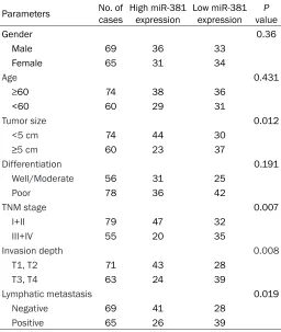
Table 1. Correlation between miR-381 expression and clini-copathological features in 134 gastric cancer patients Parameters No. of cases High miR-381 expression Low miR-381 expression valueP
Gender 0.36
Male 69 36 33
Female 65 31 34
Age 0.431
≥60 74 38 36
<60 60 29 31
Tumor size 0.012
<5 cm 74 44 30
≥5 cm 60 23 37
Differentiation 0.191
Well/Moderate 56 31 25
Poor 78 36 42
TNM stage 0.007
I+II 79 47 32
III+IV 55 20 35
Invasion depth 0.008
T1, T2 71 43 28
T3, T4 63 24 39
Lymphatic metastasis 0.019
Negative 69 41 28
Positive 65 26 39
Gaithersburg, MD). First strand cDNA was synthesized according to the manufacturer’s instructions (Invitro- gen, Carlsbad, CA). Reverse tran-scription and RTq-PCR were per-formed by using a qSYBR-green PCR kit (Qiagen, Germantown, USA). U6 snRNA and β-actin were used as endogenous controls for miR- 381 and ROCK2 relative express- ion, respectively. The primers used are as follow: ROCK2-F: 5’-CAGT- GGCATTGGGATAACATAA-3’, ROCK2- R: 5’-GGAATTGGGAAGGTTTCTACAT- 3’; β-actin-F: 5’-AGGGGCCGGACTC- GTCATACT-3’, β-actin-R: 5’-GGCGGC- ACCACCATGTACCCT-3’. The specific primers for miR-381 and U6 snRNA were purchased from GeneCopoeia. The relative levels of miR-381 and ROCK2 of each sample were mea-sured in triplicates using the 2-ΔΔCt
method. Western blot
Plasmid construction, siRNA and cell transfec -tion
ROCK2 coding region was amplified with the following primers (ROCK2-F, 5’-GAGCCTGGAT- GGACAAGAGGAA-3’; ROCK2-R, 5’-CTAGATTTT- CGAGCTAATCATC-3’) and cloned into pcDNA3.1 vector to generate ROCK2 expressing vectors. The empty pCDNA3.1 vector was used as con-trol. MiR-381 mimics, miR-381 inhibitor mimics and scramble mimics were purchased from RiboBio (Guangzhou, China). ROCK2 specific small interfering RNAs (siRNA-ROCK2, sense, 5’-GGCACGACUAGCAGAUAAAUU-3’; antisense, 5’-UUUAUCUGCUAGUCGUGCCUU-3’) and the negative control siRNA (siRNA-NC) were pur-chased from GeneChem (Shanghai, China). Cell transfection was performed using Lipo- fectamine 2000 (Invitrogen) according to the manufacture’s instruction.
Cell proliferation, invasion and migration as -says
BGC823 cells were plated in 96-well plates (5,000 per well), and allowed to grow for 24, 48 and 72 h, then cell proliferation ability was assessed by a colorimetric assay using MTT solution (5 mg/ml) at 570 nm. Cell invasion ability was assessed using transwell invasion chambers coated with mgatrigel (BD Bioscien- ces, Franklin Lakes, NJ, USA). Cells suspended in 200 µl of FBS-free medium were added into the upper chamber. 500 µl of medium with 10% FBS as the nutritional attractant was added into the lower chamber. After 24 h, the cells
that had invaded through the membrane were fixed with 20% methanol, stained with 0.1% crystal violet, imaged and counted using an inverted microscope (Olympus, Tokyo, Japan). Cell migration ability was assessed by wound healing assays. An artificial “wound” was cre-ated using a pipette tip on a confluent cell monolayer. Then, the cells were then cultured for 24 hours and the wound closure was as- sessed by Scion Image Software (Scion Cor- poration, Frederick, MD).
Statistical analysis
SPSS 19.0 software (IBM, NY, USA) was used for all statistical analysis. Student’s t-test was used to analyze the differences in the experi-ments. Chi-squared test was used to analyze differences of miR-381 expression with clinico-pathological features. P<0.05 was considered statistically significant.
Results
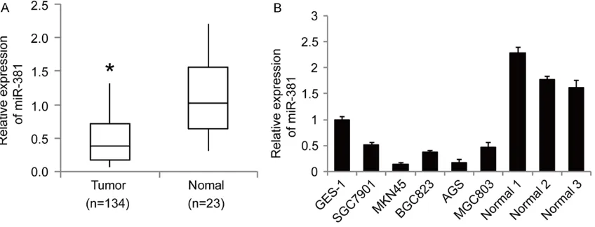
MiR-381 is frequently downregulated in gastric cancer
[image:3.612.96.520.76.238.2]GC cell lines (SGC7901, MKN45, BGC823, AGS and MGC803) than that of the gastric epithe- lial cell line GES-1 and normal gastric mucosa (Figure 1B). Using the median value of the rela-tive expression levels of miR-381 in all 134 samples as a cutoff, we classified 67 samples as the low expression group. Correlation analy-sis between miR-381 expression levels and clinicopathologic parameters revealed that the downregulation of miR-381 was significantly correlated with tumor size (P=0.012), TNM stage (P=0.007), invasion depth (P=0.008) and lymphatic metastasis (P=0.019) (Table 1). Taken together, these data suggest that de- creased expression level of miR-381 may be
MiR-381 inhibits the proliferation, invasion and migration of BGC823 cells
We then assessed the biological role of miR-381 in GC cells. We restored miR-miR-381 expres-sion in BGC823 cells (Figure 2A). MTT assays revealed that restoration of miR-381 inhibited the proliferation of BGC823 cells (Figure 2B). Transwell assays with matrigel (Figure 2C) and wound healing assays (Figure 2D) also showed that miR-381 had inhibitory effect on cell inva-sion and migration ability, respectively. In con-trast, when endogenous miR-381 was silenced with anti-miR-381 mimics in BGC823 cells, the cell proliferation (Figure 2B), invasion (Figure
These observations suggested that miR-381 might act as a tumor suppressor miRNA in GC cells.
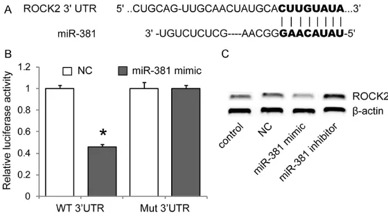
MiR-381 directly inhibits ROCK2 by targeting its 3’UTR
To elucidate the molecular mechanism by which miR-381 exerts its inhibitory effects on GC cells, miRNA target analysis tools PicTar and TargetScan were used to explore potential tar-get of miR-381. Among hundreds of potential targets, we focused on ROCK1 and ROCK2 in the study, considering the involvement of them in the pathogenesis of many human cancers including gastric cancer. To validate targeting of ROCK1 and ROCK2 by miR-381, we performed luciferase reporter assays. The wild type 3’UTR luciferase reporter plasmid (Wt-3’UTR) or the mutated (Mut-3’UTR) was cotransfected with miR-381 expressing plasmid or the control vector into BGC823 cells. The cotransfection of miR-381 expression plasmid with ROCK2 Wt-3’UTR (but not ROCK1 Wt-3’UTR, data not shown) showed significant decreased lucifer-ase activity compared with the control vector group, while ROCK2 Mut-3’UTR luciferase ac- tivity was not changed (Figure 3A, 3B). In addi-tion, western blot analysis revealed that miR-381 mimic significantly reduced the expression
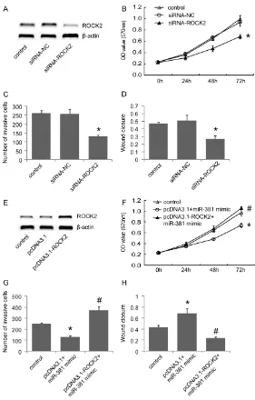
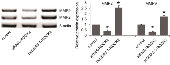
silencing ROCK2 inhibited cell proliferation, invasion and migration ability (Figure 4B-D). To further validate whether miR-381 exerts its inhibitory effect through downregulation of ROCK2 in GC cells, we overexpressed ROCK2 in miR-381 mimic transfected BGC823 cells by transfecting ROCK2 expression plasmid. As expected, ROCK2 overexpression significantly attenuated the inhibitory effects of miR-381 mimics on cellular proliferation, invasion and migration of BGC823 cells (Figure 4F-H). Col- lectively, our results revealed that ROCK2 par-ticipates in cell growth and migration of GC cells. In addition, we examined the effect of ROCK2 on the expression levels of MMP2 and MMP9, which are well known important regula-tors of cellular invasion and migration. Western blot analysis revealed that silencing of RO- CK2 significantly inhibited MMP2 and MMP9 expression while ROCK2 overexpression sig- nificantly increased MMP2 and MMP9 in BGC- 823 cells (Figure 5), suggesting that ROCK2 might promote cell invasion and migration, at least partially by upregulating MMP2 and MMP9 in GC cells.
Discussion
Emerging evidence implied that miRNAs play crucial roles in the regulation of cellular func-Figure 3. MiR-381 directly inhibits ROCK2 by targeting its 3’UTR. A. Predicted
binding sequences of miR-381 in the 3’UTR of ROCK2. B. The ROCK2 3’UTR containing wild-type (WT) or mutant (Mut) 3’UTR of ROCK2 was subcloned into the luciferase reporter vector, then luciferase assay was performed in BGC823 cells cotransfected with miR-381 mimic and a luciferase reporter containing the wild-type (WT) or mutant (Mut) 3’UTR of ROCK2. Luciferase activity was determined 48 h after transfection. C. The expression levels of ROCK2 protein in BGC823 cells after 48 h transfection with miR-381 mim-ic, miR-381 inhibitor or scramble mimic (NC) were measured western blot. *P<0.05 versus the negative control (NC), data shown are means ± S.D.
of ROCK2, while miR-381 in- hibitor increased ROCK2 ex- pression in BGC823 cells (Figure 3C). We did not found that ROCK1 protein expres-sion was significantly chang- ed by miR-381 mimic or in- hibitor treatment (data not shown). These results reveal- ed that miR-381 directly in- hibited ROCK2 expression. Silencing of ROCK2 inhibits the proliferation, invasion and migration of BGC823 cells
[image:5.612.92.372.73.230.2]tions and altered miRNA levels have been found in the initiation or development process of human cancers [10]. MiR-381 has been report-ed to be downregulatreport-ed in several human
[image:6.612.91.372.71.511.2]can-ported that miR-381 increased the in vitro and in vivo proliferation of glioma cells, and inhi- biting the endogenous expression of miR-381 decreased cell proliferation and tumor growth, Figure 4. Silencing of ROCK2 inhibits the proliferation, invasion and
migra-tion of BGC823 cells. (A) BGC823 cells were transfected with ROCK2 specific siRNA (siRNA-ROCK2) or negative control siRNA (siRNA-NC). The knockdown of ROCK2 was validated by western blot. The effects of ROCK2 knockdown on cell proliferation, invasion and migration were determined using MTT as-says (B), transwell asas-says with matrigel (C) and wound healing assay (D), respectively. (E) BGC823 cells were transfected with ROCK2 expression plasmids lacking its 3’UTR (pcDNA3.1-ROCK2) or empty pCDNA3.1 vector, and the expression of ROCK2 was examined by western blot. The effects of ROCK2 overexpression in miR-381 mimic transfected BGC823 cells on the proliferation, invasion and migration were assessed by MTT assays (F), transwell assays with matrigel (G) and wound healing assay (H), respectively. *P<0.05 versus the control, #P<0.05 versus the pcDNA3.1+miR-381 mimic group, data shown are means ± S.D.
suggesting that increased miR-381 is involved in the pathogenesis of glioma [16]. These data suggest that the roles of miR-381 might vary in different types of cancers. Here, we demon-strated that miR-381 functions as a tumor suppressor by inhibiting the proliferation and migration and invasion of GC cells.
Generally, a specific microRNA exerts its func-tion during a certain cancer development th- rough post-transcriptional regulating specific target genes [22, 23]. Thus, a specific miRNA may function as a tumor suppressor by nega-tively regulating oncogenes, or an oncogene by downregulating tumor suppressor genes [11, 24]. In the present study, we predicted and vali-dated that ROCK2 was a novel direct target of miR-381 in GC cells. Luciferase reporter as- say and western blot analysis revealed that miR-381 directly targeted and inhibited ROCK2 expression in BGC823 cells. ROCK2 plays a central role in the organization of the actin cytoskeleton [25-27] and is involved in a wide range of fundamental processes such as cell adhesion, proliferation, migration and apopto-sis [28, 29]. Emerging evidence revealed that ROCK2 level is elevated and involved in the growth and metastasis of many kinds of can-cers including GC [30, 31]. In the present study, we firstly conformed that targeting RO- CK2 by siRNA inhibited the proliferation, inva-sion and migration ability of BGC823 cells. Matrix metalloproteinases (MMPs) are modu- lators of cancer invasion and metastasis in many kinds of cancers including GC [32, 33]. We found that silencing of ROCK2 significantly inhibited MMP2 and MMP9 expression while ROCK2 overexpression significantly increased
BGC823 cells. Collectively, our data suggest a functional miR-381/ROCK2 interaction in GC cells.
In summary, our current results demonstrated that miR-381 might act as a tumor suppressor in GC cells via inhibiting ROCK2. Therefore, res-toration of miR-381 might provide a therapeu-tic strategy for GC treatment.
Disclosure of conflict of interest
None.
Address correspondence to: Guoqing Liao, Depart- ment of Gastrointestinal Surgery, Xiangya Hospi- tal of Central South University, 87 Xiangya Road, Changsha 410008, Hunan Province, P. R. China. Tel: +8613617498772; Fax: +86073184327016; E-mail: liaoguoqing2016@163.com
References
[1] Duraes C, Almeida GM, Seruca R, Oliveira C and Carneiro F. Biomarkers for gastric cancer: prognostic, predictive or targets of therapy? Virchows Arch 2014; 464: 367-378.
[2] Ferlay J, Shin HR, Bray F, Forman D, Mathers C and Parkin DM. Estimates of worldwide burden of cancer in 2008: GLOBOCAN 2008. Int J Can-cer 2010; 127: 2893-2917.
[3] Ferro A, Peleteiro B, Malvezzi M, Bosetti C, Ber-tuccio P, Levi F, Negri E, La Vecchia C and Lu-net N. Worldwide trends in gastric cancer mor-tality (1980-2011), with predictions to 2015, and incidence by subtype. Eur J Cancer 2014; 50: 1330-1344.
[image:7.612.88.373.74.184.2][4] Shibuya K, Mathers CD, Boschi-Pinto C, Lopez AD and Murray CJ. Global and regional esti-mates of cancer mortality and incidence by Figure 5. ROCK2 promoted MMP2 and MMP9 expression in BGC823 cells.
Western blot was carried out to examine the effect of ROCK2 on the expres-sion levels of MMP2 and MMP9 in BGC823 cells transfected with ROCK2 siRNA (siRNA-ROCK2) and ROCK2 expression plasmids (pcDNA3.1-ROCK2). *P<0.05 versus the control.
site: II. Results for the global burden of disease 2000. BMC Cancer 2002; 2: 37.
[5] Esteller M. Non-coding RNAs in human dis-ease. Nat Rev Genet 2011; 12: 861-874. [6] Fabian MR, Sonenberg N and Filipowicz W.
Regulation of mRNA translation and stability by microRNAs. Annu Rev Biochem 2010; 79: 351-379.
[7] Osada H and Takahashi T. MicroRNAs in bio-logical processes and carcinogenesis. Carcino-genesis 2007; 28: 2-12.
[8] Beermann J, Piccoli MT, Viereck J and Thum T. Non-coding RNAs in development and disease: background, mechanisms, and therapeutic ap-proaches. Physiol Rev 2016; 96: 1297-1325. [9] Kong YW, Ferland-McCollough D, Jackson TJ
and Bushell M. microRNAs in cancer manage-ment. Lancet Oncol 2012; 13: e249-258. [10] Sayed D and Abdellatif M. MicroRNAs in
devel-opment and disease. Physiol Rev 2011; 91: 827-887.
[11] Zhang B, Pan X, Cobb GP and Anderson TA. mi-croRNAs as oncogenes and tumor suppres-sors. Dev Biol 2007; 302: 1-12.
[12] He X, Wei Y, Wang Y, Liu L, Wang W and Li N. MiR-381 functions as a tumor suppressor in colorectal cancer by targeting Twist1. Onco Targets Ther 2016; 9: 1231-1239.
[13] Liang Y, Zhao Q, Fan L, Zhang Z, Tan B, Liu Y and Li Y. Down-regulation of MicroRNA-381 promotes cell proliferation and invasion in co-lon cancer through up-regulation of LRH-1. Biomed Pharmacother 2015; 75: 137-141. [14] Xia B, Li H, Yang S, Liu T and Lou G. MiR-381
inhibits epithelial ovarian cancer malignancy via YY1 suppression. Tumour Biol 2016; 37: 9157-9167.
[15] Ming J, Zhou Y, Du J, Fan S, Pan B, Wang Y, Fan L and Jiang J. miR-381 suppresses C/EBPal-pha-dependent Cx43 expression in breast can-cer cells. Biosci Rep 2015; 35.
[16] Tang H, Liu X, Wang Z, She X, Zeng X, Deng M, Liao Q, Guo X, Wang R, Li X, Zeng F, Wu M and Li G. Interaction of hsa-miR-381 and glioma suppressor LRRC4 is involved in glioma growth. Brain Res 2011; 1390: 21-32.
[17] Tang H, Wang Z, Liu Q, Liu X, Wu M and Li G. Disturbing miR-182 and -381 inhibits BRD7 transcription and glioma growth by directly tar-geting LRRC4. PLoS One 2014; 9: e84146. [18] Wang Z, Yang J, Xu G, Wang W, Liu C, Yang H,
Yu Z, Lei Q, Xiao L, Xiong J, Zeng L, Xiang J, Ma J, Li G and Wu M. Targeting miR-381-NEFL axis sensitizes glioblastoma cells to temozolomide by regulating stemness factors and multidrug resistance factors. Oncotarget 2015; 6: 3147-3164.
[19] Rothschild SI, Tschan MP, Jaggi R, Fey MF, Gugger M and Gautschi O. MicroRNA-381 re-presses ID1 and is deregulated in lung
adeno-carcinoma. J Thorac Oncol 2012; 7: 1069-1077.
[20] Zhang Q, Zhao S, Pang X and Chi B. MicroR-NA-381 suppresses cell growth and invasion by targeting the liver receptor homolog-1 in he-patocellular carcinoma. Oncol Rep 2016; 35: 1831-1840.
[21] Formosa A, Markert EK, Lena AM, Italiano D, Finazzi-Agro E, Levine AJ, Bernardini S, Ga-rabadgiu AV, Melino G and Candi E. MicroR-NAs, 154, 299-5p, 376a, miR-376c, miR-377, miR-381, miR-487b, miR-485- 3p, miR-495 and miR-654-3p, mapped to the 14q32.31 locus, regulate proliferation, apop-tosis, migration and invasion in metastatic prostate cancer cells. Oncogene 2014; 33: 5173-5182.
[22] Garzon R, Marcucci G and Croce CM. Targeting microRNAs in cancer: rationale, strategies and challenges. Nat Rev Drug Discov 2010; 9: 775-789.
[23] Jain CK, Gupta A, Dogra N, Kumar VS, Wadhwa G and Sharma SK. MicroRNA therapeutics: the emerging anticancer strategies. Recent Pat Anticancer Drug Discov 2014; 9: 286-296. [24] Kent OA and Mendell JT. A small piece in the
cancer puzzle: microRNAs as tumor suppres-sors and oncogenes. Oncogene 2006; 25: 6188-6196.
[25] Newell-Litwa KA, Badoual M, Asmussen H, Patel H, Whitmore L and Horwitz AR. ROCK1 and 2 differentially regulate actomyosin orga-nization to drive cell and synaptic polarity. J Cell Biol 2015; 210: 225-242.
[26] Schofield AV, Steel R and Bernard O. Rho-associated coiled-coil kinase (ROCK) protein controls microtubule dynamics in a novel sig-naling pathway that regulates cell migration. J Biol Chem 2012; 287: 43620-43629.
[27] Shi J, Wu X, Surma M, Vemula S, Zhang L, Yang Y, Kapur R and Wei L. Distinct roles for ROCK1 and ROCK2 in the regulation of cell detach-ment. Cell Death Dis 2013; 4: e483.
[28] Julian L and Olson MF. Rho-associated coiled-coil containing kinases (ROCK): structure, reg-ulation, and functions. Small GTPases 2014; 5: e29846.
[29] Wei L, Surma M, Shi S, Lambert-Cheatham N and Shi J. Novel insights into the roles of Rho Kinase in cancer. Arch Immunol Ther Exp (Warsz) 2016; 64: 259-278.
[30] Hinsenkamp I, Schulz S, Roscher M, Suhr AM, Meyer B, Munteanu B, Fuchser J, Schoenberg SO, Ebert MP, Wangler B, Hopf C and Burger-meister E. Inhibition of Rho-associated Kinase 1/2 Attenuates tumor growth in murine gastric cancer. Neoplasia 2016; 18: 500-511. [31] Jia S, Jia Y, Weeks HP, Ruge F, Feng X, Ma R, Ji
protein 2, in gastric cancer indicates lymph node metastasis and cell migration. Antican-cer Res 2014; 34: 2185-2194.
[32] Chen SX, Yin JF, Lin BC, Su HF, Zheng Z, Xie CY and Fei ZH. Upregulated expression of long noncoding RNA SNHG15 promotes cell prolif-eration and invasion through regulates MMP2/ MMP9 in patients with GC. Tumour Biol 2016; 37: 6801-6812.
[33] Wang HL, Zhou PY, Zhang Y and Liu P. Relation-ships between abnormal MMP2 expression and prognosis in gastric cancer: a meta-analy-sis of cohort studies. Cancer Biother Radio-pharm 2014; 29: 166-172.