Original Article
Depletion of NEDD9, a target gene of TGF-β, inhibits
the proliferation and invasion of ectopic
endometriotic stromal cells
Chunbo Yang, Junyan Ma, Jun Lin
Department of Gynaecology, Women’s Hospital, School of Medicine, Zhejiang University, Zhejiang, China
Received November 18, 2016; Accepted January 11, 2017; Epub March 1, 2017; Published March 15, 2017
Abstract: Endometriosis is characterized by the growth of endometrial-like tissue outside the uterus. The prolif-erative and invasive ability of endometrial cells is believed to play a key role in the ectopic growth of endometrial implants. Neural precursor cell expressed developmentally down-regulated 9 (NEDD9, also known as HEF1/Cas-L), a target gene of transforming growth factor-b (TGF-β), has been identified as a metastasis gene in several cancer type. Here, endometriotic stromal cells (ESCs) were isolated from ectopic endometrium and NEDD9 expression was knocked down by small interfering RNA (siRNA) transfection. Silencing of NEDD9 in ESCs significantly inhibited cell proliferation and invasion as indicated by Cell Counting Kit-8 and Transwell assay, respectively. TGF-β1 (2.5 ng/ mL) treatment enhanced the expression of NEDD9 in ESCs. Moreover, the mRNA and protein levels of NEDD9 and TGF-β1 were significantly increased in endometriotic tissues along with the severity of endometriosis. More impor -tantly, a positive correlation between NEDD9 and TGF-β1 mRNA expression was observed in human endometriotic tissues. In conclusion, NEDD9, whose expression was enhanced by TGF-β1, may be involved in the proliferation and invasion of ESCs, thus contributing to the pathogenesis of endometriosis.
Keywords: Endometriosis, NEDD9, TGF-β, proliferation, invasion
Introduction
Endometriosis, first microscopically discovered by Karl von Rokitansky in 1860, is a chronic disease characterized by the growth of endo-metrial-like tissue outside the uterus [1]. Endometriosis affects 6% to 10% of reproduc-tive-aged women, and 35% to 50% of women with pain or infertility [2]. It has attracted wide-spread attention because it causes chronic pel-vic pain, dysmenorrhea and subfertility [3]. The most widely accepted explanation for the devel-opment of endometriosis is Sampson’s implan-tation theory [4], which postulates that during menstruation, endometrial cells travel to the abdomen and form endometriotic lesions.The ability of endometrial cells to survive, adhere, invade tissues and proliferate is critical for this process. Epidemiologic, histopathological and molecular biology studies suggest that metriosis has a malignant potential, and endo-metriosis is classified as a tumor like lesion by the World Health Organization [5, 6].
endometriosis are significantly higher than in controls and increased along with the severity of the disease [16]. Increased TGF-β activity is observed at the sites of endometriosis [17]. Recently, studies on bone marrow macro-phages and human dermal fibroblasts demon-strated that TGF-β1 can induce the transcrip-tion of NEDD9 [18-20]. However, whether TGF-β1 is able to regulate NEDD9 expression in endometrial cells is unknown.
In the present study, we isolated endometriotic stromal cells (ESCs) from ectopic endometrium, assessed the effects of NEDD9 knockdown on the proliferation and invasion of ESCs, and explored the effects of TGF-β1 stimulation on NEDD9 expression in ESCs. Moreover, we mea-sured TGF-β1 and NEDD9 expression in eutop-ic, ectopic and control endometrial tissues, and tried to investigate the association between TGF-β1 and NEDD9.
Materials and methods
Patient recruitment and specimen collection
The study was approved by the Ethics Commi- ttee of Women’s Hospital, Zhejiang University, and written informed consent was obtained from all participants. All participants were at reproductive age (27-45 years old), had not received hormones therapy for at least 3 months before specimen collection and had no prior history of malignancy or autoimmune diseases. Eutopic endometrial tissues (n=30) and ectopic endometrial tissues (n=30) were collected from 60 women with endometriosis undergoing laparoscopy. Control endometrial tissues were from 30 women with benign gyne-cological conditions and without evident endometriosis.
Immunohistochemistry (IHC)
IHC assay was conducted to measure the pro-tein levels of TGF-β1 and NEDD9 in eutopic endometrial, ectopic endometrial and normal tissues. Briefly, the formalin-fixed and paraffin-embedded tissue sections were deparaffinized, rehydrated and incubated with 0.01 M citrate buffer (pH 6) to unmask antigen. After incuba-tion with 10% normal goat serum for 30 min to block non-specific antigens, the tissue sections were incubated with anti-TGF-β1 (Abcam, Cambridge, MA, USA; ab92486) or anti-NEDD9
(Abcam, ab18056) overnight at 4°C followed by horseradish peroxidase (HRP) conjugated secondary antibody (Beyotime, Shanghai, China) for 1 h at room temperature. Visualization was performed with the 3,3-diaminobenzidine (DAB) solution (Vector Laboratories, Burlinga- me, CA, USA), and nuclei were counterstained with hematoxylin.
Isolation and culture of human endometriotic stromal cells (ESCs)
ESCs were isolated according to the previous method [21, 22]. Under sterile conditions, ecto-pic endometrium obtained at the time of lapa-roscopy were minced into small pieces (< 1 mm3) and digested with 1% collagenase solu-tion (Serva, Heidelberg, Germany) for 2-3 hours in an atmosphere of 5% CO2 at 37°C with occa-sional vortexing every 30 minutes. Cell suspen-sion was filtered through 100 mm mesh (BD Biosciences, Franklin Lakes, NJ, USA) to remove the undigested tissues. After washing two times with culture medium, the obtained was seeded onto culture plates for 6 hours. The cul-ture medium was changed to remove non-adherent and dead cells. The remaining adher-ent stromal cells were allowed to propagate. ESCs were cultured in DMEM/F12 medium (Hyclone, Logan, UT, USA) supplementary with 10% fetal bovine serum (FBS; Gibco, Carlsbad, CA, USA), 100 U/ml penicillin and 100 µg/ml streptomycin. The purity of the ESCs were eval-uated by immunocytochemical staining with anti-vimentin (Abcam, ab8978) [23].
Small interfering RNA (siRNA) transfection
To silence the expression of NEDD9 in isolated ESCs, siRNA targeting NEDD9 (GAAGCTCTATC- AAGTGCCA) [24] and a non-specific scramble siRNA (NC) were synthesized by Genepharma (Shanghai, China). ESCs were transfected with 400 nmol/L NEDD9 siRNA or NC by using lipo-fectamineTM 2000 (Invitrogen, Shanghai, Chi- na) per the instructions. At 48 hours post trans-fection, qRT-PCR and western blot analyses were carried out to confirm the knockdown of NEDD9.
Reverse transcription and quantitative real-time PCR (qRT-PCR) assay
(Invitrogen, Carlsbad, CA, USA). Reverse tran-scription was conducted using cDNA Synthesis Kit (Fermentas, Hanover, MD, USA) according to the manufacture’s protocol. qRT-PCR was used to examine the mRNA levels of indicated genes with GADPH as internal control. The qRT-PCR reaction was performed with SYBR Green PCR kit (Fermentas) on an ABI-7300 Real-Time PCR system (Applied Biosystem, Foster City, CA). The PCR procedure was 10 min at 95°C, followed by 40 cycles of 15 s at 95°C, 45 s at 60°C. The primer sequences were as follows: NEDD9 (NM_001142393.1), 5’-TGTGAGGTCC- GTAATGTC-3’ and 5’-GCCCTTGCCATAAGATTC-3’; MMP2 (NM_004530.4), 5’-TTGACGGTAAGGAC- GGACTC-3’ and 5’-GGCGTTCCCATACTTCACAC-3’; MMP9 (NM_004994.2), 5’-AAGGGCGTCGTGG- TTCCAACTC-3’ and 5’-AGCATTGCCGTCCTGGGT- GTAG-3’; TGF-β1 (NM_000660.4), 5’-GACTACT- ACGCCAAGGAGGTC-3’ and 5’-GAGAGCAACAC-
GGGTTCAG-3’; and GADPH (NM_001256799.1), 5’-CACCCACTCCTCCACCTTTG-3’ and 5’-CCACC- ACCCTGTTGCTGTAG-3’.
Western blot analysis
For tissue samples, about 100 mg of tissue was ground with a mortar and pestle under liq-uid nitrogen. Frozen tissue powder and ESCs were lysed in ice-cold radio immunoprecipita-tion assay buffer (JRDUN, Shanghai, China) supplemented with protease inhibitor cocktail (Sigma) for 30 minutes. Total protein extracts were separated by 10% SDS-PAGE and trans-ferred electrophoretically to a nitrocellulose membrane (Millipore, Bredford, MA, USA). Anti-NEDD9 (Abcam, ab88584), anti-TGF-β1 (Ab- cam, ab92486), anti-MMP2 (Abcam, ab92- 536), anti-MMP9 (Abcam, ab119906), anti-p-smad2/3 (CST, Danvers, MA, USA; #8828), anti-smad2/3 (CST, #8685) and anti-GAPDH (CST, #5174) were used in Western analysis according to the manufacturers’ instructions. Visualization was performed using the enhan- ced chemiluminescence system (Millipore).
Cell proliferation assay
[image:3.612.94.520.73.174.2]Cell Counting Kit (CCK)-8 (Beyotime) was used to assess the proliferation property of ESCs. Briefly, ESCs were seeded in triplicate in 96-well culture plates at a density of 5,000 cells/well and transfected with NEDD9 siRNA or NC. The cultures were incubated for 0, 24, 48 and 72 hours, and then CCK-8 solution were added to each well and incubation was continued for 1 hour. The optical density of each well at 450 nm (OD450) was determined by a microplate reader (Bio-Rad, Richmond, CA, USA).
Figure 1. Suppressing of NEDD9 expression by siRNA transfection. (A) ESCs were isolated from ectopic endome-trium, and immunocytochemical staining with anti-vimentin. Magnification: 200×. (B, C) ESCs were transfected with
NEDD9 siRNA or a non-specific scramble siRNA (NC). ESCs without any treatment (Mock) were set as a control. Relative mRNA (B) and protein (C) expression levels of NEDD9 were detected at 48 hours after siRNA transfection. ****P < 0.0001.
[image:3.612.92.291.258.398.2]Transwell invasion assays
The invasive ability of ESCs was evaluated by Transwell invasion assays using chamber with 8 μm pore filters (Corning, New York, NY, USA).
[image:4.612.93.522.75.338.2]Before assays, the inserts of upper chambers were coated with Matrigel (BD Biosciences). ESCs were serum starved overnight at 24 hours post siRNA transfection, and seeded into the upper chamber with serum-free medium. Then
Figure 3. NEDD9 knockdown repressed the invasive ability of ESCs. (A) Invasion assays were performed in Matrigel-precoated Transwell chambers as described in Materials and Methods. (B, C) mRNA and protein levels of MMP2 and MMP9 were examined in ectopic ESCs by qRT-PCR (B) and Western bolt (C) at 48 hours post RNA interference. Data were presented as mean ± SD from three independent experiments. ***P < 0.001.
[image:4.612.94.522.410.601.2]medium with 10% FBS were added to the lower chamber. After 24 hours, non-migrating cells were completely removed with a cotton swab, and the migrated cells were fixed and stained with 0.5% crystal violet and counted in five ran-domly selected fields under an inverted micro-scope (Olympus, Lake Success, NY, USA).
Statistical analysis
In vitro experiments were performed for three times with each time in triplicate. The results were analyzed by Graphpad Prism 6.0 software (GraphPad, San Diego, CA, USA). Data were pre-sented as mean ± SD and analyzed with ANOVA for statistical comparisons. P-value less than 0.05 was considered statistically significant.
Results
Isolation of endometriotic stromal cells (ESCs)
In order to explore the roles of NEDD9 on the progression of endometriosis, ESCs were iso-lated from ectopic endometrial tissues. Vimen- tin was abundantly expressed in ESCs, but not in epithelial cells [23]. The immunohistochem-istry (IHC) staining demonstrated that vimentin was positively expressed in more than 90% of cultured cells (Figure 1A) and the cells can be used in the subsequent assays.
Suppressing of NEDD9 expression by siRNA transfection
We then knocked down NEDD9 expression in the isolated ESCs by siRNA transfection. After 48 hours, NEDD9 mRNA and protein levels were examined in transfected ESCs by qRT-PCR and Western blot, respectively. As shown in Figure 1B, NEDD9 mRNA expression was sig-nificantly reduced after NEDD9 siRNA transfec-tion compared to that with control siRNA trans-fection (NC) (P < 0.0001). The inhibition rate of mRNA expression was 79.8%. NC transfection had no effects on NEDD9 mRNA expression as compared with cells without any treatment (Mock). Similar results were obtained in the protein levels as illustrated by Figure 1C.
Knockdown of NEDD9 inhibited the prolifera-tion of ESCs
To test the effects of NEDD9 on the prolifera-tive capacity of ESCs, the CCK-8 proliferation assay was performed at 0, 24, 48 and 72 hours
after siRNA transfection (Figure 2). ESCs with silenced NEDD9 exhibited a significantly lower proliferation rate compared with NC or Mock cells at 48 and 72 hours post transfection (P < 0.001).
Silencing of NEDD9 inhibited the invasion of ESCs
The ability of endometrial cells to invade the extracellular matrix (ECM) is critical for the development of endometriosis. The ability of cells that digest matrigel and migrate through the membrane was then measured by Trans- well assay (Figure 3A). ESCs transfected with NEDD9 siRNA (30 ± 8) exhibited significantly lower invasive properties compared with con-trol cells (Mock, 81 ± 4; NC, 77 ± 9) (P < 0.001). Matrix metalloproteinases (MMPs) play a key role in endometrial ECM breakdown, which is required for the ectopic growth of endometrial implants [25]. We then examined the mRNA and protein levels of MMP2 and MMP9 at 48 hours post RNA interference. As shown in Fig- ure 3B, NEDD9 knockdown notably decreased the expression of both metalloproteinases in transcriptional and translational levels.
NEDD9 expression was enhanced by TGF-β1
treatment
Previous studies demonstrated that TGF-β1 exposure can induce the transcription of NED- D9 in bone marrow macrophages and human dermal fibroblasts [18-20]. In order to explore the effect of TGF-β1 on NEDD9 expression in ESCs, we stimulated ESCs with TGF-β1 (2.5 ng/ mL). As shown in Figure 4, TGF-β1 exposure caused a significant increase in smad2/3 phos-phorylation and NEDD9 expression. These data demonstrated that NEDD9 expression was enhanced by TGF-β1 treatment.
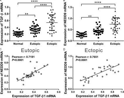
Positive correlation between mRNA levels of
TGF-β1 and NEDD9 in endometriotic tissues
endometrial tissues (P < 0.0001) than in con-trol endometrial tissues. A significant increase in the mRNA level of TGF-β1 and NEDD9 was also observed in ectopic endometriosis tissues as compared with eutopic endometriotic tis-sues (P < 0.0001).
We then assessed whether any relationship existed between mRNA levels of NEDD9 and TGF-β1 in endometriotic tissues. Pearson cor-relation analysis revealed that NEDD9 mRNA level was positively correlated with TGF-β1 mRNA level in eutopic (Figure 5C, r=0.7181, P < 0.0001) and ectopic endometrial tissues (Figure 5D, r=0.7651, P < 0.0001). These data indicated the association of NEDD9 and TGF-β1 during the pathogenesis of endometriosis.
To verify protein expression pattern of NEDD9 and TGF-β1, Western blot and IHC assays were performed. As shown in Figure 6, the same
trend was observed in the protein levels of both NEDD9 and TGF-β1 in three types of endome-trial tissues.
Discussion
[image:6.612.94.518.77.412.2]Although the detailed mechanism underlying the pathogenesis of endometriosis, a tumor like lesion [5, 6], is still unclear, the proliferative and invasive ability of endometrial cells is believed to play a key role in the ectopic growth of endometrial implants. During this process, TGF-β is suspected to be involved [15]. NEDD9, a TGF-β-smad2/3 target gene [18-20], has been regarded as a positive regulator for can-cer cell proliferation and invasion [24, 26-29]. However, there is no study investigated the role of NEDD9 in the development of endometriosis until now. In the present study, we found that NEDD9 was associated with the proliferation and invasion of ESCs, and its expression was
enhanced by TGF-β1. NEDD9 expression was increased in endometriotic tissues, which was positively related with TGF-β1 expression.
Firstly, suppressing NEDD9 expression result-ed in an obvious decrease in cell proliferation (Figure 2) and invasion (Figure 3) of ESCs, which was consistent with previous studies on cancer cells [26-29]. MMPs can facilitate the breakdown of endometrial ECM and aberrant expression of MMPs will lead to the establish-ment and progression of endometriosis. Altered expression of MMP2 and MMP9 has been reported in eutopic and ectopic endometrial tissues [25]. In breast cancer cells, ectopic expression of NEDD9 upregulated the activity of MMP9 [24]. We then tested the effects of NEDD9 knockdown on the expression of MMP2 and MMP9. NEDD9 siRNA transfection notably down-regulated the mRNA and protein levels of MMP2 and MMP9. These data suggested that NEDD9 may promote ESCs invasion by regulat-ing both proteinases.
TGF-β1, a multifunction cytokine, is suggested to play a role in the pathogenesis of endome-triosis [15]. Previous studies have investigated the effect of TGF-β1 on NEDD9 transcription in normal human cell lines and cancer cell lines [18-20]. Here, in vitro experiment demonstrat-ed that mRNA and protein expression of NEDD9 was enhanced by TGF-β1 treatment in ESCs (Figure 4). Moreover, the mRNA (Figure 5) and protein (Figure 6) levels of NEDD9 and TGF-β1 were significantly increased in endometriotic tissues along with the severity of endometrio-sis. More importantly, we found a positive cor-relation between NEDD9 and TGF-β1 mRNA expression in human endometriotic tissues (Figure 5). Our study firstly investigated the expression pattern of NEDD9 in human endo-metriotic tissues and uncovered the associa-tion between NEDD9 and TGF-β1.
[image:7.612.96.525.70.390.2]In summary, NEDD9 played a critical role in the proliferation and invasion of ESCs. NEDD9 expression was up-regulated in endometriotic
tissues, and positively correlated with TGF-β1 expression. Meanwhile, TGF-β1 could induce the expression of NEDD9 in both mRNA and protein levels. Thus, we speculate that TGF-β1 might be involved in the up-regulation of NED- D9 expression in endometriotic tissues, which would probably contribute to the development of endometriosis. Our study may provide a novel therapy target for the treatment of endometriosis.
Acknowledgements
This work was supported by the Natural Science Foundation of China (81370694).
Disclosure of conflict of interest
None.
Address correspondence to: Jun Lin, Women’s Hos- pital, School of Medicine, Zhejiang University; 1 Xueshi Road, Hangzhou 310006, China. Tel: +86 571-89992135; E-mail: linjun@zju.edu.cn
References
[1] Dan CM. A history of endometriosis. J Minim Invasive Gynecol 2012; 19: 269-270.
[2] Rogers PA, D’Hooghe TM, Fazleabas A, Gargett CE, Giudice LC, Montgomery GW, Rombauts L, Salamonsen LA and Zondervan KT. Priorities for endometriosis research: recommendations from an international consensus workshop. Reprod Sci 2009; 16: 335-346.
[3] Arya P and Shaw R. Endometriosis: current thinking. Curr Obstet Gynaecol 2005; 15: 191-198.
[4] Sampson JA. Metastatic or embolic endome-triosis, due to the menstrual dissemination of endometrial tissue into the venous circulation. Am J Pathol 1927; 3: 93-110.43.
[5] Nezhat F, Datta MS, Hanson V, Pejovic T, Ne-zhat C and NeNe-zhat C. The relationship of endo-metriosis and ovarian malignancy: a review. Fertil Steril 2008; 90: 1559-1570.
[6] Scully RE. Classification of human ovarian tu -mors. Environ Health Perspect 1987; 73: 15-25.
[7] Li P, Zhou H, Zhu X, Ma G, Liu C, Lin B and Mao W. High expression of NEDD9 predicts adverse outcomes of colorectal cancer patients. Int J Clin Exp Pathol 2014; 7: 2565-2570.
[8] Shi R, Wang L, Wang T, Xu J, Wang F and Xu M. NEDD9 overexpression correlates with the pro-gression and prognosis in gastric carcinoma. Med Oncol 2014; 31: 852.
[9] Miao Y, Li AL, Wang L, Fan CF, Zhang XP, Xu HT, Yang LH, Liu Y and Wang EH. Overexpression of NEDD9 is associated with altered expression of E-Cadherin, beta-Catenin and N-Cadherin and predictive of poor prognosis in non-small cell lung cancer. Pathol Oncol Res 2013; 19: 281-286.
[10] Wang H, Mu X, Zhou S, Zhang J, Dai J, Tang L, Xiao L, Duan Z, Jia L and Chen S. NEDD9 over-expression is associated with the progression of and an unfavorable prognosis in epithelial ovarian cancer. Hum Pathol 2014; 45: 401-408.
[11] Sima N, Cheng X, Ye F, Ma D, Xie X and Lu W. The overexpression of scaffolding protein NEDD9 promotes migration and invasion in cervical cancer via tyrosine phosphorylated FAK and SRC. PLoS One 2013; 8: e74594. [12] Stajduhar E, Sedic M, Lenicek T, Radulovic P,
Kerenji A, Kruslin B, Pavelic K and Kraljevic Pavelic S. Expression of growth hormone re-ceptor, plakoglobin and NEDD9 protein in as-sociation with tumour progression and metas-tasis in human breast cancer. Tumour Biol 2014; 35: 6425-6434.
[13] Zeinieh M, Salehi A, Rajkumar V and Barker PA. p75NTR-dependent Rac1 activation re-quires receptor cleavage and activation of an NRAGE and NEDD9 signaling cascade. J Cell Sci 2015; 128: 447-459.
[14] Ice RJ, McLaughlin SL, Livengood RH, Culp MV, Eddy ER, Ivanov AV and Pugacheva EN. NEDD9 depletion destabilizes Aurora A kinase and heightens the efficacy of Aurora A inhibitors: implications for treatment of metastatic solid tumors. Cancer Res 2013; 73: 3168-3180. [15] Omwandho COA, Konrad L, Halis G, Oehmke F
and Tinneberg HR. Role of TGF-βs in normal human endometrium and endometriosis. Hum Reprod 2010; 25: 101-109.
[16] Pizzo A, Salmeri FM, Ardita FV, Sofo V, Tripepi M and Marsico S. Behaviour of cytokine levels in serum and peritoneal fluid of women with endometriosis. Gynecol Obstet Invest 2002; 54: 82-87.
[17] Komiyama S, Aoki D, Komiyama M and Noza-wa S. Local activation of TGF-βeta1 at endome -triosis sites. J Reprod Med 2007; 52: 306-312. [18] Morimoto K, Tanaka T, Nitta Y, Ohnishi K, Ka-washima H and Nakatani T. NEDD9 crucially regulates TGF-βeta-triggered epithelial-mesen -chymal transition and cell invasion in prostate cancer cells: involvement in cancer progres-siveness. Prostate 2014; 74: 901-910. [19] Omata Y, Nakamura S, Koyama T, Yasui T,
TGF-βeta-Smad2/3 target gene involved in RANKL-in-duced osteoclastogenesis by comprehensive analysis. PLoS One 2016; 11: e0157992. [20] Zheng M and Mckeownlongo PJ. Regulation of
HEF1 expression and phosphorylation by TGF-βeta 1 and cell adhesion. J Biol Chem 2002; 277: 39599-39608.
[21] Arnold JT, Kaufman DG, Seppälä M and Lessey BA. Endometrial stromal cells regulate epithe-lial cell growth in vitro: a new co-culture model. Hum Reprod 2001; 16: 836-845.
[22] Rajaei S, Mirahmadian M, Jedditehrani M, Ta-vakoli M, Zonoobi M, Dabbagh A and Zarnani AH. Effect of 1,25(OH)vitamin Don cytokine production by endometrial cells of women with repeated implantation failure. Gynecol Endo-crinol 2012; 28: 906-911.
[23] Delbandi AA, Mahmoudi M, Shervin A, Akbari E, Jeddi-Tehrani M, Sankian M, Kazemnejad S and Zarnani AH. Eutopic and ectopic stromal cells from patients with endometriosis exhibit differential invasive, adhesive, and prolifera-tive behavior. Fertil Steril 2013; 100: 761-769. [24] Kong C, Wang C, Wang L, Ma M, Niu C, Sun X,
Du J, Dong Z, Zhu S, Lu J and Huang B. NEDD9 is a positive regulator of epithelial-mesenchy-mal transition and promotes invasion in ag-gressive breast cancer. PLoS One 2011; 6: e22666.
[25] Shaco-Levya R, Benharroch D, Piura B and Sion-Vardy N. Matrix metalloproteinases 2 and 9, E-cadherin, and beta-catenin expression in endometriosis, low-grade endometrial carci-noma and non-neoplastic eutopic endometri-um. Eur J Obstet Gynecol Reprod Biol 2008; 139: 226-232.
[26] Dai J, Van Wie PG, Fai LY, Kim D, Wang L, Poyil P, Luo J and Zhang Z. Downregulation of NEDD9 by apigenin suppresses migration, in-vasion, and metastasis of colorectal cancer cells. Toxicol Appl Pharmacol 2016; 311: 106-112.
[27] Feng J, Zhao J, Xie H, Yin Y, Luo G, Zhang J, Feng Y and Li Z. Involvement of NEDD9 in the invasion and migration of gastric cancer. Tu-mour Biol 2015; 36: 3621-3628.
[28] Singh M, Cowell L, Seo S, O’Neill G and Gole-mis E. Molecular basis for HEF1/NEDD9/Cas-L action as a multifunctional co-ordinator of in-vasion, apoptosis and cell cycle. Cell Biochem Biophys 2007; 48: 54-72.