Original Article
Expression of ZO-1 in laryngeal squamous
cell carcinoma and its prognostic value
Xiaoxia Qiu1*, Zhen Wu2*, Xu Wang3, Huaci Ma1, Shu Zhang4, Hao Wu1
1Department of Otorhinolaryngology, Affiliated Hospital of Nantong University, Nantong 226001, Jiangsu, China;
2Department of Otorhinolaryngology, Second People’s Hospital of Changshu, Changshu 213003, Jiangsu, China; 3Department of Otorhinolaryngology, Affiliated Haian Hospital of Nantong University, Nantong 226600, Jiangsu,
China; 4Department of Pathology, Affiliated Hospital of Nantong University, Nantong 226001, Jiangsu, China. *
Co-first authors.
Received May 17, 2016; Accepted June 1, 2016; Epub November 1, 2016; Published November 15, 2016
Abstract: Laryngeal cancer is one of the most common fatal cancers in head and neck carcinomas, but its mecha-nism is still unclear. The zonula occludens-1 (ZO-1) is closely related to the genesis and development of a variety of respiratory tract and gastrointestinal tumors but the expression of ZO-1 in laryngeal squamous cell carcinoma (LSCC) and its correlation with clinicopathologic features still remain undetermined. The aim to this study is to
iden-tify the clinical significance of ZO-1 in laryngeal cancer. Quantitative real-time polymerase chain reaction (qRT-PCR)
and immunohistochemistry with tissue microarrays were used to characterize the expression of the ZO-1 mRNA and protein in LSCC. The statistical analysis was carried out by combining the follow-up data and clinicopathologic features. Both the ZO-1 mRNA and protein expressions in LSCC are lower than their expressions in corresponding peritumoral tissue (P<0.05); the ZO-1 expression is related to tumor differentiation (P=0.009), clinicopathological stages (P=0.025) and lymphatic metastasis (P=0.013). As shown in the Cox regression analyses, the ZO-1 expres-sion (P=0.035), tumor location (P=0.030), lymphatic metastasis (P=0.002) and tumor differentiation (P=0.004) are independent prognostic factors. The ZO-1 can serve as an independent prognostic factor of LSCC and the high expression of ZO-1 is associated with good prognosis.
Keywords: ZO-1, laryngeal neoplasms, squamous cell carcinoma, tumor marker
Introduction
Laryngeal squamous cell carcinoma (LSCC) is a common malignant tumor of head and neck and its incidence in primary respiratory cancer ranks only second to lung cancer at about 1/4 of global annual head and neck squamous cell carcinomas [1, 2]. Tumor stage, pathologic dif-ferentiation, primary site, and lymph node metastasis of LSCC are correlated with progno-sis. These indexes, however, are insufficient for precise assessment of the tumor. Therefore, a thorough investigation of potential molecular biomarkers in predicting laryngeal SCC progno-sis is of great significance [3, 4].
Tight junctions (TJs) are a sort of top connexin which can mediate the adhesion between epi-thelial cells and endoepi-thelial cells by combining with other cellular connexins [5]. Previous
stud-ies have reported that TJs can serve as barriers for tight junctions of multiple tissues and index-es of permeability function and participate in the maintaining and adjusting the epithelial bar-rier function such as blood brain barbar-rier, entero-cyte and retinal pigment epithelium etc [6, 7]. They have many types including the Occludin, Claudins, Junctional Adhesion Molecule-A (JAM-A) and ZO family etc. Among them, the ZO family is the important factor of epithelial cell struc-ture and function regulation mainly including ZO-1, ZO-2 and ZO-3 [8].
main-taining cell polarity, and, meanwhile, has the characteristics of epithelial marker [9]. Re- searches show that decreased expression of ZO-1 in lung cancer and colon cancer tissues can facilitate the occurrence of lung cancer and colon cancer. The decrease in ZO-1 mRNA may be one of the indicators of early development of lung cancer [10, 11].
The expression of ZO-1 in LSCC and its relation-ship with the clinicopathologic features of LSCC are remain unknown. We used the qRT-PCR to detect the ZO-1 mRNA expression in 18 pairs of fresh LSCC tissues and its corresponding peritumoral tissue. In addition, we prepared tis-sue microarrays of LSCC, and determined the expression of ZO-1 in LSCC and corresponding adjacent tissues using immunohistochemistry, and analyzed its relationship with clinicopatho-logic parameters of patients with LSCC.
Materials and methods
Clinical information and preparation of tissue microarrays
All hospitalized patients between January 2002 and December 2010 were enrolled from the Department of Otolaryngology/Head and Neck Surgery, Nantong University. All patients were diagnosed with laryngeal SCC in accord with the World Health Organization (WHO) criteria [12] and TNM (tumor, node, metastasis) classi-fication (Union Internationale Contre le Cancer [UICC]) and underwent total or partial laryn- gectomy and neck dissection (unilateral or bilateral, radical or functional, based on clinical and surgical findings). Nodal metastasis was confirmed by postoperative histologic examina-tion. In this study, 98 paraffin-fixed tissue sam-ples of LSCC and 26 control samsam-ples were pre-pared as tissue microarrays. Another 18 fresh LSCCs and surrounding normal tissues were collected as controls. None of the patients had received preoperative radiotherapy or chemo-therapy. Complete follow-up data until De- cember 2015 were documented in all these cases.
Tissue microarrays were produced by Shanghai Xinchao Bio-tech Co. Ltd. The representative cancer area was labeled in specific paraffin blocks in accord with hematoxylin and eosin staining results. A tissue array needle was inserted to obtain a 2-mm-diameter tissue
sample, with 1 core for each sample. The tissue was then sequentially aligned into the prepre-pared blank paraffin blocks. The tissue micro-arrays were cut into 4-μm sections and placed on tissue microarray-specific adhesive-coated glass slides. Ethical approval to perform this study was obtained from the Human Research Ethics Committee of the local hospital.
One-step quantitative polymerase chain
reac-tion
The primers of ZO-1 (NC_000015) and endo- genous control GAPDH (NM_002046) were designed with the software Primer Premier 5.0. All primers were synthesized by Shanghai Sangon Biological Engineering Technology and Services. Total RNA was extracted from 18 laryngeal SCC samples and their adjacent peri-tumoral tissues using the TRIzol reagent (Invi- trogen, Carlsbad, CA). Expressions of ZO-1 and GAPDH were determined by real-time PCR with IQ5 (Bio-Rad, Hercules, CA) using SensiMix One-Step Kit–based SYBR Green method (Quantace, London, UK). The primers for ZO-1 were as follows: forward primer 5’-GTGTTGT- GGATACCTTGT-3’ and reverse primer 5’-GAT- GATGCCTCGTTCTAC-3’ for GAPDH, forward pri- mer 5’-TCGGAGTCAACGGATTTGGTCGT-3’, rever- se primer 5’-TGCCATGGGTGGAATCATATTGGA-3’ Amplification conditions consisted of 30 min-utes at 42°C for reverse transcription and 2 minutes at 94°C for Taq activation, followed by 35 cycles of 95°C for 20 seconds, 56°C for 20 seconds, and elongation at 72°C for 30 sec-onds. Each measurement was performed in triplicate.
Immunohistochemical staining and scoring
over-night at the temperature of 4°C and were washed with PBS every ten minutes for three times; secondary antibodies (1:50) were dropped; color development of diaminobenzi-dine (DAB) (Dako, Germany) was controlled under an optical microscope; after being re-dyed with hematoxylin, they were dehydrated with gradient alcohol and hyalinized with hyalin-ize. PBS instead of primary antibodies was used as the negative control.
The double blind method was used to judge the immunohistochemical result; two pathological doctors judged the staining result under the optical microscope. Discordant cases were reevaluated under a double-headed micro-scope to achieve a consensus. In the case of disagreement, the slides were reviewed by a third pathologist until a consensus score was established. The rate of positive cells and stain-ing strength were scored respectively; the per-centage of ZO-1 positive cells was scored as follows: 0 for 0-19%, 1 for 20%~39%, 2 for 40~59%, 3 for 60-100%. Staining strength was scored as follows: 0 for no staining; 1 for weak staining and light yellow; 2 for medium staining and brown; 3 for strong staining and dark brown. The product of the percentage of posi-tive cells and scores of staining strength is the final score, namely the staining index which was defined as follows: 6-9 scores: high expres-sion; 0-4 scores: low expression [13].
Statistical analysis
Statistical analysis was carried out with SPSS19.0 software. The paired t-test was taken for the expression of ZO-1 mRNA in LSCC and peritumoral tissue; the chi-square test was taken for the ZO-1 protein expression in LSCC tissues and peritumoral tissue. The Mann-Whitney U test in the rank sum test was con-ducted to analyze the relation between ZO-1 and all clinicopathological parameters; Survival curves were calculated using the Kaplan-Meier method, and the Log-rank test was used for analysis; Multivariate analysis was performed using Cox proportional hazards model, the risk ratio and its 95% confidence interval were recorded for each marker; P values<0.05 were considered statistically significant.
Results
Patient demographics and tumor clinicopatho-logic characteristics
Among 98 patients (96 men and 2 women; mean age, 58.4 years; age range, 29-84 years), 28 patients (28.6%) presented with supraglot-tic laryngeal SCC and 70 (71.4%) with glotsupraglot-tic laryngeal SCC. Seventeen cases (17.3%) were complicated with lymph node metastasis. Regarding the histologic differentiation, there were 48 patients (49.0%) with well-differentiat-ed, 50 (51.0%) with moderately differentiated and poorly differentiated. In terms of distribu-tion of TNM classificadistribu-tion, 62 patients (63.3%) were found with stage I-II, 36 (33.7%) with stage III-IV tumors. Patients were divided into 2 groups by the total exposure (pack-years) to cigarette smoking [14], 31 patients (31.6%) with <60 pack-years were grouped together and the other group consisted of 67 patients (68.4%) with >60 pack-years. The 5-year overall survival was calculated from the date of sur-gery until the date of death or last follow-up. Five-year overall survival is 70.4% (69/98). Results of quantitative polymerase chain
reac-tion
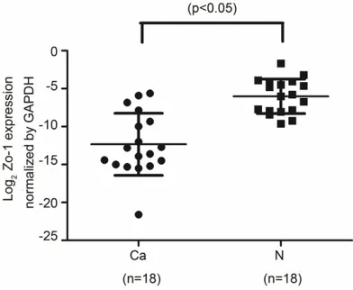
qRT-PCR was used to detected expression of ZO-1 mRNA in LSCC and corresponding adja-cent tissues. Compared with GAPDH internal control mRNA, the expression quantity of ZO-1 mRNA in LSCC tissues (0.0030±0.0014) was lower than in the adjacent nontumorous
[image:3.612.91.287.73.234.2]sues (0.0440±0.0174) (t=-2.310, P=0.034) (Figure 1).
Immunohistochemistry results
ZO-1 expressed is presented as brown parti-cles. The ZO-1 in highly differentiated LSCC cells is mainly located at the cell membranes while the expression of ZO-1 in moderately dif-ferentiated LSCC cells is mainly located in the cytoplasm. The expression of ZO-1 in poorly dif-ferentiated LSCC cells significantly decreases
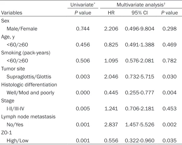
tasis (P=0.001) and ZO-1 expression level (P=0.001) with lifespan of patients with LSCC. As indicated in the analysis of Cox proportional hazard regression model, the tumor location (P=0.030), Histologic differentiation (P=0.004), lymphatic metastasis (P=0.002) and ZO-1 expression level (P=0.035) were independent factors for bad prognosis of LSCC patient (Table 2).
[image:4.612.89.379.71.504.2]In the high ZO-1 expression group among 98 LSCC patients, the total survival rate for 5 years
Figure 2. Expression of ZO-1 protein in LSCC tissues and peritumoral tissue. A1 and A2: High expression of ZO-1 in highly differentiated LSCC tissues. B1 and B2: Medium to high expression of -ZO-1 moderately differentiated LSCC tissues. C1 and C2: Low expression of ZO-1 in poorly differentiated LSCC tissues.
ZO-1 protein expression rate 51.1% (50/98) in tumor tis-sures and 100% (26/26) in ad- jacent noncancerous tissues was of statistical significan- ce (χ2=20.778, P=0.000). The rate of positive expression of ZO-1 in LSCC tissues is re- markably lower than that in the normal peritumoral tissue which was consistent with the qPCR results mentioned ear- lier.
Relationship between ZO-1 protein expression and clini-cal parameters
As for the correlation between ZO-1 expression and LSCC’s clinicopathologic factors (Ta- ble 1), it can be seen from the rank sum test and analysis that ZO-1 expression is relat-ed to the histopathological differentiation of carcinoma (P=0.009), lymphatic metas-tasis (P=0.013) and TNM cla-sification (P=0.025). In con-trast, no significant correla-tion was found between sex, age, smoking, and tumor site. Correlation of ZO-1 protein expression and clinicopatho-logic parameters with
lifes-pan
metas-than 58.3% (28/48) of the low ZO-1 expression group (P=0.015). Kaplan-Meier survival curves showed that the patients with low ZO-1 expression had a shorter survival time than those with high expres-sion (Figure 3).
Discussion
[image:5.612.91.391.95.399.2]LSCC is a common malig-nant tumor of head and neck. As the larynx is an important organ involving breath, pronunciation and deglutition, the growth and invasion of tumor will not only result in a pa- tient’s loss of the above-mentioned functions but also severely threaten a patient’s life [15]. There- fore, searching for the bio-logical markers of LSCC-related molecules for ea- rly diagnosis and accurate treatment is critically sig-nificant for raising the pa- tient’s survival rate, reduc-ing the full larynx removal rate and thus improving the patient’s post-treat-ment living quality. The wholesome inter-epi-thelial connection is an important factor to main-tain the epithelial stabili- ty. Researches show that ZO-1, which is the most important protein for ad- herent junction, has the adhesion and barrier fu- nctions [16]. In addition, ZO-1 also participates in the construction of cellu-lar coupling function, can form a complex with ZO-2 and ZO-3, and forms the major closely-connected structural and functional protein through intercon-nections between its PDZ zone and other connexins
Table 1. Relationship between ZO-1 expression and clinical param-eters
Clinical Parameter n expressionLow expressionHigh Z value* P value* Sex
Male 96 48 (50.0) 48 (50.0) -1.393 0.164 Female 2 0 (0.00) 2 (100.0)
Age, y
<60 39 19 (48.7) 20 (51.3) -0.042 0.967
≥60 59 29 (49.2) 30 (50.1)
Smoking history (pack-years)
<60 31 14 (45.2) 17 (54.8) -0.512 0.609
≥60 67 34 (50.7) 33 (49.3)
Tumor site
Supraglottis 28 21 (75.0) 7 (25.0) 0.875 0.361 Glottis 70 27 (38.6) 43 (61.4)
Histologic differentiation
Well 48 17 (35.4) 31 (64.6) -2.618 0.009 Moderately and poorly 50 31 (62.0) 19 (38.0)
TNM classification
I-II 62 25 (40.3) 37 (59.7) -2.238 0.025 III-IV 36 23 (63.9) 13 (36.1)
Lymph node metastasis
No 81 37 (45.7) 44 (54.3) -2.481 0.013 Yes 17 11 (64.7) 6 (35.3)
*The Z and p values were calculated by rank-sum test. Low and high expression were defined in immunohistochemical staining and scoring of materials and methods.
Table 2. Univariate and multivariate analysis of prognostic variables
Univariate* Multivariate analysis†
Variables P value HR 95% CI P value Sex
Male/Female 0.744 2.206 0.496-9.804 0.298 Age, y
<60/≥60 0.456 0.825 0.491-1.388 0.469
Smoking (pack-years)
<60/≥60 0.506 1.095 0.576-2.081 0.782
Tumor site
Supraglottis/Glottis 0.003 2.046 0.732-5.715 0.030 Histologic differentiation
Well/Mod and poorly 0.000 0.445 0.255-0.777 0.004 Stage
I-II/III-IV 0.005 1.241 0.706-2.181 0.453 Lymph node metastasis
No/Yes 0.001 2.837 1.457-5.526 0.002 ZO-1
High/Low 0.001 0.556 0.322-0.960 0.035
*Statistical analyses were performed by log-rank test. †Statistical analyses were
[image:5.612.91.392.458.697.2]such as actin [17-19]. Reichert M et al found that the β-catenin/Tcf/Lef signaling pathway was activated in the cells expressing the ZO-1 PDZ protein. The mutants of the ZO-1, which encode the PDZ domains (ZO-1 PDZ) but no lon-ger localize at the plasma membrane, induced a dramatic epithelial to mesenchymal transi-tion (EMT) of Madin-Darby canine kidney I (MDCKI) cells and lead to the occurrence of tumor [20].
We observed that both ZO-1 mRNA and protein levels were significantly lower in LSCC tissues than in the corresponding adjacent noncancer-ous tissues. Our study is the first to quantita-tively analyze the expression of tight junction proteins in LSCC, and demonstrates that ZO-1 expression may play an important role in the LSCC tumorigenesis. Besides, higher degree of LSCC differentiation also indicates the higher expression of ZO-1 protein and the expression is low or absent in the poorly differentiated LSCC. This result is consistent with the research of small cell lung cancer [13] and, meanwhile, also proves that ZO-1 has the characteristics of epithelial marker, so it can be used as the opin-ion of indicators of the degree of epithelial dif-ferentiation [9]. In this test, the loss of ZO-1 expression is related to the decrease in differ-entiation degree and distant metastasis of tumor. On the one hand, ZO-1 is the major
pro-tein maintaining epithelial differentiation, so decrease in its expression will lead to the frus-tration of epithelial differentiation. On the other hand, ZO-1 can facilitate the cancer cells to reduce the epithelial polarity and obtain the polarity of interstitial cells. While the integrity of tight junction and adherent junction is lost , the construction of TJs is influenced to promote the occurrence of EMT in cancer cells [21]. The cell polarity malfunction will cause changes in epi-thelial morphology, reduce the intercellular adhesive force and strengthen its invasive-ness. The permeability barrier malfunction will increase the permeability, help tumor cells to absorb abundant nutrient substances, thus promote the abnormal proliferation of tumor cells, and further increase the invasion as well as transfer ability of cancer cells [22].
In this research, the ZO-1 expression, tumor’s differentiation, clinical stages and lymphatic metastasis are independent factors affecting LSCC prognosis. As shown in Kaplan-Meier analyses, the 5-year survival rate of LSCC patients with high ZO-1 expression is notably higher than that of patients with low expression (P<0.05). This proved that ZO-1 has the func-tions to regulate the growth of tumor cells and facilitate the invasion and transfer of tumor cells [11]. Besides, the relation between the LSCC’s primary tumor location, lymphatic me- tastasis, clinicopathological stages and prog-nosis have been verified [23].
In this research, we made a relevant conclusion after making statistical analyses on the ZO-1 protein and LSCC clinical features by using the immunohistochemical method of tissue micro-arrays, so this conclusion has a certain limita-tion: firstly, there was a low quantity of samples especially the lymphatic metastasis samples; secondly, there was a lack of relevant cytologi-cal and animal in vivo experiments.
To sum up, we made a conclusion as follows: ZO-1 may be used as a prognostic factor in LSCC. ZO-1 may also represent a novel thera-peutic target in LSCC. Further studies are required to investigate the biolaogical functions of ZO-1 in LSCC.
Acknowledgements
[image:6.612.91.281.72.266.2]This research is supported by grant from the Postdoctoral funding Project of Jiangsu (130-
Figure 3. Kaplan-Meier with laryngeal SCC by expres-sion of ZO-1. Overall survival rate in patients with
low expression of ZO-1 (blue line) was significantly
2083C), Six Talent Peaks Project in Jiangsu Province (WSW-054), and medical Project of Changshu second hospital (csey201502).
Disclosure of conflict of interest
None.
Address correspondence to: Hao Wu, Department
of Otorhinolaryngology, Affiliated Hospital of
Nan-tong University, NanNan-tong 226001, Jiangsu, China. E-mail: entwuhao@163.com
References
[1] Lin HW and Bhattacharyya N. Staging and sur-vival analysis for nonsquamous cell carcino-mas of the larynx. Laryngoscope 2008; 118: 1003-1013.
[2] Marioni G, Marchese-Ragona R, Cartei G,
Marchese F and Staffieri A. Current opinion in
diagnosis and treatment of laryngeal carcino-ma. Cancer Treat Rev 2006; 32: 504-515. [3] Renkvist N, Castelli C, Robbins PF and Parmiani
G. A listing of human tumor antigens recog-nized by T cells. Cancer Immunol Immunother 2001; 50: 3-15.
[4] Takes RP, Baatenburg De Jong RJ, Alles MJ, Meeuwis CA, Marres HA, Knegt PP, De La Riviere GB, De Wilde PC, Mooi WJ, Hermans J and Van Krieken JH. Markers for nodal metas-tasis in head and neck squamous cell cancer. Arch Otolaryngol Head Neck Surg 2002; 128: 512-518.
[5] Tsukita S and Furuse M. Occludin and claudins in tight-junction strands: leading or supporting players? Trends Cell Biol 1999; 9: 268-273. [6] Jayalekshmi PA, Nandakumar A, Akiba S,
Gangadharan P and Koriyama C. Associations of tobacco use and alcohol drinking with laryn-geal and hypopharynlaryn-geal cancer risks among men in Karunagappally, Kerala, India-Karuna- gappally cohort study. PLoS One 2013; 8: e73716.
[7] Tsukita S, Furuse M and Itoh M. Multifunctional strands in tight junctions. Nat Rev Mol Cell Biol 2001; 2: 285-293.
[8] Bazzoni G, Martinez-Estrada OM, Orsenigo F, Cordenonsi M, Citi S and Dejana E. Interaction of junctional adhesion molecule with the tight junction components ZO-1, cingulin, and occlu-din. J Biol Chem 2000; 275: 20520-20526. [9] Stevenson BR, Siliciano JD, Mooseker MS and
Goodenough DA. Identification of ZO-1: a high
molecular weight polypeptide associated with the tight junction (zonula occludens) in a vari-ety of epithelia. J Cell Biol 1986; 103: 755-766.
[10] Haskins J, Gu L, Wittchen ES, Hibbard J and Stevenson BR. ZO-3, a novel member of the MAGUK protein family found at the tight junc-tion, interacts with ZO-1 and occludin. J Cell Biol 1998; 141: 199-208.
[11] Woods DF and Bryant PJ. ZO-1, DlgA and PSD-95/SAP90: homologous proteins in tight, sep-tate and synaptic cell junctions. Mech Dev 1993; 44: 85-89.
[12] Thompson L. World Health Organization
clas-sification of tumours: pathology and genetics
of head and neck tumours. Ear Nose Throat J 2006; 85: 74.
[13] Ni S, Xu L, Huang J, Feng J, Zhu H, Wang G and Wang X. Increased ZO-1 expression predicts valuable prognosis in non-small cell lung can-cer. Int J Clin Exp Pathol 2013; 6: 2887-2895. [14] Lubin JH, Gaudet MM, Olshan AF, Kelsey K,
Boffetta P, Brennan P, Castellsague X, Chen C, Curado MP, Dal Maso L, Daudt AW, Fabianova E, Fernandez L, Wunsch-Filho V, Franceschi S, Herrero R, Koifman S, La Vecchia C, Lazarus P, Levi F, Lissowska J, Mates IN, Matos E, McClean M, Menezes A, Morgenstern H, Muscat J, Eluf Neto J, Purdue MP, Rudnai P, Schwartz SM, Shangina O, Sturgis EM, Szeszenia-Dabrowska N, Talamini R, Wei Q, Winn D, Zhang ZF, Hashibe M and Hayes RB. Body mass index, cigarette smoking, and alco-hol consumption and cancers of the oral cavi-ty, pharynx, and larynx: modeling odds ratios in pooled case-control data. Am J Epidemiol 2010; 171: 1250-1261.
[15] Fu S, Guo Y, Chen H, Xu ZM, Qiu GB, Zhong M, Sun KL and Fu WN. MYCT1-TV, a novel MYCT1 transcript, is regulated by c-Myc and may par-ticipate in laryngeal carcinogenesis. PLoS One 2011; 6: e25648.
[16] Tsukita S, Yamazaki Y, Katsuno T, Tamura A and Tsukita S. Tight junction-based epithelial microenvironment and cell proliferation. Onco- gene 2008; 27: 6930-6938.
[17] Komaki R, Togashi H and Takai Y. Regulation of
dendritic filopodial interactions by ZO-1 and
implications for dendrite morphogenesis. PLoS One 2013; 8: e76201.
[18] Meerschaert K, Tun MP, Remue E, De Ganck A, Boucherie C, Vanloo B, Degeest G, Vandekerckhove J, Zimmermann P, Bhardwaj N, Lu H, Cho W and Gettemans J. The PDZ2 domain of zonula occludens-1 and -2 is a phosphoinositide binding domain. Cell Mol Life Sci 2009; 66: 3951-3966.
[20] Reichert M, Muller T and Hunziker W. The PDZ domains of zonula occludens-1 induce an epi-thelial to mesenchymal transition of Madin-Darby canine kidney I cells. Evidence for a role of beta-catenin/Tcf/Lef signaling. J Biol Chem 2000; 275: 9492-9500.
[21] Berx G, Raspe E, Christofori G, Thiery JP and Sleeman JP. Pre-EMTing metastasis? Recapi- tulation of morphogenetic processes in cancer. Clin Exp Metastasis 2007; 24: 587-597. [22] Xu J, Kausalya PJ, Phua DC, Ali SM, Hossain Z
and Hunziker W. Early embryonic lethality of mice lacking ZO-2, but Not ZO-3, reveals criti-cal and nonredundant roles for individual zonula occludens proteins in mammalian de-velopment. Mol Cell Biol 2008; 28: 1669-1678.