Original Article
Elevated Aurora B expression contributes to
chemoresistance and poor prognosis in breast cancer
Yiqian Zhang1, Chunling Jiang2, Huilan Li1, Feng Lv1, Xiaoyan Li1, Xiaolong Qian1, Li Fu1, Bo Xu2, Xiaojing Guo1
1Department of Breast Pathology and Lab, Key Laboratory of Breast Cancer Prevention and Therapy, National
Clinical Research Center of Cancer, Tianjin Medical University Cancer Institute and Hospital, Tianjin 300060, China; 2Department of Oncology, Southern Research Institute, Birmingham, AL 35205
Received October 23, 2014; Accepted December 22, 2014; Epub January 1, 2015; Published January 15, 2015
Abstract: B is a major kinase responsible for appropriate mitotic progression. Elevated expression of Aurora-B has been frequently associated with several types of cancer, including breast cancer. However, it is not clear whether the alteration contributes to tumor responses to therapies and prognosis. In this study, we conducted
im-munohistochemistry using antibodies against Aurora-B, S1981p-ATM, Ki67, and p53 in paraffin-embedded tumor
tissues from 312 invasive breast cancer patients. The correlation between disease-free-survival (DFS) andAurora-B expression was analyzed using the Kaplan-Meier method and log-rank test. A Cox proportional hazards regression analysis was used to determine whether Aurora-B was an independent prognostic factor for breast cancer. We found that Aurora-B expression was correlated with the proliferation index (P < 0.001) and p53 expression (P = 0.014) in breast cancer tissues. Further we found that Aurora-B expression was associated with lymph node metastasis (P = 0.002) and histological grade (P = 0.001). Multivariate analyses indicated that elevated Aurora-B expression pre-dicted a poor survival. In a subgroup of patients that received neoadjuvant chemotherapy, we found that elevated Aurora-B contributed to chemoresistance (P = 0.011). In conclusion, elevated Aurora-B expression in breast cancer patients contributes to chemoresistance and predicts poor prognosis.
Keywords: Aurora-B, chemoresistance, prognosis, breast cancer
Introduction
Signal transduction pathways involving in the cell cycle are typically aberrantly regulated in breast cancer. Among them is the Aurora-B kinase, a critical mitotic kinase required for the chromosome segregation, the spindle assem-bly checkpoint and cytokinesis [1]. Aurora-B is a primary kinase required for Serine 10 phos-phorylation of histone H3 [2]. Aurora-B has been shown to regulate many of the genome stability maintenance proteins. For example, recent studies have shown that Aurora-B phos-phorylates p53 on several residues (Ser183, Thr211, and Ser215) to accelerate the degra-dation of p53 through the polyubiquitination-proteasome pathway [3]. Aurora-B is also required for the mitotic activation of the ATM kinase, an essential kinase required for the DNA damage response [4]. Aurora-B phosphor-ylation of ATM on serine 1403 leads to mitotic ATM activation and required for ATM to send
signals to Bub1 and Mad1 in the spindle check-point [4-6]. These regulatory mechanisms involving Aurora-B indicate the functional
sig-nificance of Aurora-B in the maintenance of
genomic and chromosomal stability. However, Aurora-B overexpression has been shown in a variety of breast cancer cell lines [7] and tumor tissues [8], suggesting there is an aberrant reg-ulatory system in Aurora-B expression in breast cancer. The fact that overexpression of Aurora-B has been implicated in tumor formation in pre-clinical studies indicates gain-of-function when overexpressed. Despite its role in breast cancer progression, clinical implications of Aurora-B overexpression in breast cancer patients is less clear as controversial data have debated the role of elevated expression of Aurora-B and its related kinase Aurora-A in predicting survival [9, 10].
assessed 312 invasive breast cancer patients regarding expression of Aurora-B, ATM Serine 1981, p53 and Ki-67 (one of the important
pro-liferation index). Interestingly we find that
Aurora-B expression is correlated with poor sur-vival and that elevated Aurora-B is associated with chemoresistance.
Materials and methods
Human breast cancer tissue samples
Paraffin-embedded materials from 312 inva -sive breast cancer patients with operable breast cancer treated in 2005-2006 was pro-vided by the Department of Breast Cancer Pathology and Research Laboratory, Tianjin Medical University Cancer Hospital, Tianjin, China, with the approval of the Tianjin Medical University Institutional Review Board, and informed consent had been obtained. Clinical tumor sizes and nodal status were determined before treatment by physical examination (with or without axillary ultrasound) and with
diag-nostic fine-needle aspiration as required. In
these cases, there were 70 patients (clinical stage II-III) who were treated with preoperative neoadjuvant chemotherapy containing sequen-tial taxane and anthracycline-based regimens. The chemotherapeutic response was evaluat-ed by the following standard: excellent response (pathologically complete response or minimal residual cancer burden [RCB-I]), or lesser response (moderate or extensive residual can-cer burden [RCBII/III]) [11, 12].
All cases were female, 26-80 years of age (mean age 52.1 years). There were 176 cases (56.4%) were involved in lymph node metasta-sis. The follow-up time was 14-82 (mean 63.91) months. All patients were treated either with
the modified radical mastectomy (n = 186) or
breast-conserving therapy (n = 126).
Pathology
The post-surgical size of the tumor was mea-sured in fresh specimens. Tumors were cut into
0.5-cm slices, fixed in 4% buffered formalde
-hyde, and embedded in paraffin. Paraffin sec
-tions were cut into highly standardized 4-μm
sections for hematoxylin-eosin (H&E) and immunostaining. Grade and TNM stage were assessed according to World Health Organi- zation criteria.
Antibodies
Antibodies against Aurora-B, S1981p-ATM, Ki67, p53, Estrogen Receptor (ER) and Progestogen Receptor (PR) were purchased from Abcam (Cambridge, MA, USA). All horse-radish-peroxidase conjugated secondary anti-bodies were obtained from Santa Cruz biotechnology.
Immunohistochemistry
Immunohistochemistry using the avidin-biotin-immunoperoxidase technique was performed for Aurora-B, S1981p-ATM, Ki67, p53, ER and PR in the 312 cases of clinical samples.
Sections of formalin-fixed tissues from all cases
were performed using a standard protocol.
Briefly, 4 μm tissue sections on coated slides
were heated for antigen retrieval, pretreated with a 3% solution of hydrogen peroxide for 5-10 minutes, rinsed and incubated with 10% normal goat serum as a blocking agent. The sections were then incubated sequentially with the primary antibodies, a biotinylated second-ary antibody and avidin-peroxidase conjugate. All steps were preceded by rinsing of sections with PBS (pH 7.6). The chromogen was 3, 3’-diaminobenzidine (DAB). The sections were counterstained with hematoxylin, dehydrated and mounted. The immune-reaction for Aurora-B, S1981p-ATM, Ki67, p53, ER and PR in the nuclei of tumor cells was evaluated inde-pendently by two experienced pathologists. For
the expression of Aurora-B, nuclei with fine
granular staining were counted, and (-/+) was < 5% of the cells stained, (++) was 5-10% of the cells stained, and (+++) was > 10% of the cells stained. ER and PR was determined positive if
finding of ≥ 1% of tumor cell nuclei were immu -noreactive. S1981p-ATM, Ki67 and p53 were scored as previously described: (-) = no positive cells, (+) = 1-10% of the cells stained, (++) = 11-50% of the cells stained, and (+++) = 51-100% of the cells stained [13].
Antigen retrieval and antibody dilution were optimized prior to the study onset. To ensure uniform handling of samples, all sections were processed simultaneously. Histology was mounted onto Superfrost Plus slides (Sigma) for immuno-histochemical analysis.
Survival analysis
than breast cancer, local or regional recurrenc-es, or the development of a second, primary cancer, including contralateral breast cancer. If a patient’s status during follow-up indicated a
confirmed metastasis without a recurrence
date, the follow-up visit date was used. Age,
time to first recurrence, and survival time were
calculated relative to the primary diagnosis date. Kaplan-Meier survival curves were con-structed, and between-group differences were tested using the log-rank test. The relative importance of potential prognostic variables was tested using Cox-proportional hazard
anal-ysis and expressed with a 95% confidence
interval (CI).
Statistical analysis
Data were analyzed by Student’s t test and Pearson Correlation test. Patients’ follow-up data were acquired, and survival duration was calculated from the date of diagnosis to the
date of progression or last follow-up in December 2013. The correlation between dis-ease-free-survival (DFS) and Aurora-B expres-sion was analyzed using the Kaplan-Meier method and log-rank test. A Cox proportional hazards regression analysis was used to deter-mine whether Aurora-B was an independent prognostic factor for breast cancer. The statisti-cal analysis was performed with the use of soft-ware packages SPSS version 16.0 (WPSS Ltd., Surrey, United Kingdom). All statistical tests were two-sided, and a P value of < 0.05 was
considered significant.
Results
Elevated expression of Aurora B is correlated with the proliferation index and p53 expres-sion in breast cancer tissues
To investigate the clinical significance of
Aurora-B expression, we conducted
immuno-Figure 1. Immunohistochemical study revealing representative images of invasive breast cancer and normal breast
[image:3.612.92.518.70.386.2]histochemistry in 312 invasive breast cancer patients using antibodies recognizing Aurora-B, Ki-67, p53 and the activated form of ATM (ATM-S1981p) (Figure 1). These antibodies were
pre-screened and selected to ensure specificity.
The Pearson correlation test was used to ana-lyze the semi-quantitative data. We found that Aurora-B expression showed a statistically
sig-nificant correlation (P < 0.001) with that of Ki-67, an important proliferation marker (Table 1). However, there is no correlation with the expression of ATM-S1981p. In contrast, Auro- ra-B showed a strong correlation with the p53 expression level with the p-value of 0.014 (Table 2).
Aurora-B expression and clinicopathologic features
We then analyzed the expression pattern of Aurora-B with several clinicopathologic param-eters in the tissues. We found that Aurora-B expression strongly correlated with the histo-logical grade (P = 0.001) and status of lymph node metastasis (P = 0.002) (Table 3). However, ATM-S1981p expression showed a different correlation pattern as it only correlated with the lymph node metastasis (P = 0.001). This is
con-sistent with our previously findings that ATM is
hyperactive in breast cancer tissues with lymph node metastasis [13]. Unlike the total ATM
expression pattern we tested before [14], nei-ther Aurora-B nor ATM-S1981p expression
showed a statistically significant correlation
with the ER status.
Elevated Aurora-B expression is correlated with the poor prognosis in breast cancer patients
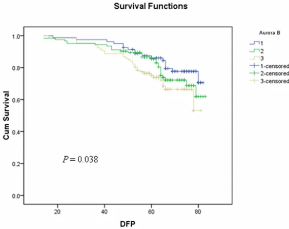
To further evaluate whether Aurora-B expres-sion can predict prognosis, we performed a sur-vival analysis of DFS in all patients. Kaplan-Meier survival curves showed that higher
expression of Aurora-B significantly correlated
with the poor survival in these cases (P = 0.038, Figure 2). A multivariate Cox regression analy-sis demonstrated that Aurora-B expression is an independent prognostic indicator of breast cancer DFS (HR = 1.39, 95% CI = 1.04-1.86) (Table 4).
Elevated Aurora-B expression and chemoresis-tance
To further investigate the clinical relevance of elevated Aurora-B expression with the poor prognosis, we hypothesized that altered expres-sion of Aurora-B might contribute to resistance to chemosensitivity. To test this notion, we
ana-lyzed the clinical data and identified 70 patients
(70/312, 22.4%) who had received pre-opera-tive neoadjuvant chemotherapy (Table 5). Because these patients have primary tumors that were measurable for their responses to the chemotherapeutic drugs (containing sequential taxane and anthracycline–based regimens), we were able to assess the chemosensitivity and its correlation with expression of Aurora-B and ATM-S1981p. We found that in 39 cases (39/70, 55.7%) who were
chemotherapy-resis-tant (as defined by RCBII/III), 82.1% (32/39)
had higher expression of Aurora-B (++~+++). The Pearson correlation test showed that the
expression of Aurora-B significantly correlated
with the chemoresistance (P = 0.011) (Table 5),
indicating a clinical significance of Aurora-B
expression. In contrast, ATM-S1981p did not show the correlation with chemoresistant. Discussion
Defining a biomarker that can predict the clini -cal response to therapies will be of criti-cal to personalized medicine for breast cancer
thera-pies. In this report, we show for the first time
that elevated Aurora-B expression in breast Table 1. Correlations of expression of Aurora
B, ATM-S1981p with Ki67 Ki67
*P
-/+ ++ +++
Aurora B -/+ 35 37 9 < 0.001
++ 24 51 49
+++ 23 35 49
ATM-S1981p -/+ 27 33 32 0.460
++ 23 47 45
+++ 32 43 30
*Pearson Correlation Test.
Table 2. Correlation of expression of p53 with Aurora B
p53 *P
-/+ ++ +++
Aurora B -/+ 25 44 12 0.014
++ 28 57 39
+++ 29 36 42
cancer tissues indicates chemoresistance and poor survival, highlighting a predictive value of Aurora-B expression.
There has been debating data regarding the role of the Aurora-kinase A and B in breast can-cer prognosis. For instance, an earlier report
when in a period that significant changes had
been seen regarding available chemotherapeu-tic drugs. Because Aurora-B’s role in
[image:5.612.91.522.83.310.2]chemore-sistance was not previously identified, chemo -therapeutic regimens that were available to the patients at the time of treatment should be carefully re-examined.
Table 3. Correlations of expression of Aurora B, p53 and S1981p-ATM with clinico-pathologic features Aurora B
*P p53 *P S1981p-ATM *P
-/+ ++ +++ -/+ ++ +++ -/+ ++ +++
Tumor Size (cm) ≤ 2 37 37 44 0.174 31 55 32 0.359 31 45 42 0.606
2-5 25 45 32 27 48 27 35 34 33
> 5 19 42 31 24 34 34 26 36 30
Nodal status - 48 50 38 0.002 38 70 28 0.006 55 40 41 0.001
+ 33 74 69 44 67 65 37 75 64
TNM stage I 21 22 21 0.165 17 32 15 0.142 23 19 22 0.540
II 50 71 59 47 83 50 53 68 59
III 10 31 27 18 22 28 16 28 24
Grade 1 26 25 11 0.001 17 29 16 0.074 17 16 29 0.553
2 31 58 49 43 58 37 47 51 40
3 24 41 47 22 50 40 28 48 36
ER status - 42 70 46 0.180 38 71 49 0.413 45 63 50 0.822
+ 39 54 61 44 66 44 47 52 55
PR status - 41 71 64 0.219 47 76 53 0.957 49 68 59 0.702
+ 40 53 43 35 61 40 43 47 46
*Pearson Correlation Test.
Figure 2. Kaplan-Meier survival curves of the 312 breast cancer patients with different expression levels of Aurora-B. Kaplan-Meier survival curves
showed that higher expression of Aurora-B significantly correlated with the
poor survival in these cases (P = 0.038).
[image:5.612.89.371.343.566.2]Table 4. Cox regression analysis of breast cancer DFS in relation to Aurora-B expression
Univariate analysis Multivariate analysis Characteristic HR (95% CI) P HR (95% CI) P Age 1.00 (0.98, 1.03) 0.793 0.99 (0.98, 1.02) 0.790 Stage 1.99 (1.39, 2.86) 1.89 × 10⁻⁴ 1.84 (1.27, 2.67) 0.001 Radiotherapy 2.05 (1.31, 3.20) 0.002 1.87 (1.19, 2.94) 0.007 Aurora B 1.39 (1.04, 1.86) 0.028 1.35 (1.00, 1.83) 0.050
How elevated Aurora-B expression contributes to chemoresistance remains to be further examined. It is likely that Aurora-B expression promotes the cellular survival in response to chemotherapeutic drugs. It is also possible that Aurora-B overexpression contributes to radiore-sistance, although the clinical responsiveness
of breast cancer radiotherapy is difficult to
assess independently in the majority of patients. Further studies in preclinical models are warranted.
Our recently findings have demonstrated
Aurora-B’s connection to ATM in mitosis as shown by the dependency of Aurora-B on mitot-ic ATM Serine 1403 phosphorylation [4]. However, we did not observe the clinical rele-vance of the two parameters. In contrast, we observed the correlation of Aurora-B expres-sion with p53 expresexpres-sion in these tissues. A recent study has shown negative regulation of Aurora-B in p53, as pharmacological inhibition of Aurora-B in cancer cells with wild-type p53 increased the p53 protein level and expression of p53 target genes to inhibit tumor growth [3]. It is possible that Aurora-B overexpression in breast cancer tissues might selectively up-reg-ulate mutant p53.
In summary, our data shown here provide strong evidence demonstrating that, in breast
Natural Science Foundation of China (Grant No. 30930038) to Li Fu, and National Natural Science Foundation of China (Grant No. 81302292) to Xiaolong Qian.
Disclosure of conflict of interest
None.
Address correspondence to: Dr. Xiaojing Guo, Department of Breast Pathology and Lab, National Clinical Research Center of Cancer, Tianjin Medical University Cancer Institute and Hospital, West Huanhu Road, Tianjin 300060, China. Tel: 86-22-27630396; Fax: 86-22-23342606; E-mail: guoxiao-jing0728@126.com
References
[1] Carmena M, Earnshaw WC. The cellular geog-raphy of aurora kinases. Nat Rev Mol Cell Biol 2003; 4: 842-54.
[2] Crosio C, Fimia GM, Loury R, Kimura M, Okano Y, Zhou H, Sen S, Allis CD, Sassone-Corsi P. Mi-totic phosphorylation of histone H3: spatio-temporal regulation by mammalian Aurora ki-nases. Mol Cell Biol 2002 ; 22: 874-85. [3] Gully CP, Velazquez-Torres G, Shin JH,
Fuentes-Mattei E, Wang E, Carlock C, Chen J, Rothen-berg D, Adams HP, Choi HH, Guma S, Phan L, Chou PC, Su CH, Zhang F, Chen JS, Yang TY, Yeung SC, Lee MH. Aurora B kinase phosphory-Table 5. Correlations of expression of Aurora B, ATM-S1981p with
chemoresistance
Neoadjuvant chemotherapy (N)
*P
chemoresistant chemosensitive
Aurora B -/+ 7 14
++ 14 12 0.011
+++ 18 5
S1981p-ATM -/+ 13 9
++ 19 10 0.137
+++ 7 12
*Pearson Correlation Test.
cancer, elevated Aurora-B expression is associated with chemoresistance and poor survival. Because several Aurora-B Inhibitors have been advanced to clinical trials, identifying Aurora-B as a pre-dicting marker of chemore-sistance should provide ratio-nale for designing combi- nation therapy using avail-able chemotherapeutic drugs with these Aurora-B inhibi- tors.
Acknowledgements
[image:6.612.90.378.221.327.2]lates and instigates degradation of p53. Proc Natl Acad Sci U S A 2012; 109: E1513-22. [4] Yang C, Tang X, Guo X, Niikura Y, Kitagawa K,
Cui K, Wong ST, Fu L, Xu B. Aurora-B mediated ATM serine 1403 phosphorylation is required for mitotic ATM activation and the spindle checkpoint. Mol Cell 2011; 44: 597-608. [5] Yang C, Wang H, Xu Y, Brinkman KL, Ishiyama
H, Wong ST, Xu B. The kinetochore protein Bub1 participates in the DNA damage re-sponse. DNA Repair (Amst) 2012; 11: 185-91. [6] Yang C, Hao J, Kong D, Cui X, Zhang W, Wang H,
Guo X, Ma S, Liu X, Pu P, Xu B. ATM-mediated Mad1 Serine 214 phosphorylation regulates Mad1 dimerization and the spindle assembly checkpoint. Carcinogenesis 2014; 35: 2007-13.
[7] Tatsuka M, Katayama H, Ota T, Tanaka T, Odashima S, Suzuki F, Terada Y. Multinucleari-ty and increased ploidy caused by overexpres-sion of the aurora- and Ipl1-like midbody-asso-ciated protein mitotic kinase in human cancer cells. Cancer Res 1998; 58: 4811-6.
[8] Keen N, Taylor S. Aurora-kinase inhibitors as anticancer agents. Nat Rev Cancer 2004; 4: 927-36.
[9] Nadler Y, Camp RL, Schwartz C, Rimm DL, Kluger HM, Kluger Y. Expression of Aurora A (but not Aurora B) is predictive of survival in
breast cancer. Clin Cancer Res 2008; 14: 4455-62.
[10] Royce ME, Xia W, Sahin AA, Katayama H, John-ston DA, Hortobagyi G, Sen S, Hung MC. STK15/Aurora-A expression in primary breast tumors is correlated with nuclear grade but not with prognosis. Cancer 2004; 100: 12-9. [11] Liedtke C, Hatzis C, Symmans WF, Desmedt C,
Haibe-Kains B, Valero V, Kuerer H, Hortobagyi GN, Piccart-Gebhart M, Sotiriou C, Pusztai L. Genomic grade index is associated with re-sponse to chemotherapy in patients with breast cancer. J Clin Oncol 2009; 27: 3185-91. [12] Symmans WF, Peintinger F, Hatzis C, Rajan R,
Kuerer H, Valero V, Assad L, Poniecka A, Hen-nessy B, Green M, Buzdar AU, Singletary SE, Hortobagyi GN, Pusztai L. Measurement of re-sidual breast cancer burden to predict survival after neoadjuvant chemotherapy. J Clin Oncol 2007; 25: 4414-22.
[13] Sun M, Guo X, Qian X, Wang H, Yang C, Brink-man KL, Serrano-Gonzalez M, Jope RS, Zhou B, Engler DA, Zhan M, Wong ST, Fu L, Xu B. Ac-tivation of the ATM-Snail pathway promotes breast cancer metastasis. J Mol Cell Biol 2012; 4: 304-15.
[14] Guo X, Yang C, Qian X, Lei T, Li Y, Shen H, Fu L,