Int J Clin Exp Pathol 2016;9(10):10563-10568 www.ijcep.com /ISSN:1936-2625/IJCEP0034487
Original Article
Nuclear expression of SOX18 in cancer cells indicates
a poor prognosis in patients with hepatocellular
carcinoma
Ya-Zhou Wang1*, Hua-Hu Guo1*, Wei-Hua Zhu2, Da-Fang Zhang2, Xiao Cui1, Xin Yu1, Xi-Sheng Leng2, Shu Li2 1Beijing Key Surgical Basic Research Laboratory of Liver Cirrhosis and Liver Cancer, Peking University People’s Hospital, Beijing, China; 2Department of Hepatobiliary Surgery, Peking University People’s Hospital, Beijing, China. *Equal contributors.
Received June 27, 2016; Accepted August 30, 2016; Epub October 1, 2016; Published October 15, 2016
Abstract: Transcription factor SOX18 is involved in the development of many tumors, but SOX18 protein expression in hepatocellular carcinoma (HCC) has not been studied. Thus, we measured SOX18 protein expression in HCC and correlated its significance for patients with HCC. After measuring expression and identifying the location of SOX18 in 153 paired HCC and adjacent non-tumor tissues, we found that SOX18 protein was chiefly expressed in the HCC cell nucleus, but was mainly expressed in normal liver cell cytoplasms. Also, nuclear expression of SOX18 protein was associated with TNM stage (P<0.01) and vascular invasion (P<0.01) of HCC. Furthermore, greater nuclear expres-sion of SOX18 protein indicated shorter 3-year overall and disease-free survival of patients with HCC. Thus, SOX18 may be a prognostic marker for HCC.
Keywords: SOX18, nuclear expression, Hepatocellular carcinoma, prognosis
Introduction
Hepatocellular carcinoma (HCC) is the fifth
mo-st common cancer and the second leading cau- se of cancer-associated deaths worldwide [1, 2]. Approximately, 675,000 new HCC cases are diagnosed annually globally and although treat-ments including hepatic resection, chemother-apy and radiotherchemother-apy offer hope, the 5-year survival for liver cancer patients is only 16.6% [3]. Thus, better understanding of underlying molecular mechanisms of hepatocarcinogene-sis is need.
The SOX (Sex-determining region on the Y chro-mosomerelated high mobility group box) genes are a well-conserved gene family that encodes a group of transcription factors and SOX18 belongs to the subgroup F of SOX family [4, 5]. Previous studies indicate that SOX18 contrib-utes to neonatal and postnatal vascularization [6, 7], and SOX18 loss function contributes to cardiovascular and hair follicle defects in ragg- ed mice and hypotrichosis-lymphodema-telean- giectasia (HLT) syndrome in humans [8, 9].
Recently, SOX18 has been reported to be invo- lved in tumor development: over-expression of SOX18 promotes migration and invasion of os- teosarcoma and cervical carcinoma cell [10, 11]. Also, over-expression of SOX18 indicates poor prognosis for non-small cell lung cancer and gastric cancer patients [12, 13]. However, SOX18 protein in HCC has not been reported. We measured the expression and location of SOX18 in HCC tissues and analyzed an associa-tion between SOX18 locaassocia-tion in HCC cells and clinicopathological features of HCC patients. We observed that the SOX18 protein mainly localized to the nucleus of HCC cells, but was
chiefly in the cytoplasm of non-tumor liver cells.
Also, nuclear SOX18 protein expression indicat-ed poor prognosis for HCC patients.
Materials and methods
Patients and tissue samples
his-topathologically at Peking University People’s Hospital from 2009 to 2012. Samples were snap-frozen in liquid nitrogen and then stored
at -80°C or fixed in formaldehyde solution. The
clinicopathological features of patients and samples were collected from the patient infor-mation management system of our hospital. The TNM system of the International Union Against Cancer and American Joint Committee on Cancer was used to judge the tumor stages and the Edmondson-Steiner grading system was used to assess the histological grade [14, 15]. This study was conducted with the approv-al of the Ethics Committee of Peking University People’s Hospital.
Immunohistochemistry
HCC tissues were fixed in 10% formaldehyde
solution after resection and then embedded in
paraffin. Next, 5-µm-thick sections were cut from the paraffin blocks and placed on poly-L-lysine-coated slides. Slides were deparaffinized
through xylene and rehydrated in graded ethyl alcohols and then rinsed in distilled water. After quenching endogenous peroxidase activity with 0.3% H2O2 for 30 min at room temperature, slides were autoclaved for 8 min in sodium citrate buffer (10 mM, pH 6.0) for antigen retrieval. Slides were blocked with 10% normal goat serum (Beyotime Biotechnology, Beijing, China) for 30 min and incubated with
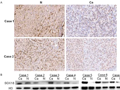
[image:2.612.93.514.70.388.2]anti-Figure 1. Expression of SOX18 in hepatocellular carcinoma (HCC) and adjacent non-tumor liver tissues. A. SOX18 protein mainly localized to the nucleus of HCC cells, but was chiefly in the cytoplasm of non-tumor liver cells. B. Western Blot analysis of 7 representative cases showed that the nuclear expression of SOX18 was increased in HCC cells compared with non-tumor liver cells. (Ca, HCC tissues; N, non-tumor liver tissues).
Table 1. SOX18 expression in HCC cells and non-tumor liver cells
Tissues SOX18 expression in nucleus P value SOX18 expression in cytoplasm P value
Low High Low High
[image:2.612.90.525.474.527.2]Nuclear expression of SOX18 in liver cancer cells indicates a poor prognosis
SOX18 antibody (1:50, Santa Cruz Biotech- nology, Santa Cruz, CA, USA) at 4°C overnight. Next, the slides were incubated with biotin-con-jugated secondary antibody (1:100, Zhongshan Jinqiao Biology Technology Company, Beijing, China) for 45 min at room temperature, and the 3, 3-diaminobenzidine tetrahydrochloride (DAB) was used as a chromogen. Finally, slides were counterstained with hematoxylin. Each slide was assessed by 2 double-blinded pathologists in 2 respects: the proportion and the staining intensity of positive cells. The proportion of po- sitive cells was graded as follows: grade 0, no positive cells; grade 1, 1%-25% positive cells; grade 2, 25%-50% positive cells; grade 3, >50% positive cells. The staining intensity, ranging from light brown to dark brown, was graded as follows: 0, negative; 1, weak intensity; 2,
mod-erate intensity; 3, strong intensity. The final
score of each slide was generated by multiply-ing the two values. A score of <6 was regarded
as low, while a score of ≥6 was regarded as
high [16].
Nuclear protein extraction and Western blot-ting
Nuclear protein of each clinical specimen was extracted with a nuclear extraction kit (Thermo
Fisher Scientific, Waltham, MA, USA) according
to the manufacture’s protocol, and the concen-tration was measured by taking bovine serum
albumin (BSA) as a standard. Equal amount of nuclear protein was separated by 10% SDS-PAGE and then transferred to PVDF membranes (PALL life science, New York, USA). After blocked with 5% skim milk, the membranes were incu-bated with anti-human SOX18 antibody (1:100, Santa Cruz Biotechnology, Santa Cruz, CA, USA) and anti-human Histone H3 antibody (1:100, Santa Cruz Biotechnology, Santa Cruz, CA, USA), which was taken as an internal control, overnight at 4°C. Next, the membranes were incubated with the corresponding secondary antibody (1:2000, Zhongshan Jinqiao Biology technology Company, Beijing, China) conjugat-ed with horseradish peroxidase (HRP) for 1 h at room temperature. Finally, the ECL Western Blotting system (Thermo, South Logan, UT, USA) was used to detect the immunolabeled proteins.
Statistical analysis
SPSS 17.0 software (SPSS Inc., Chicago, IL,
USA) was used for statistical analysis. The χ2 test was used to estimate correlations between expression of SOX18 and clinicopathologic
[image:3.612.93.525.96.322.2]fea-tures, and the χ2 test was also used for assess-ing SOX18 in HCC and non-tumor liver tissues. A Kaplan-Meier method and a log-rank test were used to calculated 3-year overall survival (OS) and disease-free survival (DFS). A multi-variate Cox model was used to for multimulti-variate Table 2. Nuclear expression of SOX18 and clinicopathological features in 153 hepatocellular carci-noma patients
Features Low sox18 expression High sox18 expression P value
Age (year) <50 48 (31.37%) 35 (22.88%) 0.531
≥50 40 (26.14%) 30 (19.61%)
Sex Female 8 (5.22%) 7 (4.58%) 0.787
Male 80 (52.29%) 58 (37.91%)
Tumor size (cm) <5 40 (26.14%) 25 (16.34%) 0.412
≥5 48 (31.37%) 40 (26.14%)
Vascular invasion None 42 (27.45%) 17 (11.11%) 0.007
Present 46 (30.07%) 48 (31.37%)
Tumor grade G1-G2 14 (9.15%) 11 (7.19%) 0.867
G3-G4 74 (48.37%) 54 (35.30%)
TNM stage I-II 72 (47.06%) 35 (22.88%) <0.001
III-IV 16 (10.46%) 30 (19.61%)
Recurrence No 42 (27.45%) 22 (14.38%) 0.099
Yes 46 (30.07%) 43 (28.10%)
AFP(μg/L) <400 45 (29.41%) 34 (22.22%) 1.000
analysis, and results are presented with 95%
confidence intervals (CI) and hazards ratio (HR).
P<0.05 was considered to be statistically sig-
nificant.
Results
Expression of SOX18 protein in HCC
Immunohistochemistry (IHC) results showed th- at SOX18 was expressed in HCC tissues and in adjacent non-tumor tissues. In HCC cells, SOX-
18 expression was chiefly nuclear and in adja -cent non-tumor cells, SOX18 staining was ma- inly localized to the cytoplasm (Figure 1A; Table 1).
To verify IHC, we extracted nuclear protein from HCC cells and matched non-tumor liver cells
and use Western blot to confirm that HCC cells
had more nuclear SOX18 expression compared with non-tumor liver cells (Figure 1B).
SOX18 protein nuclear expression and HCC clinicopathological features
Using a χ2 test, we assessed patient age, sex, tumor size, vascular invasion, TNM stage, tumor grade, recurrence and serum AFP (Table 2), and we noted that nuclear expression of SOX18
protein was significantly correlated to vascular
invasion (P<0.01) and TNM stage (P<0.01).
Prognostic value of nuclear expression of SOX18 in HCC
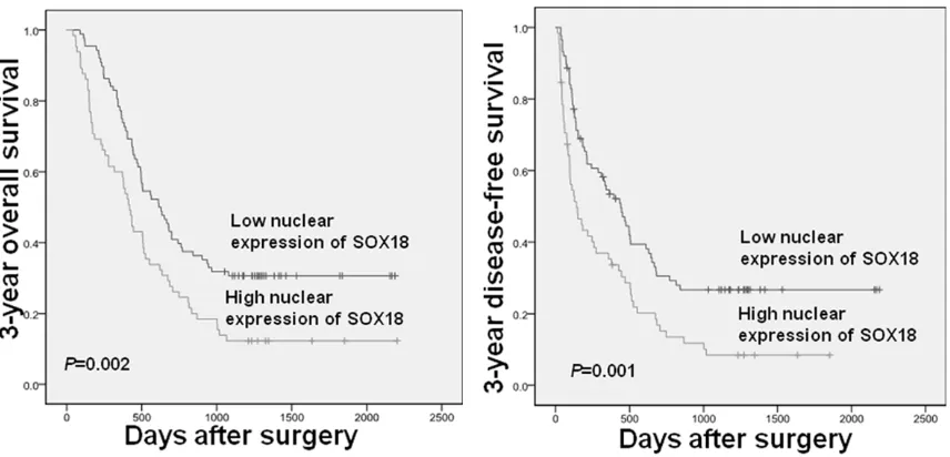
[image:4.612.93.521.72.278.2]The Kaplan-Meier method was used to investi-gate nuclear expression of SOX18 protein and overall survival (OS) and disease-free survival (DFS). Figure 2 showed that patients with more Table 3. Multivariate survival analysis of 3-year overall survival and disease-free survival in 153 HCC patients
features 3-year overall survival 3-year disease-free survival
HR 95% CI P value HR 95% CI P value
Tumor size 1.424 0.964-2.104 0.076 1.546 1.049-2.278 0.028
TNM stage 1.100 0.725-1.668 0.655 0.996 0.656-1.513 0.985
Tumor grade 1.730 0.938-3.191 0.079 2.086 1.145-3.800 0.016
Vascular invasion 1.663 1.051-2.632 0.030 1.921 1.216-3.034 0.005
Serum AFP 1.072 0.730-1.573 0.724 1.305 0.885-1.926 0.179
[image:4.612.91.523.363.470.2]Nuclear expression of SOX18 1.640 1.123-2.395 0.010 1.716 1.170-2.518 0.006
Nuclear expression of SOX18 in liver cancer cells indicates a poor prognosis
nuclear expression of SOX18 tended to have prolonged OS (P<0.01, a) and DFS (P<0.01, b) compared to those with low nuclear SOX18 expression. Then, a multivariate Cox model was used for multivariable analysis, and nuclear SOX18 expression was an independent poor prognostic factor for both 3-year OS and DFS (Table 3).
Discussion
Recently, SOX18 has been shown to be over-expressed in many tumors. Wang’s group repor- ted that the SOX18 mRNA was over-expressed in HCC compared with non-tumor tissues [17]. However mRNA does not always approximate protein. Recent studies in non-small cell lung cancer indicated non-small cell lung cancer tis-sues had more SOX18 protein but less mRNA expression compared with paired normal lung tissues [12, 18, 19].
Thus, we measured the expression and loca-tion of SOX18 protein in cells, and HCC cells had the greatest SOX18 protein in the nucleus, but non-tumor liver cells had more cytoplasmic SOX18 protein expression. Nuclear expression of SOX18 protein was associated with TNM stage and vascular invasion, and high nuclear expression of SOX18 protein indicated a poor 3-year OS and DFS according to Kaplan-Meier
analysis. A multivariate Cox model confirmed
that nuclear expression of SOX18 protein was an independent poor prognostic factor for 3-year OS and DFS.
Nuclear expression of SOX18 in HCC cells may
be explained by the specific structure of this
transcription factor. SOX18 has a high-mobility group domain, which can bind to the 5’-CAAG-3’
DNA sequence motif specifically [20, 21]. Spe-cifically, SOX18 protein can bind to the minor
groove of DNA to modify its conformation [22], thus regulating expression of other genes. Therefore, we speculate that nuclear expres-sion of SOX18 in HCC cells is an activated state of SOX18, which may regulate another gene or cell signaling pathway. For example, in human endothelial cells, SOX18 regulates the expression of MMP-7 by binding to the proximal site in the MMP-7 promoter [23]. SOX18, SOX7 and SOX17 are all subgroup F members of SOX family, and studies show that
SOX7 and SOX17 are involved in Wnt/β-catenin
signaling pathway [24, 25]. Therefore, the un- derlying association between nuclear
expres-sion of SOX18 in HCC cells and Wnt/β-catenin
signaling pathway activity will be the focus of
future studies. We have shown for the first time
that nuclear expression of SOX18 may be a new prognostic marker for HCC outcomes, but more intensive studies are needed to reveal the role of SOX18 in HCC.
Acknowledgements
We would like to thank the generous help from Dr. Fang-Fang Liu and Gong-Wei Wang, patholo-gists in the Pathology Department of Peking University People’s Hospital, for their contribu-tion to the evaluacontribu-tion of the immunohistochem-ical staining. This work was supported by the National Natural Science Foundation of China (800270).
Disclosure of conflict of interest
None.
Address correspondence to: Shu Li, Department of Hepatobiliary Surgery, Peking University People’s Hospital, Beijing 100044, China. E-mail: renmins-huli@sina.com
References
[1] Torre LA, Bray F, Siegel RL, Ferlay J, Lortet-Tieulent J, Jemal A. Global cancer statistics, 2012. CA Cancer J Clin 2015; 65: 87-108. [2] Maluccio M, Covey A. Recent progress in
un-derstanding, diagnosing, and treating hepato-cellular carcinoma. CA Cancer J Clin 2012; 62: 394-399.
[3] Siegel R, Ma J, Zou Z, Jemal A. Cancer statis-tics, 2014. CA Cancer J Clin 2014; 64: 9-29. [4] Kamachi Y, Uchikawa M, Kondoh H. Pairing
SOX off: with partners in the regulation of em-bryonic development. Trends Genet 2000; 16: 182-187.
[5] Downes M, Koopman P. SOX18 and the tran-scriptional regulation of blood vessel develop-ment. Trends Cardiovasc Med 2001; 11: 318-324.
[6] Francois M, Koopman P, Beltrame M. SoxF genes: Key players in the development of the cardio-vascular system. Int J Biochem Cell Biol 2010; 42: 445-448.
-ic defects caused by Sox18 dysfunction in mice. Development 2009; 136: 2385-2391. [8] Irrthum A, Devriendt K, Chitayat D, Matthijs G,
Glade C, Steijlen PM, Fryns JP, Van Steensel MA, Vikkula M. Mutations in the transcription factor gene SOX18 underlie recessive and dominant forms of hypotrichosis-lymphedema-telangiectasia. Am J Hum Genet 2003; 72: 1470-1478.
[9] Pennisi D, Gardner J, Chambers D, Hosking B, Peters J, Muscat G, Abbott C, Koopman P. Mutations in Sox18 underlie cardiovascular and hair follicle defects in ragged mice. Nat Genet 2000; 24: 434-437.
[10] Wu Z, Liu J, Wang J, Zhang F. SOX18 knock-down suppresses the proliferation and metas-tasis, and induces the apoptosis of osteosar-coma cells. Mol Med Rep 2016; 13: 497-504. [11] Petrovic I, Milivojevic M, Popovic J, Schwirtlich
M, Rankovic B, Stevanovic M. SOX18 Is a Novel Target Gene of Hedgehog Signaling in Cervical Carcinoma Cell Lines. PLoS One 2015; 10: e0143591.
[12] Jethon A, Pula B, Olbromski M, Werynska B, Muszczynska-Bernhard B, Witkiewicz W, Dziegiel P, Podhorska-Okolow M. Prognostic significance of SOX18 expression in non-small cell lung cancer. Int J Oncol 2015; 46: 123-132.
[13] Eom BW, Jo MJ, Kook MC, Ryu KW, Choi IJ, Nam BH, Kim YW, Lee JH. The lymphangiogen-ic factor SOX 18: a key indlymphangiogen-icator to stage gas-tric tumor progression. Int J Cancer 2012; 131: 41-48.
[14] Edge SB, Compton CC. The American Joint Committee on Cancer: the 7th edition of the AJCC cancer staging manual and the future of TNM. Ann Surg Oncol 2010; 17: 1471-1474. [15] EDMONDSON HA, STEINER PE. Primary
carci-noma of the liver: a study of 100 cases among 48,900 necropsies. Cancer 1954; 7: 462-503. [16] Goldstein NS, Armin M. Epidermal growth fac-tor recepfac-tor immunohistochemical reactivity in patients with American Joint Committee on Cancer Stage IV colon adenocarcinoma: impli-cations for a standardized scoring system. Cancer 2001; 92: 1331-1346.
[17] Wang G, Wei Z, Jia H, Zhao W, Yang G, Zhao H. Knockdown of SOX18 inhibits the proliferation, migration and invasion of hepatocellular carci-noma cells. Oncol Rep 2015; 34: 1121-1128. [18] Dammann R, Strunnikova M, Schagdarsurengin
U, Rastetter M, Papritz M, Hattenhorst UE, Hof- mann HS, Silber RE, Burdach S, Hansen G. CpG island methylation and expression of tu-mour-associated genes in lung carcinoma. Eur J Cancer 2005; 41: 1223-1236.
[19] Azhikina T, Kozlova A, Skvortsov T, Sverdlov E. Heterogeneity and degree of TIMP4, GATA4, SOX18, and EGFL7 gene promoter methylation in non-small cell lung cancer and surrounding tissues. Cancer Genet 2011; 204: 492-500. [20] Wegner M. From head to toes: the multiple
fac-ets of Sox proteins. Nucleic Acids Res 1999; 27: 1409-1420.
[21] Harley VR, Lovell-Badge R, Goodfellow PN. De- finition of a consensus DNA binding site for SRY. Nucleic Acids Res 1994; 22: 1500-1501. [22] Wegner M. All purpose Sox: The many roles of
Sox proteins in gene expression. Int J Biochem Cell Biol 2010; 42: 381-390.
[23] Hoeth M, Niederleithner H, Hofer-Warbinek R, Bilban M, Mayer H, Resch U, Lemberger C, Wagner O, Hofer E, Petzelbauer P, de Martin R. The transcription factor SOX18 regulates the expression of matrix metalloproteinase 7 and guidance molecules in human endothelial ce- lls. PLoS One 2012; 7: e30982.
[24] Liu H, Yan ZQ, Li B, Yin SY, Sun Q, Kou JJ, Ye D, Ferns K, Liu HY, Liu SL. Reduced expression of SOX7 in ovarian cancer: a novel tumor sup-pressor through the Wnt/β-catenin signaling pathway. J Ovarian Res 2014; 7: 87.

![catena poly[[[tetraaquacobalt(II)] μ 4,4′ bipyridine κ2N:N′] phthalate dihydrate]](data:image/gif;base64,R0lGODlhAQABAIAAAP///wAAACH5BAEAAAAALAAAAAABAAEAAAICRAEAOw==)