Original Article
miR-381 regulates pituitary adenoma cell growth and
migration by targeting TWIST1
Chuangxin Liao, Jinshan Wang, Wenli Chen, Dongsheng He, Haijun Wang
Department of Neurosurgery and Pituitary Tumor Center, The First Affiliated Hospital, Sun Yat-sen University,
Guangzhou, China
Received May 29, 2016; Accepted September 6, 2016; Epub January 1, 2017; Published January 15, 2017
Abstract: MicroRNAs (miRNAs) regulate tumor development, invasion and metastasis. Although miR-381 has been shown to play different roles in tumors, its function in pituitary adenoma is unknown. qRT-PCR assays revealed lower miR-381 expression in pituitary adenoma cell lines than in normal pituitary cell lines, and miR-381 overexpression using miR-381 mimics in a pituitary adenoma cell line inhibited proliferation and migration and promoted apoptosis. miR-381 is predicted to target the 3’UTR of TWIST1, which was confirmed by luciferase reporter assays, and TWIST1 overexpression abrogated miR-381 function. These results suggest that miR-381 influences pituitary tumor growth and migration by targeting TWIST1. Therefore, we propose that up-regulating miR-381 could be a potential new strategy for treating pituitary adenoma.
Keywords: microRNA, pituitary adenoma, TWIST1, migration, apoptosis, proliferation
Introduction
Pituitary adenomas are common benign tumors and account for 10-15% of all brain tumors. However, some of these tumors can be aggres-sive and spread to neighboring tissues. Agg- ressive pituitary adenomas cause severe symp-toms and are difficult to treat [1]. Furthermore, the mechanisms underlying the development and invasion of pituitary adenomas remain unknown.
MicroRNAs (miRNAs), a class of 19-25 nt-long noncoding RNAs, degrade or post-translational-ly repress a target gene by binding to comple-mentary sequences within the 3’ untranslated region (3’UTR) [2]. Many studies have shown that miRNAs can act as either oncogenes or tumor suppressors in tumor development [3]. For example, various miRNAs are deregulated in pituitary adenomas [4, 5]. Using a TaqMan miRNA array, our research team found 35 miR-NAs to be abnormally expressed in pituitary adenoma tissues, with miRNA-381 showing 3.5-fold increases [5]. Although miR-381 is up-regulated in glioma and acts as an oncogene [6], it is down-regulated in renal cancer cells, lung adenocarcinoma and glioblastoma,
exhib-iting a tumor suppressor characteristic [7-9]. In contrast, the biological function of miR-381 in pituitary adenomas remains unknown.
In a previous study based on miRNA array and qRT-PCR analyses, we found miR-381 to be expressed at low levels in pituitary adenoma tissues [5]. In the present study, we employ qRT-PCR to confirm that miR-381 shows low expression in a pituitary adenoma cell line. We increased miR-381 expression in a pituitary adenoma cell line (MMQ cells) using a miR-381 mimic and found alteration in the growth and migration of MMQ cells, and we used luciferase reporter assays to confirm that TWIST1 is a direct target of miR-381. We demonstrate here that by inhibiting TWIST1, miR-381 plays a critical role in pituitary adenoma cell growth and migration.
Materials and methods
Cell culture
Biochemistry and Cell Biology of the Chinese Academy of Science (Shanghai, China). RPC, GH3 and MMQ cells were cultured in Dulbecco’s modified Eagle’s medium (DMEM, Gibco Co., New York, USA) supplemented with 3.5% fetal bovine serum and penicillin in a 37°C incubator under a 10% CO2 atmosphere.
RNA isolation and qRT-PCR
Total RNA was isolated from cultured cells using TRIzol (Invitrogen, Carlsbad, CA, USA) according to the manufacturer’s instructions. cDNAs were synthesized using Superscript II reverse transcriptase (Invitrogen, Carlsbad, CA, USA), and qRT-PCR was performed using SYBR Green PCR master mix (Applied Biosystems) with a 7300 real-time PCR system (Applied Biosystems). For measurement of miR-381, primers were purchased from Applied Biosy- stems; the U6 gene was used as an endoge-nous control. The following primers were used for measurement of TWIST1 RNA: TWIST1 forward, 5’-ACAAGAATCAGGGCGTGGG-3’, and reverse, 5’-CGTTGCCTCTGGGAATCTCT-3’. The results were normalized to that of the 18 s rRNA gene in the same sample using the primers 18 s rRNA-F, 5’-CCTGGATACCGCAGC-TAGGA-3’, and 18 s rRNA-R, 5’-CGGCGCAATA- CGAATGCCCC-3’. Each experiment was repeat-ed three times in triplicate.
Bioinformatics analysis
Putative miRNA targets were predicted using the databases TargetScan 6.0, PicTar and miRanda. The DNA sequence of the 3’UTR region of the putative target mRNA was obtained from GenBank (http://www.ncbi.nlm. nik.gov).
Plasmid construction and luciferase reporter assay
A TWIST1 mRNA 3’UTR fragment containing the miR-381 binding site was amplified using PCR and then cloned into the XhoI/Not I sites of the psiCHECK-I vector (Promega) to generate the plasmid luc-TWIST1. The oligonucleotide primers used for TWIST1 were TWIST1 XhoI forward, 5’-CCGCTCGAGCAGGCGGAGCTCCCC-ACCCCCTC-3’, and TWIST1 NotI reverse, 5’-ATA- AGAATGCGGCCGCTAAAGTTCTTAAAATTTTTCA-ATC-3’. Seven nucleotides in the core bind- ing site of the TWIST1 3’UTR were mutated
(UUGUAU to CCACGC) to produce a mutant luc-TWIST1 plasmid.
HEK-293T cells were cultured in 24-well plates. The luc-TWIST1 plasmid (or mut-luc-TWIST1 plasmid) and miR-381 mimics (RiboBio, Guang- zhou, China) or NC mimics (RiboBio, Guang- zhou, China) were transfected into the cells using Lipofectamine 2000. Luciferase activity was measured at 48 h after cotransfection using a Dual-Luciferase kit, and activity was normalized to the activity of Renilla luciferase produced from the same vector.
Western blotting
For protein extraction, cells were washed twice in cold phosphate-buffered saline (PBS) and lysed in lysis buffer with protease inhibitors. Samples were collected after centrifugation. The protein concentration of the lysates was quantified using a BCA protein assay reagent kit (Boster, Wuhan, China). Proteins were sepa-rated by 12% SDS-PAGE and transferred to polyvinylidene difluoride (PVDF) membranes. The membranes were blocked with 5% skim milk at room temperature for 1 h and incubated at 4°C overnight with an anti-TWIST1 antibody (Santa Cruz Biotechnology, CA, USA). An anti-body against GAPDH was used to ensure equal loading. Bands were quantified using Quantity One software (Bio-Rad).
Apoptosis assay
Cells were collected, washed twice with cold PBS, centrifuged at 1,000×g for 5 min and resuspended at a concentration of 1×10⁶ cells/ml in physiological buffer (1×). The cells were then maintained in the dark for 15 min at room temperature with 5 µl fluorescein isothiocyanate-conjugated Annexin V and 5 µl propidium iodide. The percentage of apop-totic cells was analyzed using a FACSC alibur flow cytometer (BD Biosciences, San Jose, CA, USA), and the results were quantified using CellQuest Pro™ software (version 5.2.1; BD Biosciences). The experiments were repeated three times.
Cell proliferation assay
used for the comparisons of miR-381 expres-sion assay, Apoptosis assay, Cell proliferation assay, Transwell invasion assay, luciferase reporter assay and TWIST1 mRNA levels assay.
P values of less than 0.05 were considered statistically significant.
Results
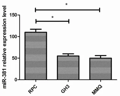
miR-381 is down-regulated in pituitary
adeno-ma cell lines
We used qRT-PCR to examine miR-381 expres-sion in pituitary adenoma cell lines, including GH3 and MMQ cells, and the normal pituitary cell line RPC. The expression levels in the two pituitary adenoma cell lines were lower than that in the RPC cell line (P=0.040 and P=0.035) (Figure 1).
Overexpression of miR-381 affects MMQ cell
proliferation and apoptosis
We transfected MMQ cells with miR-381 mim-ics or negative control mimmim-ics (NC-mimmim-ics) to assess the biological functions of miR-381 in pituitary adenoma.
A CCK-8 proliferation assay performed after transfection showed significantly suppressed proliferation in the miR-381 group compared to the NC-mimics and untransfected control groups (between the miR-381 mimics group and NC-mimics group, P=0.030 after 72 h) (Figure 2A). Furthermore, we compared apop -tosis rates among the three groups (miR-381, NC-mimics and untransfected control) using an Annexin V-FITC/PI assay. The percentage of apoptotic cells in the miR-381 mimics group was significantly increased compared to the NC-mimics and control groups (P=0.008 and P=0.007). However, there were no significant differences between the control group and the NC mimics group in the two assays (Figure 2B-E). These data suggest that overexpres- sion of miR-381 suppresses the tumorigenicity of pituitary adenoma cells.
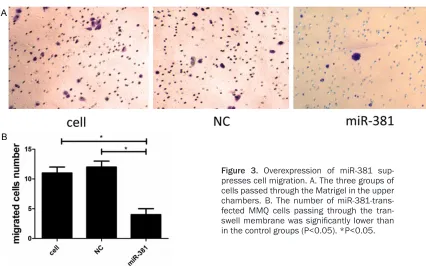
Overexpression of miR-381 suppresses cell
migration
We performed a transwell assay to assess the potential role of miR-381 in pituitary adenoma cell migration. The results showed reduced migration by the miR-381 mimics group com-h (up to 72 com-h); 10 μl CCK-8 solution was added
to the medium in each well and incubated for 4 h. Absorbance was measured at 450 nm using a microplate reader (Thermo Fisher Scientific, Grand Island, USA).
Transwell invasion assay
Transwell assays were performed using tran-swell chambers (BD Bioscience, San Jose, CA, USA). The bottom of the upper chambers was coated with 200 mg/ml Matrigel and dried overnight. At 24 h post-transfection, 1×10⁴ cells from each group were seeded into the upper chamber, and 500 µl medium contain- ing 10% FBS was added to the lower chamber. The cells were incubated at 37°C in a 5% CO2 atmosphere for 24 h. The cells remaining in the upper chamber were removed using cotton swabs, and the cells that had migrated to the other side of the membrane were fixed with methanol and stained with 0.1% crystal violet. The number of cells that invaded the Matrigel was counted in five randomly selected fields (200×).
Statistical analysis
[image:3.612.93.288.72.228.2]Figure 2. Overexpression of miR-381 suppresses proliferation and promoted apoptosis. A. The proliferation of MMQ cells was assessed by CCK8 assays. B-E. The apoptosis of MMQ cells was measured by Annexin V-FITC/PI assays. *P<0.05. **P<0.01.
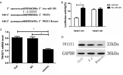
[image:4.612.92.518.450.716.2]of the miR-381 target sequence (P=0.310) (Figure 4B).
To examine the role of miR-381 on TWIST1 gene expression, we also transfected miR-381 mimics into MMQ cells and found that expres-sion at both the mRNA and protein levels was inhibited in the miR-381 mimics group com-pared with the untransfected and NC mimics groups (P=0.028 and P=0.029, respectively, for TWIST1 mRNA). These data suggest that miR-381 directly regulates TWIST1 expression (Figure 4C, 4D).
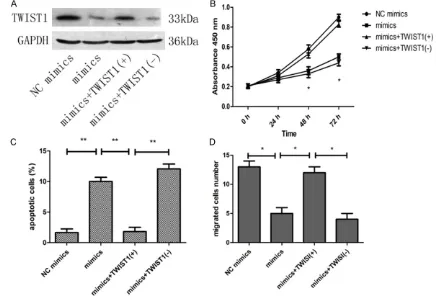
Overexpression of TWIST1 abrogates the bio-logical function of miR-381
To determine whether the in vitro phenoty- pes induced by miR-381 expression could be reversed by TWIST1 overexpression, we purchased TWIST1 plasmids (GeneCopoeia, Guangzhou, China) and cotransfected these plasmids and miR-381 mimics into MMQ cells. Western blot analysis showed that the level of TWIST1 protein in the miR-381 mimics + pared to the untransfected control group
(P=0.025) and the NC group (P=0.029) (Figure 3A, 3B).
TWIST1 is a direct target of miR-381
[image:5.612.95.521.71.330.2]Because miR-381 influences pituitary adeno -ma cell growth and invasion, we hypothesized that miR-381 might target genes related to tumor growth and migration. Therefore, we searched for potential target genes using the TargetScan, PicTar and miRanda databases and identified TWIST1, an oncogene that har -bors a putative miR-381 binding site in its 3’UTR (Figure 4A). Accordingly, the TWIST1 3’UTR was cloned into a reporter plasmid downstream of the luciferase gene, and the plasmid was transfected into HEK-293T cells for assays. Luciferase activity was reduced in cells cotransfected with miR-381 mimics and the luc-TWIST1 plasmid compared with the cells transfected with NC mimics and the luc-TWIST1 plasmid (P=0.029). However, this suppressive effect was abolished by mutation
ence tumor development and metastasis through interactions with target genes. Many miRNAs have been shown to function as either tumor suppressors or oncogenes in pituitary adenoma [3-5], and several miRNAs acting as tumor suppressors are underexpressed in this tumor type. For example, miR-21a targets the PRKCD gene and is involved in apoptosis of ACTH-secreting pituitary adenoma cells [10]. miR-132, miR-15a and miR-16 synergistically inhibit pituitary adenoma cell proliferation, invasion and migration by targeting the SOX5 gene [11]. miR-23b and miR-130b, which target the HMGA2 and CCNA2 genes, are expressed at low levels in GH gonadotroph and NFPA ade -nomas [12], and up-regulation of these two genes inhibits cell proliferation. In contrast, several miRNAs identified as up-regulated in pituitary adenomas function as oncogenes. For example, miR-200c targets the PTEN gene and is overexpressed in pituitary adenoma, and down-regulation of miR-200c can enhance TWIST1 (+) plasmid group was higher than that
in the miR-381 mimics or miR-381 mimics + NC TWIST1 (-) plasmid groups (Figure 5A). Furthe-rmore, absorbance at 450 nm in the miR-381 mimics + TWIST1 (+) group was higher than that in the miR-381 mimics group (at 48 h, P=0.030, at 72 h, P=0.037) (Figure 5B). In addition, a lower apoptosis rate was obse- rved in the miR-381 mimics + TWIST1 (+) group compared with the miR-381 mimics (P=0.008) and miR-381 mimics + NC TWIST1 (-) plasmid groups (P=0.009) (Figure 5C). The number of migrated cells in the miR-381 mimics + TWIST1 (+) group was higher than that in the miR-381 mimics (P=0.032) and miR-381 mim-ics + NC TWIST1 (-) plasmid groups (P=0.035 and P=0.029) (Figure 5D).
Discussion
MicroRNAs are estimated to regulate more than 30% of all human genes, and they influ
[image:6.612.84.520.75.376.2]In summary, our study demonstrates that miR-381 is down-regulated in pituitary adeno-ma cells and that miR-381 overexpression can suppress pituitary adenoma cell growth and migration by directly targeting TWIST1. Thus, miR-381 is a novel potential target for the treatment of pituitary adenoma.
Acknowledgements
This work was supported by the Guangdong Science Foundation of Guangdong Province (grant 2013B021800123).
Disclosure of conflict of interest
None.
Address correspondence to: Drs. Haijun Wang and Chuangxin Liao, Department of Neurosurgery and Pituitary Tumor Center, The First Affiliated Hospital, Sun Yat-sen University, 183, Huangpu Dong Road, Guangzhou 510700, China. Tel: +86-20-82393181; Fax: +86-20-82393181; E-mail: 1295354394@ qq.com (HJW); lcxsl@126.com (CXL)
References
[1] Wang S, Lin S, Wei L, Zhao L, Huang Y. Analysis
of operative efficacy for giant pituitary adeno-ma. BMC Surg 2014; 14: 59.
[2] Syro LV, Scheithauer BW, Kovacs K, Toledo RA, Londoño FJ, Ortiz LD, Rotondo F, Horvath E and Uribe H. Pituitary tumors in patients with MEN1 syndrome. Clinics (Sao Paulo) 2012; 67: 43-48.
[3] Adlakha YK, Saini N. Brain microRNAs and
in-sights into biological functions and therapeutic potential of brain enriched miRNA-128. Mol Cancer 2014; 13: 33.
[4] Iorio MV and Croce CM. MicroRNA dysregula-tion in cancer: diagnostics, monitoring and therapeutics. A comprehensive review. EMBO Mol Med 2012; 4: 143-159.
[5] Mao ZG, He DS, Zhou J, Yao B, Xiao WW, Chen
CH, Zhu YH, Wang HJ. Differential expression of microRNAs in GH-secreting pituitary adeno-mas. Diagn Pathol 2010; 5: 79.
[6] Tang H, Wang Z, Liu Q, Liu X, Wu M, Li G.
Dis-turbing miR-182 and -381 inhibits BRD7 tran-scription and glioma growth by directly target-ing LRRC4. PLoS One 2014; 9: e84146.
[7] Chen B, Duan L, Yin G, Tan J, Jiang X. miR-381,
a novel intrinsic WEE1 inhibitor, sensitizes re-nal cancer cells to 5-FU by up-regulation of Cdc2 activities in 786-O. J Chemother 2013; 25: 229-238.
apoptosis in pituitary adenoma cells [13]. miR-26 targets the PRKCD gene, and down-regula-tion of miR-26a can suppress the growth of pituitary adenoma cells [10].
Using miRNA array and qRT-PCR analyses, our research team previously found low exp- ression of miR-381 in pituitary adenoma tis-sues [5]. In the present study, we utilized qRT-PCR to confirm low expression of miR-381 in a pituitary adenoma cell line. We also overex-pressed miR-381 in MMQ cells and found that migration and proliferation were inhibited and that apoptosis was promoted. These results suggest that miR-381 is a tumor suppressor and may target genes involved in tumor growth and metastasis. Many potential target genes have been found using bioinformatics analy- sis, and three databases identified TWIST1, which contains potential miR-381 binding sites, as a target of this miRNA. TWIST1 is a highly conserved basic helix-loop-helix (bHLH) protein that regulates gastrulation and meso-dermal development. TWIST1 can induce tumor cell epithelial to mesenchymal transition (EMT), which is essential for metastasis [14].
TWIST1 is involved in the proliferation, apo- ptosis and metastasis of many cancers. For example, by targeting Twist1, miR-186 sup-presses prostate cancer G1 cell-cycle arrest and enhances apoptosis [15], miR-32 inhibits non-small-cell lung cancer cell proliferation [16], highly expressed miR-543 inhibits tumor cell proliferation, migration and invasion in aggressive endometrial cancer cell lines [17], and miR-720 inhibits tumor invasion and migration in breast cancer [18].
We verified our hypothesis using luciferase reporter assays. Cotransfection of miR-381 mimics and a luc-TWIST1 plasmid into HEK-293T cells repressed the relative luciferase activity of the reporter. These results indicate that miR-381 directly targets TWIST1.
[8] Rothschild SI, Tschan MP, Jaggi R, Fey MF, Gug-ger M and Gautschi O. MicroRNA-381 repress-es ID1 and is deregulated in lung adenocarci-noma. J Thorac Oncol 2012; 7: 1069-1077.
[9] Skalsky RL and Cullen BR. Reduced
expres-sion of brain-enriched microRNAs in glioblasto-mas permits targeted regulation of a cell death gene. PLoS One 2011; 6: e24248.
[10] Gentilin E, Tagliati F, Filieri C, Molè D, Minoia M, Rosaria Ambrosio M, degli Uberti EC and Zatelli MC. MiR-26a plays an important role in cell cycle regulation in ACTH-secreting pituitary adenomas by modulating protein kinase Cdel-ta. Endocrinology 2013; 5: 1690-1700.
[11] Renjie W and Haiqian L. MiR-132, miR-15a
and miR-16 synergistically inhibit pituitary tu-mor cell proliferation, invasion and migration by targeting Sox5. Cancer Lett 2015; 356: 568-578.
[12] Leone V, Langella C, D’Angelo D, Mussnich P, Wierinckx A, Terracciano L, Raverot G, Lachuer J, Rotondi S, Jaffrain-Rea ML, Trouillas J and Fusco A. Mir-23b and miR-130b expression is downregulated in pituitary adenomas. Mol Cell Endocrinol 2014; 390: 1-7.
[13] Liao C, Chen W, Fan X, Jiang X, Qiu L, Chen C, Zhu Y and Wang H. MicroRNA-200c inhibits apoptosis in pituitary adenoma cells by target-ing the PTEN/Akt signaltarget-ing pathway. Oncol Res 2013; 21: 129-136.
[14] Gajula RP, Chettiar ST, Williams RD, Nugent K, Kato Y, Wang H, Malek R, Taparra K, Cades J, Annadanam A, Yoon AR, Fertig E, Firulli BA, Mazzacurati L, Burns TF, Firulli AB, An SS and Tran PT. Structure-function studies of the bHLH phosphorylation domain of TWIST1 in prostate cancer cells. Neoplasia 2015; 17: 16-31. [15] Zhu X, Shen H, Yin X, Long L, Xie C, Liu Y, Hui L,
Lin X, Fang Y, Cao Y, Xu Y, Li M, Xu W and Li Y. miR-186 regulation of Twist1 and ovarian can-cer sensitivity to cisplatin. Oncogene 2016; 35: 323-332.
[16] Li L and Wu D. miR-32 inhibits proliferation, epithelial-mesenchymal transition, and metas-tasis by targeting TWIST1 in non-small-cell lung cancer cells. OncoTargets Ther 2016; 9: 1489-1498.
[17] Bing L, Hong C, Li-Xin S and Wei G. MicroR-NA-543 suppresses endometrial cancer onco-genicity via targeting FAK and TWIST1 expres-sion. Arch Gynecol Obstet 2014; 290: 533-541. [18] Li LZ, Zhang CZ, Liu LL, Yi C, Lu SX, Zhou X,