Original Article
CXCL12 induces migration of Schwann cells via p38
MAPK and autocrine of CXCL12 by the CXCR4 receptor
Dekun Gao*, Hui Sun*, Jin Zhu, Yinda Tang, Shiting Li
Department of Neurosurgery, Xinhua Hospital, School of Medicine, Shanghai Jiao Tong University, Shanghai, China. *Equal contributors.
Received February 28, 2018; Accepted March 28, 2018;Epub June 1, 2018; Published June 15, 2018
Abstract:Schwann cells (SCs) play a crucially supportive role in repair of injured peripheral nerve system (PNS). CXCL12 plays a significant role in migration of stem cells and embryonic developmental cells and CXCL12 is strongly chemotactic for a variety of cells. Our study was designed to determine the role of CXCL12 in Schwann cell prolifera-tion and migraprolifera-tion. Our study demonstrated that CXCL12 had no effect on Schwann cell proliferaprolifera-tion while signifi-cantly promoting Schwann cell migration. CXCL12-induced Schwann cell migration was signifisignifi-cantly attenuated by inhibition of its receptor CXCR4 and p38 MAPK through co-treatment with AMD3100 and SB203580, separately. Besides, Western blot, QRT-PCR, and ELISA indicated that treatment with CXCL12 enhanced expression of CXCL12 by Schwann cells. In conclusion, CXCL12-enhanced SCs migration is mediated by secreting CXCL12 and p38 MAPK via receptor CXCR4, suggesting that CXCL12 has potential application value for PNS regeneration and could serve as a new therapeutic strategy in peripheral nerve diseases.
Keywords: CXCL12, CXCR4, AMD3100, p38MAPK, migration
Introduction
Peripheral nerves are surrounded by Schwann cells which form myelinated nerve fibers [1]. Peripheral nerves easily become dysfunctional in trauma [2], autoimmune attacks [3], or neu-rotoxins [4]. There is a close relationship be- tween these deficits and Schwann cells. It has been reported that CXCL12 is a powerful che-mokine that can promote directional migration of various stem cells through interaction with its receptor CXCR4 and CXCR7 [5-7]. Furth- ermore, CXCL12 can promote regeneration of injured motor axon terminals [8]. CXCL12-CXCR4-CXCR7 chemokine axis are therapeutic targets for remyelination in demyelinating dis-eases [9]. It is still unclear, however, whether CXCL12 can promote migration of Schwann cells. In this study, we investigated the effect of CXCL12 on Schwann cells and its possible molecular mechanism. Our results show that CXCL12 promoted Schwann cell migration by acting on the receptor CXCR4 and p38MAPK and enhancing CXCL12 secretion of Schwann cells. Our research provides a new direction
for the study of migration and repair of Schwann cells after peripheral nerve injuries.
Material and methods
Culturing SCs
changed every 3 days. Tissues were incubat- ed for 1 week and passaged for 3 genera- tions. This step was done to remove over- grown fibroblast cells and ensure pure and viable Schwann cells.
Immunofluorescence analysis
Immunofluorescent staining was used for char-acterization of the Schwann cells after 3 gen-erations. After cell fixation with 4% paraformal-dehyde (PFA; Sigma, USA) for 10 minutes at room temperature and permeabilization with 0.15% Triton-X 100 (Sigma, USA) for 15 min-utes, the cells were blocked with 0.1% BSA (Sigma, USA) for 30 minutes and incubated
Transwell assay
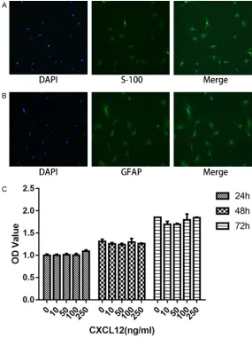
To test the migratory ability of SCs, SCs were resuspended in serum-free medium and then seeded in the upper chambers of Trans- well system to a number of 40,000 cells per chamber, with or without AMD3100. The low- er chambers contained medium with 10% fetal calf serum (FCS), with or without CXCL- 12. After 12 hours of incubation at 37°C, cells remaining on the upper surface were removed using cotton swabs whereas cells that migrat- ed to the lower surface were fixed with 4% PFA for 15 minutes and stained with 0.1% crystal violet solution for 10 minutes. Cells were count-ed in at least 5 random fields under micro-Figure 1. Fluorescent immunocytochemistry of cultured Schwann cells
stained by S-100 (A) and GFAP (B), with nuclei counterstained with DAPI. (C) Effects of different CXCL12 concentration on Schwann cell proliferation performed by CCK-8 assay. Data are presented as mean ± SD, n = 3. One-way ANOVA.
at 37°C for 1 hour with rabbit monoclonal antibody against S-100 protein (1:1000; Ab- cam, USA) and rabbit poly-clonal antibody against GFAP protein (1:500; Boster, China). After rinsing in PBS 3 times, goat anti-rabbit secondary antibody (1:1000; Jackson, USA) was applied in a dark place at room temperature for 1 hour. Cells were then stained with 4’,6’ -diamidino-2-phenylindole hydro-chloride (DAPI; Sigma, USA). Images were photographed using a fluorescence microscope (Ol- ympus BX51, Japan).
Determination of cell viability with CCK-8 assay
[image:2.612.90.375.71.452.2]scope. Assay was repeated three times for quantitative analysis. The amount of cells was quantified by Image J software (USA).
Western blotting
Cell samples were collected 24 hours after CXCL12 was added to SCs and then lysated in RIPA (Byetime, China). Protein concentrations were determined using bicinchoninic acid (B- CA) protein assay kit (Byetime, China). Equal amounts of total protein (30 ug) were loaded onto 12% gradient gels for electrophoresis, fol-lowed by transfer to polyvinylidene difluoride (PVDF) membranes. Membranes were blocked in 5% milk followed by incubation with anti-CXCL12 and p-p38 primary antibodies (1:1000; Abclonal).
Real-time reverse transcription-polymerase chain reaction (RT-PCR) analysis
Total RNA was extracted from SCs using TRI- zol Reagent (Invitrogen, Carlsbad, CA, USA) and treated with RNase-free DNase I. Total RNA (1000 ng) was used as a template for reverse transcription and q-PCR (Novoprotein, China).
were cleaned with PBS three times. Fresh cul-ture medium was then added for 12 hours. Cytokines in the supernatants (CXCL12) were detected by ELISA (MEIMIAN, Jiangsu, China), according to manufacturer instructions. Triplica- tes were included in each sample and experi-ments were repeated three times.
Statistical analysis
Data are presented as mean ± standard devia-tion (SD), obtained from at least three indepen-dent experiments. Statistical analysis was con-ducted by one-way ANOVA and P < 0.05 was regarded to be statistically significant.
Results
Cellular features and identification of Schwann cells
Most cultured Schwann cells displayed a spin-dle-like or multi-angle shape, with a protuber-ance growing from both sides, and a spherical nuclei. Almost all of these cultured Schwann cells expressed S-100 and GFAP, two specific biomarkers of Schwann cells (Figure 1A and
1B).
The sequences of primers were as follows: rat CXCL12 upstream, 5’-GTGACGGTAAG- CCAGTCAGC-3’; and down-stream, 3’-TGCACACTTGTCTG- TTGTTGC-5’; rat GAPDH up-stream, 5’-CAGTGCCAGCCTC- GTCTCAT-3’; and downstream, 3’-AGGGGCCATCCACAGTCT- TC-5’. RT-PCR conditions were as follows: 5 min. at 95°C, fol-lowed by 40 cycles of 30 sec. at 95°C, 30 sec. at 57°C, and 30 sec. at 72°C. CT values were normalized to the GAP- DH gene and RQ values were viewed as our final results. Enzyme-linked immunosor-bent assay (ELISA)
[image:3.612.94.371.68.316.2]SCs were cultured in 6-well plates, before treatment of CXCL12, at concentrations of 100 ng/mL for 24 hours. Af- ter that, culture supernatants were abandoned and cells Figure 2. Migratory SCs stained with crystal violet 12 h after induced by
CXCL12 has no effect on Schwann cell prolif-eration
The effects of CXCL12 on SCs proliferation were examined using CCK-8 assay. It can be seen that the number of cells increased as time went by. However, there was no significant dif-ference between CXCL12-treated group and blank control group. These data show that CXCL12 has no significant impact on prolifera-tion of SCs (Figure 1C).
CXCL12 promotes Schwann cell migration The function of CXCL12 to Schwann cell migra-tion ability was analyzed by Transwell migramigra-tion assay. After 12 hours of cell spreading, migra-tory cells were stained with crystal violet. As shown in Figure 2, the number of migratory cells increased at first and then decreased with an increase of CXCL12 concentration. The largest migration concentration was 100 ng/
mL. These results suggest that CXCL12 dose-dependent promotes migration of SCs, in a cer-tain concentration range, and inhibits migra-tion of SCs at higher concentramigra-tions.
Contribution of CXCR4 to CXCL12-induced mi-gration in SCs
[image:4.612.90.523.69.407.2]To analyze the contribution of CXCR4 to CXCL- 12-dependent migration in SCs, we employed CXCR4 antagonist AMD3100 (100 ng/mL), which selectively inhibits binding of CXCL12 to CXCR4 without affecting other combinations in SCs. Blocking CXCR4 with AMD3100 com-pletely abolished CXCL12-induced migration (Figure 3A and 3C). Additionally, compared with control group, the number of cell migration was significantly reduced by the addition of AMD- 3100 group alone. These data show that CX- CL12 plays a role in migration relying on its receptor CXCR4.
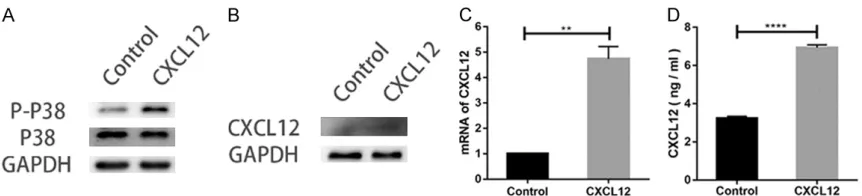
CXCL12 supports and protects activation of P38 MAPK signaling cascades
As demonstrated in Figure 3B and 3D, treat-ment with SB203580, a p38 inhibitor, de- creased the migrated cells in groups co-treat- ed with CXCL12. Furthermore, levels of p-p38 in groups treated with CXCL12 was higher than that of incubation with blank groups alone. Expression of total p38 remained unchange-able across different groups (Figure 4A). These data suggest that CXCL12 functions to protect or restore the activation level of p38 MAPK sig-naling cascades blocked by SB203580 in SCs. CXCL12 promotes autocrine of CXCL12 in SCs To study why the use of AMD3100 alone could reduce mobility of SCs, we performed Western blot, RT-PCR, and ELISA to detect the level of CXCL12. All three methods showed that expres-sion of CXCL12 was significantly increased after chemokine treatment (Figure 4B-D).
Discussion
Schwann cells play a crucial role in the regen-eration of PNS [1]. A full understanding of the molecular regulation mechanism of Schwann cells would contribute to treatment of PNI. Our experiments, for the first time, demonstrated that CXCL12 promotes migration of Schwann cells by positively affecting secretion of CXCL- 12 via acting on the CXCR4 receptor and p38MAPK.
RSC96 SCs are cell lines that evolved from rat Schwann cells and have been used in sev-eral experiments to study Schwann cell migra-tion [10-14]. However, there are many differ-ences between cell lines and primary cells, especially in migration direction [15], so we
chose the primary extraction of Schwann cells for experimental study. Herein, we found that CXCL12 attracts SCs in a dose-dependent manner and has the greatest effect at a con-centration of 100 ng/mL, consistent with a previous study at 100 ng/mL for optimal physiological concentrations [16, 17]. When the concentration reached 250 ng/mL, howev-er, the number of cell migration decreas- ed. We speculated that this may be related to the saturation of CXCR4 receptor on Schwann cell membranes. In addition, CXCR7, another receptor of CXCL12, may also play an important regulatory role. CXCL12 has an increased aff- inity for CXCR7 when CXCL12 concentrations are too high whereas CXCR7 operates as a sen-sor for CXCL12 and adjusts CXCR4 protein lev-els [18, 19]. The detailed relationship between these two receptors in SCs requires further elucidation.
As a chemokine, CXCL12 is generally consid-ered to promote cell proliferation, particularly significant in stem cell and tumor research [20, 21]. Our study found that the proliferation activ-ity of Schwann cells is almost invariably affect-ed by CXCL12 for up to three days. Tumor cells and stem cells grow rapidly and cell prolifera-tion is their main phenotype, but this is easily changed by external intervention. The main characteristic of SCs is the formation of myelin. Proliferation is not the main phenotype and, thus, is not easily affected.
[image:5.612.91.522.70.168.2]through other pathways of MAPK, such as ERK1/2, JNK, and ERK5, remains unknown and further experimentation is needed.
During our experiment, we found that, com-pared with the control group, AMD3100 group significantly reduced the number of SCs mi- gration. Further experiments found that CX- CL12 can promote secretion of CXCL12 after CXCL12 acts on SCs, thus, forming a positive feedback regulation. In this process, the re- gulatory signal can be amplified in a short time, promoting migration of more SCs to CXCL12. This is of major importance in the study of repair of peripheral nerve injury. If CXCL12 is slowly released at the lesion, only a small amount of CXCL12 can play a pivotal role. In summary, our findings demonstrate that CXCL12 recognizes CXCR4 receptors in SCs and promotes cell migration through the p38MAPK pathway. This indicates that the CXCL12/CXCR4 system is likely to become a new therapeutic target for clinical application, promoting regeneration and repair of peripher-al nerve injuries.
Acknowledgements
This study was funded by the National Natur- al Science Foundation of China (No. 816705- 0708) and Medical Cross Research Fund Pro- ject of Shanghai Jiao Tong University (No. YG2016ZD11).
Disclosure of conflict of interest
None.
Address correspondence to: Dr. Shiting Li, Depart- ment of Neurosurgery, Xinhua Hospital, School of Medicine, Shanghai Jiao Tong University, 1665 Kongjiang Rd, Shanghai 200092, China. Tel: +86-2125078001; E-mail: lishiting@xinhuamed.com.cn
References
[1] Heinen A, Lehmann H, Küry P. Negative regula-tors of schwann cell differentiation-novel tar-gets for peripheral nerve therapies? J Clin Immunol 2013; 33 Suppl 1: S18-26.
[2] Gomez-Sanchez J, Pilch K, van der Lans M, Fazal S, Benito C, Wagstaff L, Mirsky R, Jessen K. After nerve injury, lineage tracing shows that myelin and remak schwann cells elongate extensively and branch to form repair schwann
cells, which shorten radically on remyelination. J Neurosci 2017; 37: 9086-9099.
[3] Hou X, Liang Q, Wu Y. Transplantation of Schwann cells co-cultured with brain-derived neurotrophic factor for the treatment of experi-mental autoimmune neuritis. J Neuroimmunol 2013; 263: 83-90.
[4] Ciutat D, Calderó J, Oppenheim R, Esquerda J. Schwann cell apoptosis during normal devel-opment and after axonal degeneration in-duced by neurotoxins in the chick embryo. J Neurosci 1996; 16: 3979-3990.
[5] Li M, Hale J, Rich J, Ransohoff R, Lathia J. Chemokine CXCL12 in neurodegenerative dis-eases: an SOS signal for stem cell-based re-pair. Trends Neurosci 2012; 35: 619-628. [6] Chen Q, Zhang M, Li Y, Xu D, Wang Y, Song A,
Zhu B, Huang Y, Zheng J. CXCR7 mediates neu-ral progenitor cells migration to CXCL12 inde-pendent of CXCR4. Stem Cells 2015; 33: 2574-2585.
[7] Moridi I, Mamillapalli R, Cosar E, Ersoy G, Taylor H. Bone marrow stem cell chemotactic activity is induced by elevated CXCl12 in endo-metriosis. Reprod Sci 2017; 24: 526-533. [8] Negro S, Lessi F, Duregotti E, Aretini P, La Ferla
M, Franceschi S, Menicagli M, Bergamin E, Ra-dice E, Thelen M, Megighian A, Pirazzini M, Mazzanti C, Rigoni M, Montecucco C. CXCL- 12α/SDF-1 from perisynaptic Schwann cells promotes regeneration of injured motor axon terminals. EMBO Mol Med 2017; 9: 1000-1010.
[9] Chu T, Shields LB, Zhang YP, Feng SQ, Shields CB, Cai J. CXCL12/CXCR4/CXCR7 chemokine axis in the central nervous system: therapeutic targets for remyelination in demyelinating dis-eases. Neuroscientist 2017; 23: 627-648. [10] Huang CY, Kuo WW, Shibu MA, Hsueh MF,
Chen YS, Tsai FJ, Yao CH, Lin CC, Pan LF, Ju DT. Citrus medica var. sarcodactylis (Foshou) acti-vates fibroblast growth factor-2 signaling to in-duce migration of RSC96 Schwann cells. Am J Chin Med 2014; 42: 443-452.
[11] Ju DT, Kuo WW, Ho TJ, Paul CR, Kuo CH, Viswanadha VP, Lin CC, Chen YS, Chang YM, Huang CY. Protocatechuic acid from alpinia oxyphylla induces schwann cell migration via ERK1/2, JNK and p38 activation. Am J Chin Med. 2015; 43: 653-665.
[12] Ju DT, Liao HE, Shibu MA, Ho TJ, Padma VV, Tsai FJ, Chung LC, Day CH, Lin CC, Huang CY. Nerve regeneration potential of protocatechuic acid in RSC96 schwann cells by induction of cellular proliferation and migration through IGF-IR-PI3K-Akt signaling. Chin J Physiol 2015; 58: 412-419.
cells via p38 MAPK and PI3K-Akt signaling pathway mediated by the UNC5B receptor. Biochem Biophys Res Commun 2015; 464: 263-268.
[14] Ma Y, Ge S, Zhou D, Zhang H, Sun L, Wang X, Su J. [Rat bone marrow mesenchymal stem cells promote apoptosis and inhibit prolifera-tion and migraprolifera-tion of RSC96 cells]. Xi Bao Yu Fen Zi Mian Yi Xue Za Zhi 2017; 33: 190-195. [15] Ji Y, Shen M, Wang X, Zhang S, Yu S, Chen G,
Gu X, Ding F. Comparative proteomic analysis of primary schwann cells and a spontaneously immortalized schwann cell line RSC 96: a com-prehensive overview with a focus on cell adhe-sion and migration related proteins. J Proteome Res 2012; 11: 3186-3198.
[16] Liu YJ, Wang J, Chi ZB. [Comparison of chemo-taxis of different concentrations of SDF1, bFGF, IL-8 on bone marrow mesenchymal stem cell]. Shanghai Kou Qiang Yi Xue 2015; 24: 275-279.
[17] Pasquier J, Abu-Kaoud N, Abdesselem H, Madani A, Hoarau-Vechot J, Thawadi HA, Vidal F, Couderc B, Favre G, Rafii A. SDF-1alpha con-centration dependent modulation of RhoA and Rac1 modifies breast cancer and stromal cells interaction. BMC Cancer 2015; 15: 569.
[18] Wang Y, Li G, Stanco A, Long JE, Crawford D, Potter GB, Pleasure SJ, Behrens T, Rubenstein JL. CXCR4 and CXCR7 have distinct functions in regulating interneuron migration. Neuron 2011; 69: 61-76.
[19] Sánchez-Alcañiz JA, Haege S, Mueller W, Pla R, Mackay F, Schulz S, López-Bendito G, Stumm R, Marín O. Cxcr7 controls neuronal migration by regulating chemokine responsiveness. Neuron 2011; 69: 77-90.
[20] Gravina GL, Mancini A, Colapietro A, Vitale F, Vetuschi A, Pompili S, Rossi G, Marampon F, Richardson PJ, Patient L, Patient L, Burbidge S, Festuccia C. The novel CXCR4 antagonist, PRX177561, reduces tumor cell proliferation and accelerates cancer stem cell differentia-tion in glioblastoma preclinical models. Tumour Biol 2017; 39: 1010428317695528.