Supported by the Ministry of Education, Science, Research and Sport of the Slovak Republic and the Slovak Academy of Sciences (Project “VEGA” 2/0006/12) and the Slovak Research and Development Agency of the Slovak Republic (Project APVV-0137-10).
Distribution of tetraspanin family protein CD9 in bull
reproductive system
P. Cupperová
1, M. Simon
1, J. Antalíková
1, K. Michalková
1, Ľ. Horovská
1,
S. Hluchý
21Institute of Animal Biochemistry and Genetics, Slovak Academy of Sciences,
Ivanka pri Dunaji, Slovak Republic
2Faculty of Agrobiology and Food Resources, Slovak University of Agriculture in Nitra,
Nitra, Slovak Republic
ABSTRACT:TheCD9 cell surface molecule has been found to be important for the fertilization process of mammals. The aim of this study was to investigate, whether the molecule CD9 is expressed on bull sperm during the spermatogenesis and maturation of spermatozoa as well as in bull reproductive organs and their secreta. The expression of bovine CD9 was examined by immunohistochemistry, immunofluorescence, and immunoblotting. The histochemical studies using an anti-CD9 monoclonal antibody showed strong staining in the myeloid and collagenous tissue layer of testis and epididymis. Strong reaction was observed in the lumen of epididymal duct (the fluid of the duct) but the clumped spermatozoa in the lumen of the duct remained unstained. Intensive tissue staining was observed in the range of epithelial microvilli of epididymis (body and tail) and in the fluid content of tubules. The Western blot analysis showed the 24kDa molecule in sperm protein extracts of ejaculated sperm and also in the protein extracts of the sperm obtained from the head, body, and tail of epididymis but the localization of CD9 on the sperm was not directly confirmed. However, the obtained data could be considered in the interpretation of the role of CD9 in spermatogenesis and sperm-oocyte interactions.
Keywords:monoclonal antibody; immunohistochemistry; sperm; tissue
The membranes of spermatozoa play a crucial role in sperm-oocyte interaction. CD9 is a tetra- spanin family membrane protein widely expressed on animal cell membranes (Berditchevski, 2001; Boucheix and Rubinstein, 2001; Hemler, 2001) and implicated in many cellular functions such as adhesion, migration, co-stimulation, signal transduction, and sperm egg fusion (Charrin et al., 2003; Hemler, 2003). The presence of CD9 on the egg membrane was found to be essential for sperm-egg fusion (Kaji et al., 2000; Le Naour et al., 2000; Miyado et al., 2000). However, ex-pression profile of CD9 in spermatozoa and its role in reproduction (fertilization) is still unclear.
retained in the inner acrosome membrane while a portion of CD9 was translocated to the plasma membrane covering the equatorial segment. CD9 was maintained on the sperm head after reaching the perivitelline space of eggs (Ito et al., 2010). The expression of CD9 on mouse sperm has been later confirmed by Barraut-Lange et al. (2007). In boar, significant expression levels of CD9 have been detected in Leydig cells, Sertoli cells, and germ cells within testis, in the epithelial cells of epididymis, vas deferens, and prostate gland and in spermatozoa within the lumen of epididymis (Kaewmala et al., 2011). However, in earlier study Li et al. (2004) were not able to detect the CD9 molecule on ejaculated boar sperm preserved in liquid nitrogen.
To summarize, recent studies have shown the presence of CD9 on mouse and boar spermatozoa. Results demonstrating the function of CD9 on spermatozoa have not been presented yet. The aim of this study was to investigate whether the molecule CD9 is present on the bull sperm during the spermatogenesis and maturation of sperma-tozoa as well as in the bull reproductive organs and their secreta and to compare the bull CD9 distribution profiles with those of other species.
MATERIAL AND METHODS
Production of mAbs. Hybridoma cell lines
pro-ducing mAbs directed against the bovine CD9 molecule were obtained after immunization of BALB/c mice with bovine platelets using standard hybridoma production protocol (Dušinský et al., 1988). The specificity of CD9 mAb (IVA-50) used in this study was verified in the 3rd International Workshop on Leukocyte Antigens of Ruminants in Davis (USA) 1997 and by Tomášková et al. (1999). Sperm, tissues, and fluids. Fresh ejaculates and frozen-thawed sperm obtained from Slovak Breeding Services, Inc. were used for all experiments. Ejacu-lated spermatozoa were separated from seminal plasma via centrifugation at 600 g for 10 min. The frozen sperm thawed from pellets were diluted in phosphate buffered saline (PBS) and washed twice at 600 g at room temperature. Seminal plasma was centrifuged at 10 000 g to remove amorphous substances and used for Western blot analysis.
The bull testis and epididymis obtained at local slaughterhouses were dissected into the following segments: testis, rete testis, epididymis (head, body, tail), and vas deferens. To study the male
accessory reproductive glands, the seminal vesicles and prostate were also dissected. Samples for im-munohistology were dissected into small pieces, embedded on cork blocks, and stored frozen at –80oC. The testis and epididymis tissue segments were used for preparation of suspensions of tes-ticular or epididymal spermatozoa. To obtain the spermatozoa and fluids, each segment was minced in 1 ml of PBS. The cloudy suspension was filtered through cheesecloth to remove pieces of tissue. The filtrate was centrifuged at 600 g for 10 min and the pellets containing spermatozoa were used for immunofluorescence, immunoperoxidase as-says, and Western blot analysis. The supernatant fluid, which contained few spermatozoa, was cen-trifuged at 10 000 g and used for further Western blot analysis. After removing the sperm and fluid, the tissue segments were used for total protein extracts for Western blot analysis.
Immunohistochemical staining. For
histochemi-cal studies, 5 µm frozen sections were cut using a Leica Cryocut 1800 cryostat (Leica Microsystems, Wetzlar, Germany), fixed for 5 min in a cold 1 : 1 ethanol-acetone mixture, air-dried, washed in PBS, treated with 0.6% H2O2 in PBS and stained using an indirect immunoperoxidase test. The sections were treated with IVA-50 for 1 h at 37°C, incubated with peroxidase-conjugated pig anti-mouse Ig (Sevac, Prague, Czech Republic), stained in 0.06% (wt/vl) 3,3'-diaminobenzidine tetrahydrochloride (Sigma, St. Louis, USA) in PBS containing 0.05% (v : v) H2O2 for 10 min at room temperature and slightly counterstained with Harris’s hematoxylin. Samples were evaluated by the microscopic system Nikon Eclipse E 600 (Nikon, Tokyo, Japan) and a PixeLINKTM PL-A642 Camera (PixeLINK, Ottawa,
Canada) in connection with Lucia software (Ver-sion 4.8, 2002).
Indirect immunofluorescence. Two different procedures were used to identify CD9 in the sperm. In the first, the suspension of sperm (1.106/ml) was treated for 45 min in a culture medium containing mAb IVA-50 or mouse IgG2a(Exbio, Prague, Czech Republic) as isotype control (10 µg/200 µl sperm suspension) in the humidified atmosphere of 5% CO2 at 39°C in Eppendorf tube. After washing, the sperm suspension was incubated with FITC-conjugated pig anti-mouse IgG (Sevac) (1 : 30 in PBS) for 30 min at room temperature in the dark. The smears of the sperm were fixed on slides with 3.8% paraformaldehyde (Sigma), mounted with Vectashield-DAPI (Vector Laboratories, Burlingame, USA), and evaluated under a Leica DM5500 B epi-fluorescence microscope at ×400 magnification. In the second procedure, spermatozoa were first per-meabilized with ice cold methanol before primary antibody treatment on slides. Further procedures were the same as on the sperm suspension.
RESULTS
Expression of CD9 on tissue and fluids. The
expression of CD9 molecule on the bull repro-ductive tissues (testis, epididymis, vas deferens, prostate, and seminal vesicles) was detected using IVA-50 mAb. In the testis the staining intensity was the strongest in the collagen layer and myeloid cell layer of the wall of seminiferous tubules and in the intertubular space. No staining was found in the germinal epithelium including developing spermatozoa or in the intratubular fluid (Fig-ure 1a). A similar reaction pattern was observed in rete testis (intensive reaction with fibrous stroma) (Figure 1b). In the head of epididymis,
the IVA-50 staining was observed on the connec-tive tissue and on the surface of epithelial tissue (the range of stereocilia) (Figure 1c). In the body of epididymis the immunostaining extended to the secreta in the epididymal duct. The staining with high intensity appeared in the fluid filling the epididymal duct. The clumps of spermatozoa cumulated in the lumen of the duct remained unstained (Figure 1d). Similar histochemical pattern was found in the tail of epididymis (Fig-ure 1e). The intensive reaction with IVA-50 was observed along the epididymal duct, in the range of epithelial microvilli. The scattered spermatozoa found in the ductus deferens were free of IVA-50 staining (Figure 1f ).
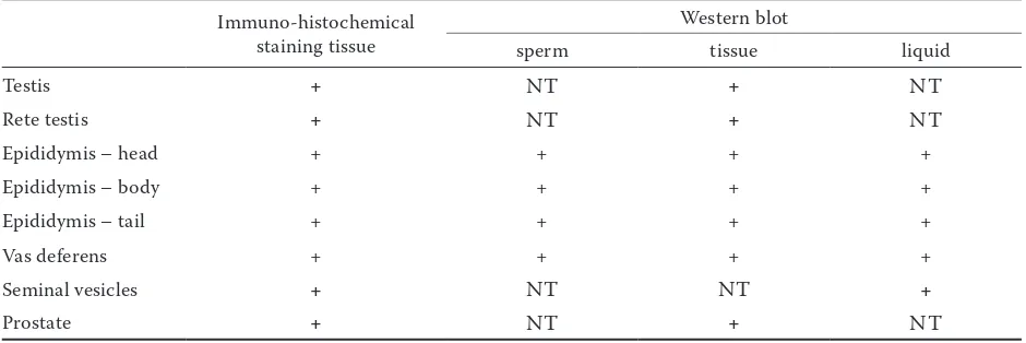
The CD9 molecule is widely distributed in the tissue of the bull accessory glands. In seminal vesicles the fibrocollagenous and the columnary secretory epithelium are stained (Figure 1g). In the prostate gland, the CD9 staining was found in the discrete part of the epithelial cells, smooth muscular cells, fibrocollagenous stroma, and in the tubular lumen of secretory epithelium (Figure 1h). The presence of CD9 (24 kDa) molecule was also detected by immunoblotting with IVA-50 in all the tested reproductive tissues and fluids (Table 1) and seminal plasma as well.
Expression of CD9 on sperm. No staining of
[image:3.595.63.533.582.739.2]CD9 on the sperm membrane was found, even though two different immunofluorescent proce-dures were used. On the other hand, by Western blot analysis with IVA-50 under non-reduced conditions a 24kDa protein was detected in the detergent extract of ejaculated sperm and also in the protein extracts of the sperm obtained from the head, body, and tail of epididymis (Figure 2).
Table 1. Expression of CD9 tetraspanin in the bull reproductive tissues, fluid, and sperm
Immuno-histochemical staining tissue
Western blot
sperm tissue liquid
Testis + NT + NT
Rete testis + NT + NT
Epididymis – head + + + +
Epididymis – body + + + +
Epididymis – tail + + + +
Vas deferens + + + +
Seminal vesicles + NT NT +
Prostate + NT + NT
Figure 1. Localization of CD9 in the bull reproductive tissues
DISCUSSION
This study provides the evidence of the pres-ence of the CD9 molecule in the bull reproductive tract. Histochemical assay has shown the high expression of CD9 in the collagenous and smooth muscle tissues. Furthermore, the CD9 is highly expressed in the accessory glands (prostate and seminal vesicles). Besides the connective tissue, also the secreta of epididymal lumen contain the CD9 molecule. The presence of CD9 in the secreta of epididymis but not in fluid of the testis (testis plasma), and the strong tissue staining in the range of epithelial microvilli of the epididymis (espe-cially in body and tail of epididymis) suggests the
secretion of CD9 by the epididymal epithelium. However, the CD9 is not bound to the plasma membrane of spermatozoa or the quantity is too small and the used histochemistry method is not sensitive enough to detect the “micro-quantity” of CD9. The “partial” sperm membrane expres-sion is characteristic for the mouse CD9 mol-ecule. According to Ito et al. (2010), it is an inner acrosomal membrane associated protein, not a plasma membrane related protein. The expression is restricted to the inner acrosomal membrane of acrosome reacted sperm and in a small quantity to plasmatic membrane of equatorial segment (Ito et al., 2010). The presented results of the Western blot analysis of tissue samples and also spermatozoa (epididymal and ejaculated spermatozoa) suggest the presence of CD9; nevertheless, this study has not revealed unequivocally the localization of CD9 on the sperm head. In order to localize this molecule, immunofluorescence was performed on non-permeabilized and permeabilized plasma membrane of the sperm but no CD9 staining on the sperm membrane was detected. Antibody IVA-50 probably does not recognize the native epitope of CD9 on the sperm membrane unlike the denatured protein in the detergent extract. These results correspond with the findings of Li et al. (2004) on boar sperm. On the other hand, Kaewmala et al. (2011) demonstrated localization
Explanation to Figure 1
(a) testis: 1 = seminiferous tubule wall (basement membrane, lamina propria), 2 = epithelium consists of Sertoli cells and germinative cells in various stages of development. The CD9 molecule was detected in the collagenous and muscular layer of the wall of tubules (b) rete testis: 1 = lamina propria of tubules, 2 = fibrous stroma, 3 = cuboidal epithelium. Observable reactions with fibrous stroma and lamina propria of tubules
(c) head of epididymis: 1 = stereocilia (microvilli), 2 = area of interstitial tissue with circular muscle fibres, 3 = pseudostratified columnar epithelium with stereocilia (microvilli). Observable reaction of CD9 molecules on stereocilia and circular muscle fibres, note the scattered spermatozoa in the lumen are contrastained with hematoxilin (blue colour)
(d) body of epididymis: 1 = clump of spermatozoa in the lumen, 2 = interstitial tissue with circular muscle fibres, 3 = pseudostrati-fied columnar epithelium with stereocilia (microvilli), 4 = stereocilia (microvilli). Very intensive DAB staining seen with the fluid of the lumen and mild staining with stereocilia and muscle fibres, scattered spermatozoa on the periphery of the clump are not stained with DAB
(e) tail of epididymis:1 = lumina of duct contains clumps of spermatozoa, 2 = stereocilia (microvilli), 3 = pseudostratified columnar epithelium with stereocilia (microvilli), 4 = interstitial tissue with circular muscle fibres. Observable CD9 staining in the region of stereocilia, connective tissue, and local reactions with luminar fluid in the lumen of tubules
(f) vas deferens: 1 = spermatozoa in the lumen of the duct (detail), 2 = inner longitudinal smooth muscle layer with lamina propria mucosae lined by tall columnar epithelium, 3 = lumen of the tubule. Mild DAB staining was found in the connective tissue, note the three spermatozoa stained with hematoxilin in the lumen of the tubule
(g) seminal vesicles: 1 = fibro-collagenous tissue containing elastic fibres and smooth muscle cell, 2 = mucosa folds form numerous channels covered with columnar epithelium. DAB stained the both tissue structures
[image:5.595.79.272.584.654.2](h) prostate gland: 1–3 = luminal epithelial cells, 4 = smooth muscular and fibrocollagenous stroma. Staining with the basal (1), lateral (2), and apical (3) cell borders of the luminal epithelial cells, also with smooth muscular and fibrocollagenous stroma
Figure 2. Reaction of IVA-50 with bovine sperm
of CD9 on the acrosomal membrane of spermato-zoa located within different parts (seminiferous tubules of testis and epididymis) of boar reproduc-tive tract. These experiments were performed on cryostat tissue sections (not isolated sperm) in the presence of secreta, detected with anti-CD9 goat polyclonal antibody by immunofluorescence. Be-cause we were not able to precisely localize CD9 on the bull sperm, it is difficult to clarify the specific function of the sperm CD9 during fertilization in cattle. Mice CD9-deficient sperm are still capable of fertilization (Kaji et al., 2000; Le Naour et al., 2000; Miyado et al., 2000, 2008). Thus, in mice the sperm CD9 is not essential for sperm-egg fusion, but it may be facilitated via the homophilic inter-action between sperm CD9 and egg CD9 under physiological conditions (Ito et al., 2010).
In conclusion, the present study reported the presence of CD9 in the bull reproductive tissues and sperm. Localization of CD9 on bull sperm and its exact role remain to be elucidated.
Acknowledgement. We would like to thank Zuzana Nádaždyová for her excellent technical assistance.
REFERENCES
Barraud-Lange V., Naud-Barriant N., Bomsel M., Wolf J.P., Ziyyat A. (2007): Transfer of oocyte membrane fragments to fertilizing spermatozoa. FASEB Journal, 21, 3446–3449. Berditchevski F. (2001): Complexes of tetraspanins with in-tegrins: more than meets the eye. Journal of Cell Science, 114, 4143–4151.
Boucheix C., Rubinstein E. (2001): Tetraspanins. Cellular and Molecular Life Science, 58, 1189–1205.
Charrin S., Le Naour F., Labas V., Billard M., Le Caer J.P., Emile J.F., Petit M.A., Boucheix C., Rubinstein E. (2003): EWI-2 is a new component of the tetraspanin web in hepatocytes and lymphoid cells. Biochemical Journal, 373, 409–421. Dusinsky R., Simon M., Nouzovská D. (1988): Production of
monoclonal antibodies against cell surface antigens in cattle. Veterinarni Medicina, 33, 135–141.
Hemler M.E. (2001): Specific tetraspanin function. Journal of Cell Biology, 155, 1103–1107.
Hemler M.E. (2003): Tetraspanin proteins mediate cellular penetration, invasion, and fusion events and define a novel
type of membrane microdomain. Annual Review of Cellular and Developmental Biology, 19, 397–422.
Ito C., Yamatoya K., Yoshida K., Maekawa M., Miyado K., Toshimori K. (2010): Tetraspanin family protein CD9 in the mouse sperm: unique localization, appearance, behaviour and fate during fertilization. Cell Tissue Research, 340, 583–594.
Kaewmala K., Uddin M.J., Cinar M.U., Grose-Brinkhaus C., Jonas E., Tesfaye D., Phatsaraa C., Tholena E., Loofta C., Schellander K. (2011): Association study and expression analysis of CD9 as candidate gene for boar sperm quality and fertility traits. Animal Reproduction Science, 125, 170–179. Kaji K., Oda S., Shikano T., Uematsu Y., Sakagami J., Tada N.,
Miyazaki S., Kudo A. (2000): The gamete fusion process is defective in eggs of CD9-deficient mice. Nature Genetics, 24, 279–282.
Kanatsu-Shinohara M., Toyokuni S., Shinohara T. (2004): CD9 is a surface marker on mouse and rat male germine line stem cells. Biology of Reproduction, 70, 70–75.
Kierszenbaum A.L., Rosselot C., Rivkin E., Tres L.L. (2006): Role of integrins, tetraspanins, and ADAM proteins during the developing of apoptotic bodies by spermatogenic cells. Molecular Reproduction and Development, 73, 906–917. Le Naour F., Rubinstein E., Jasmin C., Prenant M., Boucheix
C. (2000): Severely reduced female fertility in CD9-deficient mice. Science, 287, 319–321.
Li Y.H., Hou Y., Ma W., Yuan J.X., Zhang D., Sun Q.Y., Wang W.H. (2004): Localization of CD9 in pig oocytes and its ef-fects on sperm-egg interaction. Reproduction, 127, 151–157. Miyado K., Yamada G., Yamada S., Hasuwa Y., Nakamura Y.,
Ryu F., Suzuki K., Kosai K., Inoue K., Onura A., Okabe M., Mekada, E. (2000): Requirement of CD9 on the egg plasma membrane for fertilization. Science, 287, 321–324. Miyado K., Yoshida K., Yamagata K., Sakakibara K., Okabe M.,
Wang X., Miyamoto K., Akutsu H., Kondo T., Takashasi Y., Ban T., Ito C., Toshimori K., Nakamura A., Ito M., Miyado M., Mekada E., Umezawa A. (2008): The fusing ability of sperm is bestowed by CD-9 containing vesicles released from eggs in mice. Proceedings of the National Academy of Sciences of the United States of America, 105, 12921–12926. Tomášková J., Dušinský R., Horovská Ľ., Simon M. (1999): A
set of monoclonal antibodies specific for bovine cell surface molecule CD9. Folia Biologica, 45, 225–226.
Received: 2013–09–30 Accepted after corrections: 2013–12–28
Corresponding Author
Ing. Petra Cupperová, Slovak Academy of Sciences, Institute of Animal Biochemistry and Genetics, Moyzesova 61, 900 28 Ivanka pri Dunaji, Slovak Republic