doi:10.1093/biolre/iox082 Research Article Advance Access Publication Date: 26 July 2017
Research Article
Genomic identification, expression profiling,
and functional characterization of CatSper
channels in the bovine
†
Gillian P. Johnson
1,2,3, Anne-Marie English
1,2, Sinead Cronin
1,
David A. Hoey
3,4,5, Kieran G. Meade
5and Sean Fair
1,∗1Laboratory of Animal Reproduction, Department of Biological Sciences, School of Natural Sciences, Faculty of
Science and Engineering, University of Limerick, Limerick, Ireland;2Department of Mechanical and Manufacturing Engineering, School of Engineering, Trinity College Dublin, Dublin 2, Ireland;3Trinity Centre for Bioengineering, Trinity Biomedical Sciences Institute, Trinity College Dublin, Dublin 2, Ireland;4Advanced Materials and Bioengineering Research Centre, Trinity College Dublin and RCSI, Dublin 2, Ireland and5Animal and Bioscience Research Department, Animal and Grassland Research and Innovation Centre, Teagasc, Grange, Dunsany, Meath, Ireland
∗Correspondence:Laboratory of Animal Reproduction, Department of Biological Sciences, School of Natural Sciences,
Faculty of Science and Engineering, University of Limerick, Limerick V94 PH61, Ireland. Tel:+353 61 202548; Fax:+353 61 331490; [email protected]
†Grant Support:The authors gratefully acknowledge the financial support from the Irish Research Council (Dublin, Ireland;
GOIPG/2014/1463), European Research Council (ERC) Starting Grant (#336882), and Science Foundation Ireland ERC Support Grant SFI 13/ERC/L2864 (to D.A.H.).
Received 28 March 2017; Revised 22 June 2017; Accepted 25 July 2017
Abstract
Cation channels of sperm (CatSper) are sperm-specific calcium channels with identified roles in the regulation of sperm function in humans, mice, and horses. We sought to employ a compara-tive genomics approach to identify conservedCATSPERgenes in the bovine genome, and profile their expression in reproductive tissue. We hypothesized that CATSPER proteins expressed in bull testicular tissue mediates sperm hyperactivation and their rheotactic response in the reproductive tract of the cow. Bioinformatic analysis identified all four knownCATSPERgenes (CATSPER 1–4) in the bovine genome, and profiling by quantitative real-time polymerase chain reaction identified site-specific variation in messenger ribonucleic acid (mRNA) expression for all four genes along the reproductive tract of the bull. Using a novel antibody against CATSPER 1, protein expression was confirmed and localized to the principal piece of bull sperm, in agreement with what has been reported in other species. Subsequent treatment of bull sperm with either the calcium chelator ethylene glycol tetraacetic acid; mibefradil, a specific blocker of CatSper channels in human sperm; or CATSPER1 antibody all significantly inhibited caffeine-induced hyperactivation and the rheotac-tic response, supporting the concept that the calcium influx occurs via CatSper channels. Taken together, the work here provides novel insights into expression and function of CatSper channels in bull testicular tissue and in the function of ejaculated sperm.
Summary Sentence
The effect of blocking calcium channels on hyperactivation and rheotactic response.
Key words:sperm, calcium, rheotaxis, hyperactivation, fertility, comparative reproduction, bull.
Introduction
Despite tens of millions to billions (species dependent) of sperm being deposited in the vagina/cervix after ejaculation, only a few hundred sperm are thought to be present in the ampulla of the oviducts at the time of fertilization [1]. It is now apparent that sperm transport in the female reproductive tract is facilitated by a range of physio-logical mechanisms including rheotaxis, chemotaxis, thermotaxis as well as smooth muscle contractions of the female reproductive tract [2–4]. One of the least studied of these is rheotaxis, which is a cell’s preference to swim with or against fluid flow, and has recently been reported as a mechanism for directing sperm towards the oocyte [3]. Positive rheotaxis is the tendency of sperm to swim against the flow and has been lightly observed over the past 50 yr, but only recently has rheotaxis gained attention as a guiding mechanism [3,5]. Rheo-taxis plays a fundamental role in directing sperm towards the site of fertilization, as sperm swim against the retrograde flow of mucus secreted under the influence of oestrogen in the lead up to ovulation [3,6,7]. In mice, it has been shown that positive rheotaxis occurs as a result of the spiral rotation of the sperm tail, leading to an in-creased amplitude of the tail waves and flagellar force, orientating the sperm upstream. This mechanism has been shown to be depen-dent on the influx of extracellular calcium (Ca2+) [3]. Bull sperm have been shown to exhibit positive rheotaxis and can change their trajectory with respect to a change in fluid flow direction [7]. While bull sperm and sperm of other species have been shown to display positive rheotaxis, the mechanisms by which this is mediated are unclear.
Hyperactivation is characterized by a high amplitude, asymmet-rical beating pattern (whip-like movement) of the sperm tail [8]. It is hypothesized that hyperactivation assists sperm in pulling away from the oviductal epithelium and increases swimming efficiency in viscous mucus [8,9]. As with rheotaxis, the exact molecular mecha-nisms by which hyperactivation is mediated has yet to be elucidated; however, it has been demonstrated that Ca2+and cyclic adenosine monophosphate (cAMP) are two key factors in the regulation of hyperactivation of mammalian sperm [10]. Intracellular increases in Ca2+have been reported in hyperactivated flagella of hamster and bull sperm [11], where a high concentration of caffeine was used to increase intracellular Ca2+[12].
In most cells, entry of external Ca2+ occurs through several types of Ca2+channels: mainly voltage-activated channels and store-operated channels [13,14], while intracellular Ca2+may be released from internal stores via receptor-operated channels. Inositol-1,4,5-trisphosphate receptors and ryanodine receptors represent the two main intracellular Ca2+channels responsible for releasing stored Ca2+. In sperm of many mammals, members of a specific transmem-brane Ca2+channel family, cation channels of sperm (CatSper) also play an important role [15]. CatSper are weakly voltage-dependent, Ca2+-selective, and pH-sensitive ion channels that control the en-try of positively charged Ca2+ions into the sperm [16]. CatSper is composed of four separate pore-forming α(alpha) subunits; these are CatSper 1–4 and three additional auxiliary subunits: CatSperβ
(beta), CatSperγ (gamma), and CatSperδ(delta). The complexity of the channel due to the several subunits, seems to be necessary for its functional co-ordination, localization to the flagella, and sen-sitivity to intracellular pH, progesterone, prostaglandins, odorants, and to a potential range of other proteins and signaling molecules [16,17]. CatSper channels have been shown to localize to the princi-pal piece of the flagellum and are involved in the regulation of sperm function and male fertility in humans, mice, and horses [8,18,19].
In mice, hyperactivated sperm motility is dependent upon the pres-ence of CatSper channels, where sperm lacking in any one of the CatSper subunits fail to develop functional CatSper Ca2+currents and therefore are unable to hyperactivate [8,17,20]. Mutations in hu-manCATSPERgenes are associated with infertility and abnormal sperm motility [21]. While CatSper channels have been identified and characterized in humans, stallions, and mice [8,18,19], they have yet to be identified in the bulls.
We hypothesized that CatSper channels are present in bull sperm and they play a role in hyperactivation and rheotactic response. Therefore, the aim of this study was to use a comparative genomics approach to identify and characterize the evolutionary orthologs of CATSPERgenes in the bovine genome and to investigate the effect of CatSper agonists and antagonists as well as extracellular calcium on bull sperm hyperactivation and rheotaxis.
Materials and methods
Bioinformatic identification of bovineCATSPER orthologs
Orthology searches using the basic local alignment search tool BLAST [22] were performed for four known human and mouse CATSPERgene sequences (CATSPER 1–4) in the bovine genome (version: bosTau8). The bioinformatic tool, BLAST-Like Alignment Tool (BLAT; UCSC Genome Browser), was used to determine the chromosomal position of the four orthologs bovine genes identified. Phylogenetic analysis was also performed to investigate the evolu-tionary relationships between the four novel bovineCATSPERgenes and their evolutionary orthologs using Molecular Evolutionary Ge-netics Analysis (MEGA) software [23,24]. Bootstrap resampling was carried out 1000 times. BovineCATSPER genes were annotated on the basis of sequence similarity and phylogenetic relationships to previously describedCATSPERsequences in humans and mice to maintain consistency in the comparative analysis ofCATSPERs with other species. A multiple sequence alignment for all genes was performed using T-coffee [25] and annotated using Jalview [26].
Reproductive tissue collection
To characterize the expression profile ofCATSPERgenes, bull re-productive tract segments (tissues and sperm), including parenchyma testis, rete testis, and the different segments of the epididymis (caput, corpus, and cauda) were collected from sexually mature beef bulls (n=4) within 20 min of slaughter. All segments were immediately snap frozen in liquid nitrogen and transported to the laboratory for RNA extraction. The rationale for this experiment was simply to assess ifCATSPERgenes were expressed and, if so, which of the CATSPERgenes were most highly expressed in the bull.
RNA extraction and cDNA synthesis
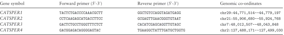
Table 1.Oligonucleotide sequences for primers as well as the genomic coordinates of theCATSPERgenes (UCSC version bosTau8).
Gene symbol Forward primer (5-3) Reverse primer (5-3) Genomic co-ordinates
CATSPER1 TACTCTGACCCCAAACGCTT GGCTGTCCAGGTAGATGAGG chr29:44,771,514--44,779,197
CATSPER2 CCTCAAGAGCATGACCTTCC GCGAGTTGAACGGGTGTAAT chr21:55,906,680--55,924,768
CATSPER3 GACTCTGCCTGGGTTTCTCT CACATCGAGCAGGTTGTAGC chr7:48,012,507--48,043,848
CATSPER4 GACGGAGACAGGGGAGTAC TGAAGGCTATTTGATGCTGGTG chr2:127,488,171--127,499,030
CA, USA) and an Eppendorf Mastercycler (Eppendorf, Hamburg, Germany).
Primer design, quantitative real-time polymerase chain reaction
Nucleotide sequences were retrieved from the University of Cali-fornia, Santa Cruz (UCSC) Genome Browser and Primer3 used for primer design (Table1). Primers were designed to be intron spanning and were commercially synthesized (Sigma Aldrich, St Louis, MO, USA). Quantitative real-time polymerase chain reaction (qRT-PCR) was performed using a 20μL reaction mix containing 10μL SYBR green PCR MasterMix (Invitrogen Ltd, Paisley, UK), 2.5μL primer and dH2O mix, 5.5μL dH2O, and 2μL sample. Plates were run in
an ABI 7500 Fast Thermocycler (Life Technologies, Carlsbad, CA, USA). The cycle parameters were as follows: Uracil N-glycosylase activation was run for 2 min at 50◦C, DNA polymerase activation for 10 min at 95◦C, the melt cycle was run for 15 s at 95◦C, and the annealing–extending cycle for 1 min at 60◦C. A no-template con-trol was run in each 96-well plate to confirm the absence of gDNA contamination. Levels of expression of the gene of interest were com-pared with the average of the two reference genes, glyceraldehyde 3-phosphate dehydrogenase (GAPDH) and beta actin (ACTb). Nor-malizing gene expression to multiple reference genes in order to give a more reliable baseline for the calculation of relative gene expression using qRT-PCR is common practice especially when small changes in gene expression are being reported [27].
In situ immunocytochemistry for theCATSPER1protein Fresh bull semen from a commercial bull stud was collected using an artificial vagina and diluted to 2×10⁶sperm/mL in
phosphate-buffered saline (PBS; Sigma Aldrich, St Louis, MO, USA). Cells were fixed in 4% paraformaldehyde, and washed with PBS before be-ing permeabilized with Triton X-100 (0.2%) for 15 min at room temperature (RT). After rinsing in PBS, cells were blocked using blocking buffer (0.4 g bovine serum albumin (BSA)+ 2μL Tri-ton X-100+ 10 mL PBS) for 1 h at RT. Immunostaining was carried out using primary antibody targeted against CATSPER1 (1:100; ab203626, Abcam, Cambridge, United Kingdom) at 4◦C overnight. The immunogen sequence of the CATSPER1 antibody used was MDSRAQGAWY. The homology between this and the bovine CATSPER1 sequence was 90%. After three 5 min washes in PBS, sperm were incubated for 1 h with goat antimouse IgG Alexa Fluor 488 (1:500; Invitrogen, California, United States) as a sec-ondary antibody. Nuclei were counterstained with 4 ,6-diamidino-2-phenylindole (DAPI). Cells were imaged with an Olympus IX83 (Norfolk, USA) inverted microscope equipped with a 40×objective.
Sperm preparation
Frozen-thawed sperm from Holstein bulls (n=3) of proven fertil-ity were used in all the functional experiments. Semen straws were thawed in a water bath at 39◦C for 30 s. For each functional
assess-ment, one straw per bull, of which there where three, were thawed. The resulting volume to run the assessment consisted of a pool of three straws coming from three bulls to eliminate the interbull vari-ability. All sperm were diluted in TALP media [28].
Effect of extracellular Ca2+on hyperactivation and rheotactic response
The aim of this experiment was to investigate the role of extracellular calcium on sperm hyperactivation and rheotaxis. Preliminary exper-iments were carried out to assess the optimum concentration of all agonists/antagonists (data not shown). To investigate this, extracel-lular Ca2+was ablated using ethylene glycol tetraacetic acid (EGTA) that chelates Ca2+in the media [29]. Motility of the frozen-thawed sperm was analyzed objectively by computer-assisted sperm analy-sis (CASA; Sperm Class Analyzer, Microptic, Viladomat, Barcelona, Spain). A droplet of diluted sperm (10μL) from each sample de-scribed above was placed on a prewarmed slide; a prewarmed cover slip was added and analyzed for sperm motion and kinematic pa-rameters using factory CASA (bull) settings. At least three randomly selected microscopic fields and a minimum of 100 sperm per treat-ment were assessed. Any samples with post-thaw progressive motility less than 30% were not used in experiments. Thawed sperm were incubated with either (i) 2 mM EGTA, (ii) 5 mM caffeine (phospho-diesterase inhibitor but also an agonist for extracellular Ca2+influx [30], (iii) 2 mM EGTA in combination with 5 mM caffeine, or (iv) no treatment, for 10 min before hyperactivation and rheotaxis were assessed. Hyperactivation was assessed using a phase-contrast mi-croscope (CX41; Olympus, Hamburg, Germany) at a magnification of 40×. A droplet of diluted sperm (10μL at a concentration of 25
×106sperm/mL) was placed on a prewarmed slide, covered with
a prewarmed cover slip and assessed subjectively by counting 100 motile sperm for each treatment. Hyperactivation was expressed as the percentage of motile sperm that displayed hyperactivated motil-ity. Hyperactivation was characterized by high amplitude, asymmet-rical beating pattern of the sperm tail [31]. This manifested in a characteristic figure of eight swimming trajectory. To assess sperm rheotactic response, sperm were loaded into the starting well (50μL at a concentration of 25×106sperm/mL) of a specialized
microflu-idic device (microchannel size of 300μm wide, 100μm deep, and 30 mm in length) with a flow rate of 30μm/s. The number of sperm that swam passed the 10 mm mark in the microchannel at 10 min were assessed. This experiment was replicated three times.
Effect of blocking calcium channels on hyperactivation and rheotactic response
induce hyperactivation as a positive control in all experiments. Sperm were incubated with (i) mibefradil (5μM), (ii) caffeine (5 mM), (iii) mibefradil (5μM) in combination with caffeine (5 mM), or (iv) no treatment, for 10 min prior to the assessment of rheotactic response and hyperactivation as described above. This experiment was repli-cated three times. Motility of the frozen-thawed sperm was analyzed as described above.
Effect of CatSper channels on hyperactivation and rheotactic response
The aim of this experiment was to assess the effect of CatSper chan-nels on hyperactivation and rheotactic response of bull sperm. To achieve this, CATSPER1 antibody (Ab) was used to specifically block CatSper1. An initial dose response hyperactivation test was carried out using: 0.8, 4, and 20μg/mL CATSPER1 Ab, and a concentration of 20μg/mL was selected for use in this study. Sperm were incubated with either (i) CATSPER1 Ab (20μg/mL), (ii) caffeine (5 mM), (iii) CATSPER1 Ab (20μg/mL) in combination with caffeine (5 mM), or (iv) with no treatment, for 10 min following which they were as-sessed for hyperactivation and rheotactic response. This experiment was replicated three times. Motility of the frozen-thawed sperm was analyzed as described above.
Data analysis
For gene expression data, the formula E=10(−1/slope)−1 was used
where slope refers to the slope of the linear curve of cycle threshold (CT) values plotted against log dilution. Only primers with PCR
efficiencies between 90% and 110% were used. Gene expression was analyzed using GenEx software (www.multid.se/genex.html), which allowed for compensation of PCR efficiencies, before averaging for RT-qPCR replicates. A normalization factor, calculated based on the geometric mean of the two reference genes, GAPDH and ACTb, was used to normalize the expression of each gene of interest. Functional data were checked for normality of distribution, transformed where appropriate using a log transformation, and analyzed using one-way Analysis of Variance (ANOVA), while qPCR data were analyzed using univariate ANOVA in the Statistical Package for the Social Sciences (SPSS, Version 21.0; IBM, Armonk, NY, USA). Posthoc tests were carried out using the Bonferroni correction and aPvalue
<0.05 was considered to be statistically significant.
Results
Bioinformatic identification of bovineCATSPER orthologs
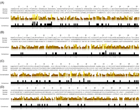
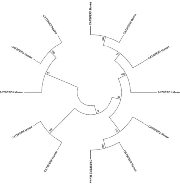
All four of the CATSPER genes reported in mice, humans, and horses were found to be present on chromosome 29, 21, 7, and 2, respectively, in the bovine genome (Table1). Multiple-sequence alignment was performed on novelCATSPERsequences with a com-plete second exon. Despite slight sequence variation between species, conserved areas were clearly identified across the three species for each protein (Fig.1). Sequence similarity for CATSPER 1–4 pep-tides between bovine, murine, and humans were 90%, 85%, 72%, and 88%, respectively. Phylogenetic analysis showed that orthologs genes (identical by descent from the common ancestor of bovine, murine, and human, and in a conserved syntenic location) can be con-fidently predicted with bootstrap values generally in excess of 90% (Fig.2). The phylogenetic relationships among different CATSPERs are much less certain. This is to be expected for genes that are so
short that the phylogenetic signal is noisy and that are also known to be under selective pressure for different structure and function. A bootstrap value of 100 indicates that the sequences below that node consistently cluster together even with multiple resamplings of the data. The proteins are thus likely to be orthologs because their similarity is systemic and internally consistent rather than dependent on a few similar sites in the alignment.
Expression ofCATSPER 1–4genes along the
reproductive tract of the bull
The expression of all fourCATSPERgenes varied depending on the location along the reproductive tract of the bull. There was an effect of tissue location for expression of all four of theCATSPERgenes, withCATSPER1–4upregulated in the parenchyma testis compared to the three segments of the epididymis (P<0.01; Fig.3). The rete testis had higher expression of allCATSPER1–4genes compared to the caudal and corpus epididymis (P<0.01); however, there was no difference in expression level between it and the caput epididymis or the parenchyma testis (P>0.05).CATSPER1expression in the parenchyma testis was upregulated 6-fold, compared to the average of the housekeeping genes, which was the highest fold change of all theCATSPERgenes.CATSPER 2–4were upregulated 4.5-, 2.5-, and 2.7-fold, respectively.
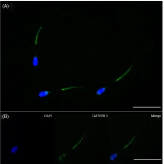
In situ immunocytochemistry for theCATSPER1protein Fluorescence labeled to the principal piece was evident in frozen-thawed bull sperm stained with the antirabbit CATSPER 1 antibody (Fig.4). CATSPER1 staining was primarily found along the principal piece of the flagella; however, light staining was also found on the postacrosomal region. No staining was evident when the primary antibody was withheld (data not shown).
Effect of extracellular Ca2+on hyperactivation and rheotactic response
There was no effect of EGTA treatment on the percentage of motile sperm. Caffeine increased hyperactivation but when extracellular calcium was removed by the addition of EGTA, there was a re-duction in hyperactivation when compared to both the control and caffeine-treated sperm (P<0.001). Interestingly, when caffeine was added back to the media containing EGTA, there was no increase in hyperactivation (Fig.5A). A mirrored response was found for the rheotactic response, where caffeine significantly increased the number of sperm to progress along the channel, while chelating ex-tracellular Ca2+inhibited this behavior (P<0.01). As before, adding caffeine back into the media did not result in a significant increase in rheotactic response (Fig.5B). These data demonstrate that extra-cellular calcium is required for hyperactivation and rheotaxis of bull sperm. Treatment with EGTA or caffeine did not have an effect on the percentage of motile sperm.
Effect of blocking calcium channels on hyperactivation and rheotactic response
Figure 1.Multiple sequence alignment of CATSPER1–4 (A–D) proteins in bovine, human, and mouse showing conserved regions (highlighted in color). Conserved regions highlight that the CATSPER proteins inBos taurus,Homo sapiens, andMus musculushave an evolutionary relationship by which they share a lineage and are descended from a common ancestor.
the microchannel, compared to the control group (P<0.01; Fig.6B); however, when sperm were treated with mibefradil prior to caffeine, there was no significant increase in sperm progression (P<0.01). These data demonstrate that an influx of calcium via mibefradil-sensitive channels is required for bull sperm hyperactivation and rheotaxis. Mibefradil treatment did not have an effect on the per-centage motile sperm.
Effect of CatSper channels on hyperactivation and rheotactic response
Blocking CatSper channels via CATSPER1 antibody reduced the lev-els of hyperactivity in the control sample and also inhibited the ac-tion of caffeine (P<0.01; Fig.7A). Similarly, CATSPER1 antibody reduced the ability of sperm to display rheotaxis compared to the control group and when added prior to caffeine it inhibited hyperac-tivation (P<0.01; Fig.7B). Despite the actions of both caffeine and the CATSPER1 antibody on hyperactivation and rheotactic response there was no effect of treatment on the percentage of motile sperm. Furthermore, sperm treated with CATSPER1 antibody displayed an increase in nonprogressive motility compared to no treatment sperm and swam in anticlockwise circles, as reported in CatSper null mice
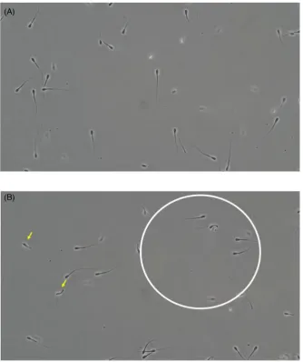
(Fig.8; Supplemental Videos S1 and S2). These data demonstrate that an influx of extracellular calcium via CatSper channels is re-quired for bull sperm hyperactivation and rheotaxis. Sperm treated with CATSPER1 antibody also displayed bending of the tail over the head.
Discussion
Figure 2.Phylogenetic tree showing the evolutionary relationship between the fourCATSPERgenes in the bovine genome and their human and mouse orthologs. The numbers at the nodes are % bootstrap values.
CATSPER 1–4 are conserved in the Bos taurus genome. Us-ing a comparative genomics approach, we searched theBos taurus genome for homologs of the fourCATSPERgenes that have recently been described [8,9,19]. We found homologs of all fourCATSPER subunit genes in theBos taurusgenome on various chromosomes, and revealed that the bovineCATSPERgenes are present in similar synthetic sequence to those of humans and mice [9,20]. Multiple-sequence alignment showed the conserved regions over the three species for each of the CATSPER protein sequences. Conserved re-gions highlight that the CATSPER proteins inBos taurus, Homo sapiens, andMus musculus have an evolutionary relationship by which they share a lineage and are descended from a common ances-tor. Phylogenetic analysis of these bovine genes in conjunction with their human and mouse orthologs showed a high degree of sequence similarity, which suggests functional conservation of these genes over the course of evolution. Where both a first and a second exon could be recovered for the bovine ortholog, the percentage of sequence identity ranged from 72% (CATSPER3) to 90% (CATSPER1). The high degree of similarity of the bovine genes to their human and mouse orthologs prompted us to delve further into the specific ex-pression and function of CatSper channels in bull sperm.
Expression analysis at the gene level showed thatCATSPER1–4 messenger RNA (mRNA) was expressed in the reproductive tract of
the bull with highest expression in the parenchyma testis, indicating that they are incorporated into sperm during spermatogenesis. Of the fourCATSPERgenes,CATSPER1was found to have the high-est expression across all tissue segments and is in agreement with studies in mice and humans [19,33]. The expression ofCATSPER1 was 2-fold that ofCATSPER3and4in the parenchyma testis. This is of particular interest as recently the profiles ofCATSPER1mRNA expression in testis biopsy of subfertile human male patients were in-vestigated [34]. Compared with patients whose infertility cannot be ascribed to a deficiency in motility, a significant reduction in the level ofCATSPER1gene expression among patients with asthenospermia was observed leading the authors to propose thatCATSPER1 ex-pression be used as a noninvasive screening method for male infer-tility [35]. Additionally,CATSPER1-deficient mice are infertile as a result of an impairment of sperm motility and an inability to fertilize oocytes [18]. Immunocytochemical analysis of the CATSPER1 pro-tein revealed CATSPER1 to be localized to the principal piece of the flagellum that is in agreement with the staining pattern reported for mouse, human, and stallions [8,18,19]. This same expression and localization of CATSPER1 in bulls as in humans, mice, and stallions points to a similar role of CATSPER1 all species.
Figure 3.Expression of four selectedCATSPERgenes across the reproductive tracts of three mature bulls. Expression was normalized to the average gene expression of bothGAPDHandACTβ. Tissue sections analyzed were: parenchyma testis, rete testis, caput epididymis (caput), corpus epididymis (corpus), and cauda epididymis (cauda). Values are means±SEM for three independent replicates.abcdDifferent superscripts differ significantly within each gene (P<0.05).
the function of this channel in bull sperm. We found that caffeine was an effective inducer of hyperactivation in bull sperm, as has been reported in other studies, and thus was used as a positive control for all functional studies [30,36]. While caffeine is known to induce hy-peractivation in both capacitated and noncapacitated bull sperm by inhibiting phosphodiesterase and thus increasing cAMP [37], stud-ies have also shown that caffeine-induced hyperactivation requires extracellular Ca2+in bull sperm [30,36], which is in agreement with this study as sperm treated with caffeine did not hyperactivate while in Ca2+-deficient medium. We investigated if bull sperm require ex-tracellular Ca2+for hyperactivated motility and the ability to display a sufficient rheotactic response to fluid flow. The results from this study, in which EGTA was used to chelate calcium, indicate that bull sperm require extracellular calcium for both hyperactivation and rheotactic responses. While the need for extracellular calcium for hyperactivation has been reported, this is the first study to our knowledge to report that bull sperm requires extracellular Ca2+to effectively induce rheotaxis. The typical characteristics of bull sperm hyperactivation, when assessed in a static droplet, is an increase in swimming speed of sperm but in a nonprogressive fashion; however, although not assessed directly in this study, it appears that when hy-peractivated bull sperm are exposed to a fluid flow they are able to
maintain the increase in swimming speed but alter their swimming pattern to linear.
Figure 4.Immunostaining for CATSPER1 (green) and nuclei counterstained with DAPI (blue; A). Fluorescent imaging showing colocalization of CATSPER1 to bull sperm flagellum. CatSper1 is specifically labeled in the principal piece of the tail (B). Bar=20μm.
Figure 5.The effect of removing extracellular calcium using EGTA treatment on (A) hyperactivation and (B) rheotactic response of bull sperm. Number of sperm refers to the number of sperm to swim past the 10 mm mark of the microfluidic channel. Values are means±SEM for three independent replicates.abcdDifferent superscripts differ significantly within panel (P<0.05).
[image:8.612.141.510.582.686.2]Figure 7.The effect of CATSPER1 antibody on (A) hyperactivation and (B) rheotactic response of bull sperm. Number of sperm refers to the number of sperm to swim past the 10 mm mark of the microfluidic channel. Values are means±SEM for three independent replicates.abcdDifferent superscripts differ significantly within panel (P<0.05).
Figure 8.Stills form supplementary videos showing the swimming patterns of control (A) and sperm treated with CATSPER1 antibody (B). Sperm swimming in an anticlockwise direction (circle) and bending of the sperm tail over the head (arrows) was observed in the Ab-treated group (B).
an anticlockwise circular plane, which does not change regardless of fluid flow [3]. We observed a similar swimming pattern in the bull sperm treated with CATSPER1 antibody. We observed a small proportion of sperm that had been treated with CATSPER1 Ab dis-played bending of the tail over the sperm head. The reason for this
[image:9.612.122.457.223.622.2]In conclusion, this study is the first to identify and characterize CATSPER genes in the bull. The testes expression and location-specific changes in mRNA abundance support an evolutionary con-served role for these channels in bull reproduction. This study demonstrates that CatSper channels play a critical role in hyper-activation and rheotactic response in bull sperm. The location and functionality of these channels points to their use as potential male infertility markers and should provide a basis for much future re-search to define the factors associated with effective sperm–oocyte interactions in this species.
Supplementary data
Supplementary data are available atBIOLREonline.
Supplemental Video S1.Video showing normal motility of control sperm. Sperm are observed to be swimming in a progressive manner.
Supplemental Video S2.Sperm treated with 20μg/mL CATSPER1 antibody swim in an anticlockwise direction (blue circle). Bending of the sperm tail over the head of sperm was observed (arrows).
Acknowledgment
The authors gratefully acknowledge the financial support from the National Cattle Breeding Centre (Naas, Co Kildare, Ireland) for the donation of bull semen.
References
1. Ishimoto K, Gaffney EA. Fluid flow and sperm guidance: a simula-tion study of hydrodynamic sperm rheotaxis.J R Soc Interface2015;
12:20150172.
2. Kaupp UB, Kashikar ND, Weyand I. Mechanisms of sperm chemotaxis. Annu Rev Physiol2008;70:93–117.
3. Miki K, Clapham DE. Rheotaxis guides mammalian sperm.Curr Biol 2013;23:443–452.
4. Bahat A, Eisenbach M. Sperm thermotaxis.Mol Cell Endocrinol2006;
252:115–119.
5. Martinez-Fresneda L, Costelloe J, O’Hara A, Lynch A, Monsonis-Centelles S, Newport D, Fair S. Characterization of the rheotaxis response of bull sperm using a microfluidic device.Anim Reprod Sci2016;169:109– 110.
6. Kantsler V, Dunkel J, Blayney M, Goldstein RE. Rheotaxis facilitates upstream navigation of mammalian sperm cells.Elife2014;3:e02403. 7. El-Sherry TM, Elsayed M, Abdelhafez HK, Abdelgawad M.
Characteri-zation of rheotaxis of bull sperm using microfluidics.Integr Biol2014;
6:1111–1121.
8. Loux SC, Crawford KR, Ing NH, Gonzalez-Fernandez L, Macias-Garcia B, Love CC, Varner DD, Velez IC, Choi YH, Hinrichs K. CatSper and the relationship of hyperactivated motility to intracellular calcium and pH kinetics in equine sperm.Biol Reprod2013;89:123.
9. Qi HY, Moran MM, Navarro B, Chong JA, Krapivinsky G, Krapivin-sky L, Kirichok Y, Ramsey IS, Quill TA, Clapham DE. All four CatSper ion channel proteins are required for male fertility and sperm cell hyperactivated motility. Proc Natl Acad Sci USA 2007;
104:1219–1223.
10. Yanagimachi R. Fertility of mammalian spermatozoa: its development and relativity.Zygote1994;2:371–372.
11. Ho HC, Suarez SS. An inositol 1,4,5-trisphosphate receptor-gated intracel-lular Ca(2+)store is involved in regulating sperm hyperactivated motility.
Biol Reprod2001;65:1606–1615.
12. Zucchi R, Ronca-Testoni S. The sarcoplasmic reticulum Ca2+ chan-nel/ryanodine receptor: modulation by endogenous effectors, drugs and disease states.Pharmacol Rev1997;49:1–51.
13. Catterall WA. Structure and function of voltage-gated ion channels.Annu Rev Biochem1995;64:493–531.
14. Darszon A, Nishigaki T, Beltran C, Trevino CL. Calcium channels in the development, maturation, and function of spermatozoa.Physiol Rev 2011;91:1305–1355.
15. Nguyen TMD, Duittoz A, Praud C, Combarnous Y, Blesbois E. Calcium channels in chicken sperm regulate motility and the acrosome reaction. FEBS J2016;283:1902–1920.
16. Singh AP, Rajender S. CatSper channel, sperm function and male fertility. Reprod Biomed Online2015;30:28–38.
17. Lishko PV, Kirichok Y, Ren D, Navarro B, Chung JJ, Clapham DE. The control of male fertility by spermatozoan ion channels.Annu Rev Physiol 2012;74:453–475.
18. Ren D, Navarro B, Perez G, Jackson AC, Hsu S, Shi Q, Tilly JL, Clapham DE. A sperm ion channel required for sperm motility and male fertility. Nature2001;413:603–609.
19. Li HG, Liao AH, Ding XF, Zhou H, Xiong CL. The expression and significance of CATSPER1 in human testis and ejaculated spermatozoa. Asian J Androl2006;8:301–306.
20. Tamburrino L, Marchiani S, Vicini E, Muciaccia B, Cambi M, Pellegrini S, Forti G, Muratori M, Baldi E. Quantification of CatSper1 expression in human spermatozoa and relation to functional parameters.Hum Reprod 2015;30:1532–1544.
21. Jarow JP. A sperm ion channel required for sperm motility and male fertility.J Urol2002;168:2309.
22. Altschul SF, Gish W, Miller W, Myers EW, Lipman DJ. Basic local align-ment search tool.J Mol Biol1990;215:403–410.
23. Stecher G, Liu L, Sanderford M, Peterson D, Tamura K, Kumar S. MEGA-MD: molecular evolutionary genetics analysis software with mu-tational diagnosis of amino acid variation. Bioinformatics2014; 30: 1305–1307.
24. Ram H, Kumar A, Thomas L, Singh VP. In silico approach to study adaptive divergence in nucleotide composition of the 16S rRNA gene among bacteria thriving under different temperature regimes.J Comput Biol2014;21:753–759.
25. Notredame C, Higgins DG, Heringa J. T-Coffee: a novel method for fast and accurate multiple sequence alignment.J Mol Biol2000;302: 205–217.
26. Waterhouse AM, Procter JB, Martin DM, Clamp M, Barton GJ. Jalview Version 2—a multiple sequence alignment editor and analysis workbench. Bioinformatics2009;25:1189–1191.
27. Vandesompele J, De Preter K, Pattyn F, Poppe B, Van Roy N, De Paepe A, Speleman F. Accurate normalization of real-time quantitative RT-PCR data by geometric averaging of multiple internal control genes.Genome Biol2002;3:RESEARCH0034.
28. Parrish JJ, Susko-Parrish J, Winer MA, First NL. Capacitation of bovine sperm by heparin.Biol Reprod1988;38(5):1171–1180.
29. Hong CY, Chiang BN, Ku J, Wei YH, Fong JC. Calcium antagonists stimulate sperm motility in ejaculated human semen.Br J Clin Pharmacol 1985;19:45–49.
30. Colas C, Cebrian-Perez JA, Muino-Blanco T. Caffeine induces ram sperm hyperactivation independent of cAMP-dependent protein kinase.Int J An-drol2010;33:e187–197.
31. Rahman MS, Kwon WS, Pang MG. Calcium influx and male fertil-ity in the context of the sperm proteome: an update.BioMed Res Int 2014, 841615. doi:10.1155/2014/841615.
32. Carlson AE, Westenbroek RE, Quill T, Ren D, Clapham DE, Hille B, Garbers DL, Babcock DF. CatSper1 required for evoked Ca2+entry and control of flagellar function in sperm.Proc Natl Acad Sci USA2003;
100:14864–14868.
33. Avidan N, Tamary H, Dgany O, Cattan D, Pariente A, Thulliez M, Borot N, Moati L, Barthelme A, Shalmon L, Krasnov T, Ben-Asher E et al. CATSPER2, a human autosomal nonsyndromic male infertility gene.Eur J Hum Genet2003;11:497–502.
34. Kirichok Y, Navarro B, Clapham DE. Whole-cell patch-clamp measure-ments of spermatozoa reveal an alkaline-activated Ca2+channel.Nature 2006;439:737–740.
subfertile men with deficient sperm motility. Hum Reprod2004; 19: 124–128.
36. Marquez B, Suarez SS. Different signaling pathways in bovine sperm regu-late capacitation and hyperactivation.Biol Reprod2004;70:1626–1633. 37. Barakat IAH, Danfour MA, Galewan FAM, Dkhil MA. Effect of various concentrations of caffeine, pentoxifylline, and kallikrein
on hyperactivation of frozen bovine semen. BioMed Res Int 2015, 948575.
38. Strunker T, Goodwin N, Brenker C, Kashikar ND, Weyand
I, Seifert R, Kaupp UB. The CatSper channel mediates
progesterone-induced Ca2+ influx in human sperm. Nature 2011;