ORIGINAL RESEARCH
Diffusion Tensor Imaging of the Normal Pediatric
Spinal Cord Using an Inner Field of View
Echo-Planar Imaging Sequence
N. Barakat F.B. Mohamed L.N. Hunter P. Shah S.H. Faro A.F. Samdani J. Finsterbusch R. Betz J. Gaughan M.J. Mulcahey
BACKGROUND AND PURPOSE: DTI in the brain has been well established, but its application in the spinal cord, especially in pediatrics, poses several challenges. The small cord size has inherent low SNR of the diffusion signal intensity, respiratory and cardiac movements induce artifacts, and EPI sequences used for obtaining diffusion indices cause eddy-current distortions. The purpose of this study was to 1) evaluate the accuracy of cervical spinal cord DTI in children using a newly developed iFOV sequence with spatially selective 2D-RF excitations, and 2) examine reproducibility of the DTI measures.
MATERIALS AND METHODS:Twenty-five typically developing subjects were imaged twice using a 3T scanner. Axial DTI images of the cervical spinal cord were acquired with this sequence. After motion correction, DTI indices were calculated using regions of interest manually drawn at every axial section location along the cervical spinal cord for both acquisitions. Various DTI indices were calculated: FA, AD, RD, MD, RA, and VR. Geometric diffusion measures were also calculated: Cp, Cl, and Cs.
RESULTS: The following average values for each index were obtained: FA⫽0.50⫾0.11; AD⫽0.97⫾ 0.20⫻10⫺3mm2/s; RD⫽0.41⫾0.13⫻10⫺3mm2/s; MD⫽0.59⫾0.15⫻10⫺3mm2/s; RA⫽0.35⫾ 0.08; VR⫽0.03⫾0.00; Cp⫽0.13⫾0.07; Cl⫽0.29⫾0.09; and Cs⫽0.58⫾0.11. The reproducibility tests showed moderate to strong ICC in all subjects for all DTI parameters (ICC⬎0.72).
CONCLUSIONS:This study showed that accurate and reproducible DTI parameters can be estimated in the pediatric cervical spinal cord using an iFOV EPI sequence.
ABBREVIATIONS:2D-RF⫽2-dimensional radio frequency; AD⫽axial diffusivity; Ci⫽confidence interval; Cl⫽linear index; Cp⫽planar index; Cs⫽spherical index; FA⫽fractional anisotropy; ICC ⫽ intraclass correlation coefficient; iFOV ⫽ inner field of view; ISNCSCI ⫽ International Standards for Neurological Classification of Spinal Cord Injury; MD⫽mean diffusivity; RA⫽relative anisotropy; RD⫽radial diffusivity; SCI⫽spinal cord injury; VR⫽volume ratio
E
ach year, an estimated 11,000 new spinal cord injuries oc-cur in the United States. Among these injuries, 1%–5.4% are reported in children.1Although spinal trauma is relativelyinfrequent in pediatric patients compared with adults, disabil-ity and mortaldisabil-ity rates are higher: 40% in pediatric patients versus 20% in adults.2,3Due to the distinctive biomechanics and anthropometrics of children’s spinal cords, 60%– 80% of all pediatric cord injuries are cervical.4Despite the continued
improvements in treatment that help many children survive SCI, most injuries will cause permanent disability. Therefore, continued research is needed to assess neurologic damage af-ter traumatic SCI and to improve care, treatment, and reha-bilitation methods.
Currently, the evaluation and classification of neurologic impairment in adults and children with SCI is assessed using
the ISNSCI published by the American Spinal Injury Associa-tion.5These standards involve the testing of sensory and
mo-tor function of the limbs, trunk, and anorectal area, and are used to predict recovery of neurologic function, plan treat-ment, and determine treatment effectiveness. Although inter-nationally recognized, the ISNSCI standards have low utility in the pediatric population and can lead to unreliable assess-ment of neurologic abnormalities. A recent study has shown that children younger than 6 years of age were unable to un-derstand and follow test instructions, children as old as 10 years were extremely anxious during the examination, and adolescents as old as 15 years withdrew from the study due to discomfort with the anal examination.6In addition, these
ex-aminations do not provide a direct assessment of damage to white matter tracts within the spinal cord. This can present a considerable challenge against the development of effective treatment approaches designed specifically at the cord level. Therefore, an objective assessment of the spinal cord may pro-vide important information complementary to conventional clinical assessment and may lead to accurate prognostic eval-uation of recovery from SCI.
DTI is a promising technique of examining the spinal cord in vivo. It quantifies the diffusion of water molecules in each voxel of an image in directions parallel and transverse to the plane of neuronal axons. The quantitative characteristic of DTI allows the characterization of physical properties of tis-sues. The unique characteristic architecture of the spinal cord
Received July 11, 2011; accepted after revision September 5.
From the Departments of Bioengineering (N.B.) and Radiology (N.B., F.B.M., L.N.H., P.S., S.H.F.), Temple University, Philadelphia, Pennsylvania; Shriners Hospital for Children (A.F.S., R.B., M.J.M.), Philadelphia, Pennsylvania; Department of Systems Neuroscience (J.F.), University Medical Center Hamburg-Eppendorf, Hamburg, Germany; and Biostatistics Consulting Center (J.G.), Temple University School of Medicine, Philadelphia, Pennsylvania.
This study was funded by the Shriners Hospitals for Children Grant 8956 (Mulcahey, PI).
Paper previously presented in part at: the International Society of Magnetic Resonance in Medicine, Montreal, Quebec, Canada, May 11, 2011.
Please address correspondence to Feroze B. Mohamed, Department of Radiology, Temple University School of Medicine, 3401 N. Broad St, Philadelphia, PA 19140; e-mail: [email protected]
http://dx.doi.org/10.3174/ajnr.A2924
PEDIATRICS
ORIGINAL
may allow DTI to localize white matter, separate white from gray matter, and assess structural damage of the cord. Previous studies reported the success of DTI in characterizing different tissues of the spinal cord and demonstrated changes in diffu-sion characteristics along injured spinal cords.7-9
DTI of the spinal cord is technically limited by various fac-tors. The small cord volume (approximately 1 cm in diameter) leads to a low SNR. CSF pulsation and blood flow can produce prominent ghosting artifacts and degrade image quality. The spinal cord is also subject to respiratory and cardiac move-ments, which cause image blurring and increased or decreased signal intensity. Different tissue interfaces (bone, soft tissue, or fluid) can create susceptibility artifacts. Swallowing or related motion artifacts are mostly seen when imaging the cervical spinal cord.10,11Finally, in pediatric imaging especially, the
possibility of increased subject motion makes obtaining accu-rate and reproducible DTI parameter values difficult.12,13
Ar-tifact-reducing techniques can be used to overcome some of these issues. However, particularly in pediatric imaging, these techniques are not without challenges. Cardiac gating and re-spiratory compensation may increase acquisition time, and sedation is typically not desirable in children. Thus, fast, reli-able, and high-resolution imaging is needed to image pediatric subjects.
Ideally, the goal is to obtain high-resolution artifact-free and reproducible images within minimal scan time. The choice of optimal pulse sequences in pediatric spinal cord DTI is therefore critical. The pulse sequences of choice have been EPI-based designs. While these sequences offer short acquisi-tion times, which reduce moacquisi-tion-related artifacts, they are highly sensitive to susceptibility artifacts and eddy-current distortions.14,15Recent work using reduced FOV imaging se-quences showed great reduction in geometric distortions when imaging the spinal cord.16-19This approach takes
advan-tage of the small-diameter spinal cord morphology and ap-plies a relatively small rectangular FOV to the area of interest. In addition to pulse sequence choice, correcting motion-re-lated artifacts may be a useful step in data analysis and can improve image quality. Artifact-reduction techniques can be applied during data acquisition, which may increase imaging time or the postprocessing stage.
While studies on diffusion imaging of the spinal cord in adults,7,20,21as well as in animal models22-25have been
re-ported, a comprehensive study of the pediatric spinal cord that examines the accuracy and reproducibility of DTI measures has not yet been reported. The investigation of normative data of spinal cord DTI represents an additional important area of inquiry. The clinical significance of this study is 2-fold. First, as in any imaging study, a thorough understanding of the diffu-sion characteristics of the healthy spinal cord is essential for the clinical interpretation of images of the injured spinal cord. If DTI proves to be a feasible, accurate, and reliable method for quantifying diffusion changes in the pediatric spinal cord, it may be a useful neurodiagnostic supplement to conventional MR imaging. Second, normative DTI values will provide a basis for which to compare DTI values from children with spinal injuries, allowing for classification of the consequence of injury based on deviation from normal values. Therefore, the purpose of this study is to investigate DTI of the pediatric cervical spinal cord in healthy controls by 1) evaluating the
accuracy of DTI in children using a newly developed iFOV sequence with spatially selective 2D-RF excitations, and 2) ex-amining the reproducibility of the DTI measures.
Materials and Methods
Subjects
Twenty-five subjects (6 males and 19 females), with a mean age of 13.28 years (range 7–21 years), were recruited. The age range up to and including 21 years of age represents the ages of youth typically cared for by centers specializing in SCI. The subjects had no evidence of spinal cord injury or pathology. Subjects and their parents pro-vided written informed assent and consent of the institutional review board–approved protocol. Data from 3 subjects were excluded from the analysis—2 due to excessive motion artifacts and 1 with an inci-dental finding of Chiari I malformation in the cervical spinal cord. Data from the remaining 22 control subjects were included in the analysis.
Imaging
The scans were performed using a 3T Verio MR scanner (Siemens, Erlangen, Germany) with a 4-channel neck matrix and an 8-channel spine matrix coil. All subjects underwent 2 identical scans (mean time⫽9 hours). The protocol consisted of conventional sagittal FSE T1- and T2-weighted scans, axial FSE T2-weighted scans, as well as axial DTI acquisition with the iFOV sequence. Anesthesia was not administered to the subjects. Cardiac and respiratory gating were not used.
DTI Acquisition Using iFOV Sequence
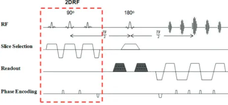
The iFOV sequence used in this study was based on a single-shot EPI sequence for diffusion-weighted imaging with spatially 2D-RF exci-tations. Details regarding this pulse sequence can be found in Finster-busch’s article.18In summary, the section-select radio-frequency
ex-citation of a standard EPI sequence was replaced by 2D-RF exex-citation targeting a rectangular profile, the inner FOV, using a blipped-planar trajectory applied in the phase-encoding direction (Fig 1). The side excitations were positioned outside of the imaged object. To avoid aliasing of the profile’s transition region, oversampling of the iFOV in the phase encoding was performed (Fig 2). The iFOV sequence was implemented on the scanner and, during a pilot phase, was optimized for imaging the pediatric spinal cord. Imaging was performed using several diffusion directions (6, 12, 20, and 30), multiple signal inten-sity averages (1 to 4), different b-values (0, 700, 800, 900, 1000, and 1200 seconds/mm2), and with cardiac gating. The optimization
[image:2.594.301.532.44.151.2]the same anatomic location prescribed for the T2-weighted images to cover the entire cervical spinal cord (C1 to T1 levels). The final imag-ing parameters included 20 diffusion directions,b⫽1000s/mm2,
voxel size⫽1.2⫻1.2⫻3 mm3, axial sections⫽35– 45 (depending
on the subject’s height), TR⫽6100 – 8000 ms, TE⫽115 ms, number of averages⫽3, and acquisition time⫽7 minutes.
Preprocessing
Initially, the diffusion datasets were corrected for motion-induced artifacts using the AIR image registration package (http://bishopw. loni.ucla.edu/AIR5/index.html) implemented in DTIstudio (http:// www.mristudio.org). The target images (20 directional images) were aligned with the reference image (B0) using a rigid registration algo-rithm and scaled-least-squares cost function.26
Tensor Estimation
After motion correction of the diffusion-weighted images, the eigen-vectors and eigenvalues of the diffusion tensor matrix were computed on a voxel-by-voxel basis from the axial DT images using MedINRIA software (http://www.sop.inria.fr/asclepios/software/MedINRIA/). Various DTI indices were calculated and tabulated, namely, FA, AD, RD, MD, RA, and VR.10,27,28
Tensor Visualization
In addition to the DTI metrics, the following geometric diffusion measures were also extracted: Cp, Cl, and Cs. These visualization indices relate to the geometric shape of the diffusion tensor and may help examine the 3D process of diffusion and fully characterize tissue microstructure.29-31
Regions of Interest
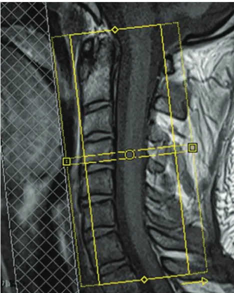
For both acquisitions, regions of interest were manually drawn on color FA maps at every axial section location along the cervical spinal cord and were validated by a neuroradiologist to ensure proper ana-tomic localization. There was a consistent sparing of the outer margin of the cervical cord that represented approximately 1 voxel width to minimize volume averaging with the CSF (Fig 3). Regions of interest were consistently defined throughout all sections. DTI indices were reported at each disk level of the cervical spinal cord as well as the middle (mid) portion of the cervical vertebral body (Fig 4). The values Fig 2.Localization image from which the axial cervical spinal cord image sections were
prescribed. The inner solid yellow rectangular line represents the iFOV, which was oversampled (dotted yellow line) to avoid aliasing. A saturation band was applied to reduce flow-related artifacts.
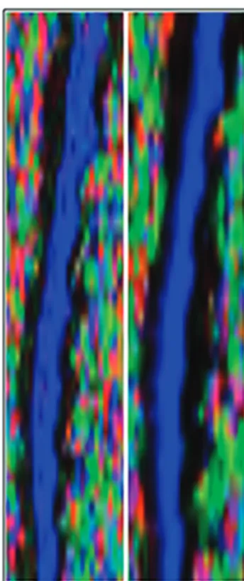
Fig 3.Axial FA color image of a control subject’s cervical spinal cord (left). Manual placement of region of interest (middle). Diffusion tensor ellipsoid representation in white and gray matter (right). There was clear discrimination between the butterfly-shaped gray matter and surrounding white matter.
[image:3.594.53.287.43.336.2] [image:3.594.301.534.43.241.2] [image:3.594.66.532.579.701.2]of each DTI parameter were averaged per cord level across all subjects.
Statistics
Statistical analysis was performed to test the reliability of the datasets from the 2 separate scans per subject. This was assessed by calculating the ICC coefficients32for each DTI parameter for the entire cervical
spinal cord as well as per cord level.
Results
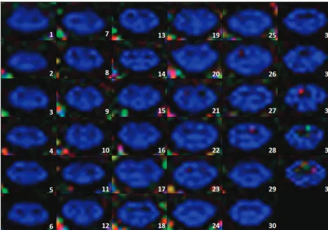
Imaging with the iFOV sequence resulted in high spatial reso-lution of DT images. Minimal eddy-current distortions and distinctive delineation of gray and white matter structures were observed in most axial sections (Fig 5). Qualitative ex-amination of image registration results showed reduction in motion-induced artifacts and well-defined CSF and cord structures (Fig 6).
The subjects showed mean⫾ standard deviation FA⫽ 0.50⫾0.11, AD⫽0.97⫾0.20⫻10⫺3mm2/s, RD⫽0.41⫾
0.13⫻10⫺3mm2/s, MD⫽0.59⫾0.15⫻10⫺3mm2/s, RA⫽ 0.35⫾0.08, VR⫽0.03⫾0.00, Cp⫽0.13⫾0.07, Cl⫽0.29⫾ 0.09, and Cs⫽0.58⫾0.11. The mean⫾standard deviation of each DTI parameter, as a function of cord level, is illustrated in Figs 7 and 8. There was a variation in the parameter values along the spinal cord. Between C1 and T1 levels, there was a progressive decrease in FA (15%), AD (16%), MD (9%), RA (23%), and Cl (27%), and a demonstrated increase in RD (1%), VR (11%), Cp (36%), and Cs (7%).
Reliability tests showed moderate to strong agreement be-tween the 2 scans in all the subjects for all DTI parameters. For the averaged DTI parameter values across all levels, the ICCs and their 95% Cis were FA [ICC⫽0.87, Ci (0.78 – 0.97)], AD [ICC⫽0.97, Ci (0.92– 0.99)], RD [ICC ⫽0.91, Ci (0.83– 0.98)], MD [ICC⫽0.95, Ci (0.92– 0.98)], RA [ICC⫽0.93, Ci (0.88 – 0.98)], VR [ICC⫽0.82, Ci (0.69 – 0.95)], Cp [ICC⫽ 0.77, Ci (0.58 – 0.95)], Cl [ICC⫽0.91, Ci (0.85– 0.97)] and Cs [ICC⫽0.72, Ci (0.50 – 0.94)]. The ICCs for each DTI param-eter as a function of cord level were also assessed and summa-rized in the Table.
Discussion and Conclusion
DTI can offer a comprehensive understanding regarding structural anisotropy of axonal white matter in any tissue. Dif-fusion measurement is particularly promising in the spinal cord because the inherent direction of anisotropy is aligned with the cord. In the pediatric spinal cord, the utility of DTI in quantifying diffusion changes has not been well studied. The small volume of the cord, CSF flow, cardiac/respiratory activ-ity, and susceptibility artifacts from adjacent bony structures limit the accuracy and reproducibility of DTI values. Addi-tional challenges include keeping a child motionless for several minutes without the use of sedation and the absence of nor-mative DTI data for comparison. Although diffusion changes in the pediatric brain have been reported,33-35no
comprehen-sive study of the pediatric spinal cord has been conducted.
[image:4.594.54.532.44.379.2]Despite these difficulties, DTI parameters were successfully obtained in this study using a well-optimized scanning proto-col. The protocol consisted of T1- and T2-weighted sequences, as well as an iFOV single-shot EPI sequence for DTI acquisi-tion. The iFOV was installed on the MR imaging scanner and pilot tested on phantom models and adult subjects, as well as children to optimize scanning parameters. The scans were per-formed using several diffusion directions, multiple signal in-tensity averages, different b-values, and with cardiac gating. Qualitative assessments of the images and SNR calculations were performed for each combination of parameters. The op-timal pulse sequence parameters were chosen based on maxi-mum spatial resolution and maximaxi-mum SNR while maintain-ing good image quality and minimum scan time. For axial acquisition of the pediatric cervical spinal cord (35– 45 sec-tions), the scan time was determined to be approximately 8 minutes.14When cardiac gating was used, there was no
signif-icant improvement in either SNR or image quality. There was, however, a significant increase in scan duration (⬎60%), which exceeded acceptable scan time limit. Therefore, cardiac
gating was not incorporated in this study. The optimization and testing of the iFOV sequence parameters led to an increase in image resolution while maintaining a short scan time. As a result, the gray and white matter structures could be clearly visualized across cord sections for most subjects (Fig 5).
Incorporating motion correction at the postprocessing stage before DTI analysis also proved to be valuable in data analysis. This step was accomplished via image registration using AIR with a rigid body transformation technique. This registration method uses information taken from the refer-ence image (B0 in this case) and the target images (20 gradient directions) and creates a cost function, which quantifies the amount of mutual information between the images. Rigid body registration assumes the movement of the imaged area is rigid (free of deformation). It is commonly used in medical applications where the imaged structure is assumed to be en-cased within a bony structure (eg, the brain).36As a result, the
rigid body registration method was chosen in this study be-cause the spinal cord is also encased and protected by a bony structure—the vertebral column. Furthermore, a study inves-tigating quantitative and qualitative analysis of several image registration methods showed that the rigid registration tech-nique was superior to other registration methods.26Although
the image registration approach was effectively capable of re-ducing artifacts caused by rigid displacement of the spinal cord, particularly motion in the anteroposterior direction, some motion-related artifacts remained: CSF pulsation, spo-radic swallowing, and subject bulk motion.
With the absence of any studies or results of DTI in the pediatric spinal cord, this test–retest methodology was used to ensure accurate and reproducible quantification of diffusion changes in the spinal cord. Reliability of a quantitative imaging test is a critical feature that may allow for monitoring in vivo changes due to disease progression and treatment effective-ness. Poor reproducibility limits the potential clinical applica-tion of any medical imaging technique. To evaluate the repro-ducibility of these results, all subjects were scanned twice, using the same acquisition protocol, within an average time interval of 9 hours. No scanner upgrades were performed dur-ing the length of the study. Image registration and DTI analysis methods were identical in both datasets, and the regions of interest were drawn the second time by the same observer. The reproducibility of the different DTI parameters was evaluated using the ICC and showed moderate to strong agreement be-tween the repeated-measurements scans (0.72– 0.97). How-ever, examining the ICC values as a function of cord level revealed slight fluctuations in lower levels. Signal intensity drop-offs at the edge of the neck coil may have been a contrib-uting factor to these differences because the lower cervical and upper thoracic levels were positioned at the extremities of the neck coil, where SNR decreases.
The results of this study showed a gradual decrease in FA, AD, MD, RA, and Cl values, and a gradual increase in VR, Cp, and Cs values along the length of the spinal cord in the supe-rior to infesupe-rior direction (C1 to T1), while RD remained rela-tively constant. This variations pattern can be attributed to the following effects:
1) In the upper cervical spinal cord, there is a higher atten-uation of large-diameter axons compared with the upper tho-racic region and, consequently, an increased axial (or
[image:5.594.82.254.41.450.2]dinal) diffusivity (AD).34,35Subsequently, all the DTI indices
(FA, MD, RA, and Cl) that are proportionally related to AD also showed a decrease in the thoracic region, and the param-eters that are inversely proportional to AD showed an increase. This pattern is similar to that reported in other studies involv-ing adult subjects.20,37
2) The geometric shape of the diffusion tensor is assumed to be ellipsoid and the 3 eigenvalues (1,2, and3)
corre-sponding to diffusivities along the principal axes of the diffu-sion tensor are sorted in the order1⬎⬎2⬃3. In a highly
anisotropic axonal-shaped medium, AD represents diffusivity along the primary axis of the diffusion tensor parallel to the
spinal cord tracts (AD⫽1). RD represents diffusivity
per-pendicular to the spinal cord tracts and is the average of2and
3. It was therefore expected that RD would remain constant
across cord levels.
3) Cardiac gating was not used in this study. The lowest cervical levels (C4 –C7) are the most sensitive to cardiac mo-tion.10,11Therefore, some cardiac-related artifacts may have biased the quantification of DTI parameters. While this may be considered a limitation in the study, it is important to note that gating increases acquisition time and does not prevent subject motion. Keeping the acquisition time relatively short was a priority for imaging the pediatric population.
Another potential limitation in the study is the sex- and age-dependent variances in the data. Previous studies on DTI in brain white matter reported differences in parameter values
Fig 7.DTI parameters values averaged across all the controls and plotted as a function of cord levels. Data from 1 measurement are shown. The error bars represent the standard deviations.
Fig 8.Tensor visualization indices (Cs, Cl, and Cp) averaged across all the controls and plotted as a function of cord levels. Data from 1 measurement are illustrated. The error bars represent the SDs.
ICCs of all DTI indices per cord level
[image:6.594.131.452.48.337.2] [image:6.594.53.284.381.537.2]related to sex38and age.39-41In addition, the use of manually
drawn regions of interest in the small pediatric spinal cord may have introduced partial volume contamination, espe-cially in the lower cervical regions.
In this study, youths up to 21 years of age were included to obtain normative values for ages that parallel age groups typ-ically seen in pediatric SCI centers. Although they are not chil-dren, youth between 18 and 21 years of age are often in tran-sition from pediatric to adult health care and, in the presence of spinal cord injury, are often cared for in specialized pediat-ric centers.42
Conclusions
This study demonstrated the feasibility of pediatric spinal cord imaging using DTI and produced accurate and reliable DTI measures. Both goals were successfully assessed using an iFOV sequence, which made high-resolution imaging possible, and by including motion correction in the data analysis steps. In future work, the DTI data from the control subjects need to be compared with those of children with SCI and correlated to clinical measures.
Acknowledgments
The authors thank Ms. Laure Rutter for her assistance with subject recruitment.
Disclosures: Scott Faro—UNRELATED: Grants/Grants Pending:Shriners.* MJ Mulcahey—
UNRELATED: Grants/Grants Pending:Shriners Hospitals for Children, Comments: Grant pending and under review. (* Money paid to institution).
References
1. National Spinal Cord Injury Statistical Center.2009 annual statistical report.
University of Alabama at Birmingham, February 2010
2. Brown RL, Brunn MA, Garcia VF.Cervical spine injuries in children: a review of 103 patients treated consecutively at a level 1 pediatric trauma center.J Pe-diatr Surg2001;36:1107–14
3. Osler TM, Vane DW, Tepas JJ, et al.Do pediatric trauma centers have better survival rates than adult trauma centers? An examination of the national pe-diatric trauma registry.J Trauma2001;50:96 –101
4. Cirak B, Ziegfeld S, Knight VM, et al.Spinal injuries in children.J Pediatr Surg
2004;39:607–12
5. Waring WP, Biering-Sorensen F, Burns S, et al.2009 review and revisions of the international standards for the neurological classification of spinal cord in-jury.J Spinal Cord Med2010;33:346 –52
6. Mulcahey MJ, Gaughan J, Betz RR, et al.The international standards for neu-rological classification of spinal cord injury: reliability of data when applied to children and youths.Spinal Cord2007;45:452–59
7. Rossi C, Boss A, Lindig TM, et al.Diffusion tensor imaging of the spinal cord at 1.5 and 3.0 Tesla.Rapid Commun2007;179:219 –24
8. Akter M, Hirai T, Minoda R, et al.Diffusion tensor tractography in the head-and-neck region using a clinical 3-T MR scanner.Acad Radiol2009;16:858 – 65 9. Ducreux D, Fillard P, Facon D, et al.Diffusion tensor magnetic resonance imaging and fiber tracking in spinal cord lesions: current and future indica-tions.Neuroimaging Clin N Am2007;17:137– 47
10. Kharbanda HS, Alsop DC, Anderson AW, et al.Effects of cord motion on dif-fusion imaging of the spinal cord.Magn Reson Med2006;56:334 –39 11. Figley CR, Stroman PW.Investigation of human cervical and upper thoracic
spinal cord motion: implications for imaging spinal cord structure and func-tion.Magn Reson Med2007;58:185– 89
12. Rosenberg DR, Sweeney JA, Gillen JS, et al.Magnetic resonance imaging of children without sedation: preparation with simulation.J Am Acad Child Ado-lesc Psychiatry1997;36:853–59
13. Malisza KL, Martin T, Shiloff D, et al.Reactions of young children to the MRI scanner environment.Magn Reson Med2010;64:377– 81
14. Mohamed FB, Hunter LN, Barakat N, et al.Diffusion tensor imaging of the pediatric spinal cord at 1.5T: preliminary results.AJNR Am J Neuroradiol
2011;32:339 – 45
15. Clark CA, Werring DJ.Diffusion tensor imaging in spinal cord: methods and applications—a review.NMR Biomed2002;15:578 – 86
16. Wilm BJ, Svensson J, Henning A, et al.Reduced field-of-view MRI using outer volume suppression for spinal cord diffusion imaging.Magn Reson Med
2007;57:625–30
17. Saritas EU, Cunningham CH, Lee JH, et al.DWI of the spinal cord with reduced FOV single-shot EPI.Magn Reson Med2008;60:468 –73
18. Finsterbusch J.High-resolution diffusion tensor imaging with inner field-of-view EPI.J Magn Reson Imaging2009;29:987–93
19. Rieseberg S, Frahm J, Finsterbusch J.Two-dimensional spatially-selective RF excitation pulses in echo-planar imaging.Mag Reson Med2002;47:1186 –93 20. Ellingson BM, Ulmer JL, Kurpad SN, et al.Diffusion tensor MR imaging of
the neurologically intact human spinal cord. AJNR Am J Neuroradiol
2008;29:1279 – 84
21. Wheeler-Kingshott CAM, Hickman SJ, Parker GJM, et al.Investigating cervical spinal cord structure using axial diffusion tensor imaging.Neuroimage
2002;16:93–102
22. Elshafiey I, Bilgen M, He R, et al.In vivo diffusion tensor imaging of rat spinal cord at 7 T.Magn Reson Imaging2002;20:243– 47
23. Ford JC, Hackney DB, Alsop DC, et al.MRI characterization of diffusion coef-ficients in a rat spinal cord injury model.Magn Reson Med1994;31:488 –94 24. Franconi F, Lemaire L, Marescaux L, et al.In vivo quantitative microimaging of
rat spinal cord at 7T.Magn Reson Med2000;44:893–98
25. Gulani V, Iwamoto GA, Jiang H, et al.A multiple echo pulse sequence for diffusion tensor imaging and its application in excised rat spinal cords.Magn Reson Med1997;38:868 –73
26. Barakat N, Middleton D, Hunter L, et al.An investigation of motion correction algorithms for pediatric spinal cord DTI in normals and patients with SCI.In
Proceedings of the 19th Annual Meeting of ISMRM Montreal, Quebec, Canada, 2011;6069
27. Le Bihan D, Mangin JF, Poupon C, et al.Diffusion tensor imaging: concepts and applications.J Magn Reson Imaging2001;13:534 – 46
28. Clark CA, Werring DJ, Miller DH.Diffusion imaging of the spinal cord in vivo: estimation of the principal diffusivities and application to multiple sclerosis.
Magn Reson Med2000;43:133–38
29. Tievsky AL, Ptak T, Farkas J.Investigation of apparent diffusion coefficient and diffusion tensor anisotropy in acute and chronic multiple sclerosis le-sions.AJNR Am J Neuroradiol1999;20:1491–99
30. Westin CF, Peled S, Gudbjartsson H, et al.Geometrical diffusion measures for MRI from tensor basis analysis.InProceedings of the 5th Annual Meeting of ISMRM Vancouver, British Columbia, Canada1997:1742
31. Alexander AL, Hasan K, Kindlmann G, et al.A geometric analysis of diffusion tensor measurements of the human brain.Magn Reson Med2000;44:283–91 32. Shrout PE, Fleiss JL.Intraclass correlations: uses in assessing rater reliability.
Psychol Bull1979;86:420 –28
33. Cascio CJ, Gerig G, Piven J.Diffusion tensor imaging: application to the study of the developing brain.J Am Acad Child Adolesc Psychiatry2007;46:213–23 34. Feldman HM, Yeatman JD, Lee ES, et al.Diffusion tensor imaging: a review for
pediatric researchers and clinicians.J Dev Behav Pediatr2010;31:346 –56 35. Lebel C, Walker L, Leemans A, et al.Microstructural maturation of the human
brain from childhood to adulthood.Neuroimage2008;40:1044 –55 36. Hill D, Batchelor P, Holden M, et al.Medical image registration.Phys Med Biol
2001;46:R1– 45
37. Song T, Chen WJ, Yang B, et al.Diffusion tensor imaging in the cervical spinal cord.Eur Spine J2011;20:422–28
38. Hsu J, Leemans A, Bai C, et al.Gender differences and age-related white matter changes of the human brain: a diffusion tensor imaging study.Neuroimage
2008;39:566 –77
39. Barnea-Goraly N, Menon V, Eckert M, et al.White matter development during childhood and adolescence: a cross-sectional diffusion tensor imaging study.
Cereb Cortex2005;15:1848 –54
40. Bonekamp D, Nagae LM, Degaonkar M, et al.Diffusion tensor imaging in children and adolescents: reproducibility, hemispheric, and age-related dif-ferences.Neuroimage2007;34:733– 42
41. McGraw P, Liang L, Provenzale JM.Evaluation of normal age-related changes in anisotropy during infancy and childhood as shown by diffusion tensor imaging.AJR Am J Roentgenol2002;179:1515–22