Biologically Active Peptide Analogues
A thesis
submitted in partial fulfilment of the requirements for the Degree
of
Doctor of Philosophy in Chemistry in the
University of Canterbury by
Glenn James Foulds ---
Acknowledgments
I have thoroughly enjoyed the time I've spent pursuing my research. This is a reflection ofthe encouragement and good humour of my supervisor Dr. Andrew Abell. I thank him for his support. I am also extremely grateful to my associate supervisors Dr. John Blunt and Dr. Murray Munro for their guidance and enthusiasm. Many other staff and students in the Department of Chemistry have contributed to my time at Canterbury University, with their advice and friendship.
I would like to dedicate this thesis to my parents Cilia and Maurice and my sister Nicky, for their fantastic support.
Contents
Chapter Page
Abstract
4
Abbreviations
6
1
Introduction7
2
Design of a cis-hydroxyethylamine isostere37
3
Synthesis of the cis-hydroxyethylamine isostere66
4
NMR spectroscopic analysis and HIV -1 protease inhibition studiesof the cis-hydroxyethylamine isostere
94
5
Synthesis of the mycalamide analogues107
6
Biological activity and assignment of configurationof the mycalamide analogues
130
7
X-ray crystallography of an unusual crystal ofDMAP/benzoic acid146
Experimental: cis-Hydroxyethylamine isostere
152
Experimental: Mycalamide analogues
184
4
Abstract
Biologically active peptides and their receptors are involved in many of life's essential processes. They are of enormous medical interest as many diseases and illnesses can be treated by agents which block or imitate the function of specific peptides and their receptors.
The research described in this thesis encompasses two distinct approaches towards the development of peptidomimetics, analogues which mimic specific biologically active peptides. Chapter one gives a general introduction to the role of peptides in drug discovery and design and some representative examples.
Chapters two deals with the rational design of a new cis-hydroxyethylamine peptidomimetic isostere and its application to the development ofHIV drugs. Chapter three describes the synthesis of 1,5-disubstituted tetrazole dipeptide compounds, based on the designed isostere, and their extension in the N and C directions. The HIV -1 protease inhibition results of a series of tetrazoles are presented in chapter four. Compounds based on the designed isostere gave reasonable JlM Ki values, compared with the nM values of the most potent known HIV protease inhibitors. The hydroxymethylene group was found to be crucial to activity and the QC ligand was shown to be favoured at the P3 site over Cbz. The C3 and C6 configurations adjacent to the tetrazole ring were also shown to influence the activity of the inhibitors. These design principles on which the cis-hydroxyethylamine isostere was based were justified and have given a basis for future development of the structure.
5 The level of activity appears to be more sensitive to changes at Rz than R1 and to favour a (1 ~)-configuration. The amino acid based analogues (R1 = NHR) showed comparable or improved activity over the R1 = OR analogues, and supported the
proposal that the series of analogues act as peptidomimetics.
Chapter seven details the X-ray crystallographic analysis of a crystal of (DMAP)5(benzoic acid)3(H20)10, with a unique layered structure in which benzoate
Abbreviations
ACE angiotensin converting enzyme
AIDS acquired immune deficiency syndrome
BOP benzotriazolyltris( dimethylamino )phosphonium hexafluorophosphate Cbz benzyloxycarbonyl
DCC Diq DMAP DMF DMSO GSH HIV HOBT HPLC ICso NCI nOe Pth
QC
0
uoJly
dicyclohexylcarbodiimide decahydroisoquinoline 4-dimethylaminopyridine N,N-dimethylformamide dimethylsulfoxide glutathione
human immunodeficiency virus 1-hydroxybenzotriazole
high pressure liquid chromatography
concentration required to inhibit cell growth by 50% National Cancer Institute
nuclear Overhauser effect phthalyl
2-quinolinylcarbonyl
0
((ft
TF A trifluoroacetic acid THF tetrahydrofuran TMSCN trimethylsilylcyanide
Chapter One
Introduction
7
Peptides are essential to sustaining life in nature. In humans biologically active peptides are involved in vital processes such as blood pressure regulation, respiration, digestion, metabolism, reproduction, immune defence and sensitivity to pain. These processes involve the interactions of specific peptides, hormones, neurotransmitters and enzyme substrates, with receptors or enzymes. Thus, peptides are of enormous medical interest, being implicated in many diseases and illnesses and having applications in the development of therapeutic agents. Medical conditions including hypertension, emphysema, gastrointestinal diseases, diabetes, fertility disorders, Acquired Immuno Deficiency Syndrome (AIDS), prostate cancer, neuropsychiatric disorders, pain and inflammation can be treated with therapeutic agents which block or imitate the function of specific biologically active peptides and their receptors or enzymes.
What follows in this introduction is a brief discussion of the structure and properties of peptides and some representative examples. Finally, a discussion of the role of peptides in the process of drug discovery and design, with a particular emphasis on natural product-based systems and peptidomimetics.
Peptides and Proteins
Chapter One Introduction 8
Twenty primary amino acids exist in proteins in nature, with several hundred more non-protein amino acids having evolved from the diverse metabolism of species, mainly from the plant world and micro-organisms. Humans and all animals must obtain the essential primary amino acids in their diets from the plant kingdom. The biosynthesis of the primary amino acids in plants starts with ammonia, itself formed by the fixation of nitrogen gas from the air or the reduction of nitrate from the soil. In contrast to animal metabolism, plants conserve all the available nitrogen that they intake. The identification of the primary amino acids from various natural sources occurred over a century1 from 1820, when Braconnet characterized glycine and leucine, until threonine was isolated in 1925. a-Amino acids contain an amino (NH2)
group, a carboxylic acid (COOH) and a sidechain group R attached to a central carbon (figure 1.1). The common amino acids differ only in the nature of the R sidechain.
L-a-amino acid Figure 1.1: General structure ofthe primary amino acids.
Chapter One Introduction 9 means that the peptide backbone is quite polar and suitable for hydrogen bonding. The positively charged hydrogen on nitrogen readily interacts with negatively charged carbonyl oxygens (figure 1.2).
o
eo
0 0R)lN_..R..._...
R~_,R
R)lN_,R~
R)lN_,HA
A
A
R
trans-
cis-conjugation of the amide bond geometry of the amide bond
-.39
0 H R
~NXy
R H
A
+.28
typical charges on an amide bond
Figure 1.2: Properties of an amide bond.
08-AN/
I
H8+
hydrogen bonding
Peptide chains are folded or coiled in an ordered way to give secondary and tertiary structure. Ordered three-dimensional structures such as a-helices and
f3-pleated sheets are exhibited with a high degree of regularity in proteins despite the huge number of possible conformations? The biological properties of peptides and proteins vary greatly with sequence and ultimately three-dimensional structure. The prediction of three-dimensional protein structures from amino acid sequence, and the understanding of the relationships to biological function, has been the focus of much research.3Chapter One Introduction
10
As advances in chemistry, medicine, and molecular biology are made, more biologically active peptides and proteins continue to be discovered and characterized, and their functions and interactions more clearly understood.
Free peptides in large concentrations are rare in humans. For peptide hormones to carry messages between remote cells it seems that a large amount of hormone would need to be produced. This is avoided by the extremely specific recognition of the hormone by receptors only at the target site. Other receptors along the way do not recognize and bind incompatible ligands. Peptides can also act as neurotransmitters. They are released at nerve endings and evoke a response after interacting with receptors on a nerve cell.
Proteases are an important class of enzyme which catalyze the hydrolysis of amide bonds in peptides. Their biological activities are closely interrelated with a number of peptides. Certain proteases can liberate peptide hormones and neuromodulators from inactive precursors, while others repress biological response by degrading these peptide messengers. They are strictly regulated by endogenous peptide inhibitors.
Enormous interest has developed in the discovery of naturally occurring peptides and the elucidation of their biological activities. What follows is a brief discussion of some biologically important peptides.
Glutathione (GSH)
1.1
Chapter One Introduction 11
due to the thiol functional group and include protecting cells against the toxic, radical and non-radical products of oxygen.
An interesting effect of GSH, not related to the thiol group but to the recognition of the peptide backbone,4 is observed in hydra (small, freshwater hydrozoan polyps). A contraction of the polyps' stinging tentacles to trap its prey is biochemically induced by 1
o-
6 M GSH.Oxytocin and Vasopressin
I
I
Cys-Tyr-lle-Gin-Asn-Cys-Pro-Leu-Giy-NH2 1.2
cts-Tyr-Phe-Gin-Asn-CJs-Pro-Arg-Giy-NH2 1.3
I
I
Cys-Tyr-lle-Gin-Asn-Cys-Pro-Arg-Giy-NH2 1.4 (oxytocin ring) (vasopressin sidechain)
The peptide hormones5 oxytocin 1.2 and vasopressin 1.3 are released by the pituitary gland, an important regulatory gland found at the base of the brain. Oxytocin causes rhythmic contractions of the uterus to initiate childbirth and the release of milk from the mammary gland, whereas vasopressin raises blood pressure and has an antidiuretic effect. The discovery of such hormones at the turn of the century led to the realization that physiological responses can be initiated by biochemistry. All physiological responses were previously thought to be caused by electrical impulses through the nervous system.
A deficiency of vasopressin causes excessive excretion of water and leads to the unpleasant condition diabetes insipidus. Large scale synthesis of vasopressin6 has allowed control of the disease in patients.
Chapter One Introduction 12
residues, despite their differences in activity. A number of receptors are involved in their biological activities. A conceivable common evolutionary ancestor of the two principle pituitary hormones, vasotocin 1.48 was synthesized and later found in extracts of the pituitary glands of birds and reptiles, plus in the pineal gland of mammals. It
contains the ring of oxytocin and side chain of vasopressin, and exhibits the biological activity of both.
Renin-Angiotensin System
Asp-Arg-Vai-Tyr-lle-His-Pro-Phe 1.5
The renin-angiotensin system plays an important role in the regulation of blood pressure.9 An increase in cases of cardiovascular diseases in humans, such as hypertension and heart failure (responsible for 10% of deaths in western society\ lead to enormous interest in this system.
Chapter One Introduction 13
Insulin12
I I
GIVEQCCTSICSL YQLENYCN
FVNQHLbGSHLVEALYLVbGERGFFYTPKT
1.6
Insulin 1.6 is the primary peptide hormone responsible for glucose metabolism.
It is released by specialized cells in the pancreas and acts on nearby liver cells to suppress the breakdown of glycogen to glucose. A deficiency of 1.6 causes the disease diabetes mellitus which affects millions of people around the world. Treatment of
patients is possible through industrial synthesis of human insulin by biogenetic engineering. This involves harnessing the protein synthesizing machinery of microorganisms with the appropriate DNA sequences incorporated into the cells. Insulin has a complex three-dimensional structure consisting of two peptide strands joined by disulphide linkages.
Somatostatin
Ala-Giy-Cys-Lys-Asn-Phe-Phe-Trp
\
I
HO-Cys-Ser-Thr-Phe-Thr-Lys
1.7
Somatostatin13 1.7 is a cyclic peptide formed in the hypothalamus. Its biological activity is as a release-inhibiting factor, preventing the release of growth hormone (GH) from the pituitary gland. It also acts on the pancreas, preventing the release of both insulin and glucagon, leading to a lowering of blood glucose concentration and is thus interesting in developing possible treatments for diabetes mellitus. In the digestive tract, motility is inhibited by somatostatin, the blood supply
Chapter One Introduction
14
Enzymes and Receptors14
Enzymes are proteins which act specifically on peptide or non-peptide substrates catalyzing many chemical transformations. Almost every biological bond-breaking and bond-forming reaction, including the breakdown of food and its reconstruction into other biomolecules, is catalyzed by one or more enzymes.
Enzymes are characterized by the chemical reactions they catalyze and their substrate specificity. Proteases are an important class of enzyme which catalyze the hydrolysis of amide bonds in peptides and proteins. This class of enzyme is the topic of some of the research presented in this thesis. Proteases are divided into types (four common types are shown in table 1.1) according to their mechanism of action (an example is the aspartic protease mechanism, scheme 1.1 ).
Table 1.1: Types ofproteases.
Protease Significant Examples Normal Function type Active site
Aspartic renin blood pressure regulation
HIV protease HIV replication
pepsin digestion
Serine Ser chymotrypsin digestion
His
Asp thrombin blood coagulation
post protein cleaving hormone metabolism enzyme
Metallo zinc ion angiotensin converting blood pressure regulation enzyme
carboxypeptidase digestion
Cysteine Cys cathepsins B, H, L, protein turnover, bone calcium activated neutral resorption
Chapter One Introduction
0
)l._.OH
1l
Scheme 1.1: Aspartic protease catalyzed hydrolysis of an amide bond.
15
Chapter One Introduction 16
82, 83 ... 8n and 81', 82', 83' ... 8n', respectively. The specificity pockets/subsites of the enzyme determine the amino acid residues accepted by the active site and define the bioactive conformation.
peptide substrate
enzyme binding sites
Figure 1.3: 8chechter15 notation for protease-substrate binding.
As the substrate binds in the active site, there is a structural change in the overall protease-substrate complex which brings a suitable catalytic group (an example is the aspartate residues in scheme 1.1) of the enzyme to bear on the substrate. The amide bond is cleaved and the products dissociate from the active site. Enzymes bind most tightly to the transition state of the reaction they catalyze, stabilizing it and lowering its free energy. This means that the activation energy is lowered relative to the equivalent uncatalyzed reaction, and rates of reaction can be greater than 1012 times faster.
The stereospecificity of the active site has great possibilities for the use of enzyme catalysis in organic synthesis and has recently been reviewed. 16
Chapter One Introduction
17
enzymatic reaction might be affected. The insulin hormone receptor is an integral membrane glucoprotein which binds insulin 1.6, migrates and makes the hormone available to be metabolized within the cell.
Drug discovery and design
There are two basic approaches to drug discovery, rational design and mass screening. The expanding knowledge of important peptide-receptor complexes and their specific function has lead to a more rational approach in the design of therapeutic agents (from this point on, the term drug receptor will be used in a generic sense to encompass both enzymes and receptors, as discussed above). Computer assisted molecular modelling, X-ray crystallographic analysis and NMR spectroscopic methods have been instrumental in the elucidation of important biological processes. The shift in philosophy towards rational design should continue until successful drugs are able to be designed de novo with predictable biological activity. At present mass screening programs for lead ligands is a valuable process to give starting points in drug development.
A major goal in medicinal chemistry is the identification of potent and specific ligands to biological receptors. Identification of such ligands is a fundamental step in probing ligand-receptor function and developing therapeutic agents. Advances in genetic engineering in the 1970s lead to the availability of large quantities of many biologically important peptides and proteins. As a result the identification of target receptors has accelerated and demands on screening for ligands has greatly increased. Random and mass screening of natural products and existing compound databases are extremely labour and time intensive. Therefore recently, enormous efforts have gone into the synthesis of combinatorial libraries where in excess of 50,000 different compounds can be synthesized rapidly and screened for biological activity.
Chapter One Introduction 18
were synthesized then 160,000 (204) products would be possible. The libraries can be
based on a random strategy of screening every possible combination of building block, or a directed strategy of screening mutations of a lead compound.
The preparation of such combinatorial libraries18 can be achieved by the split synthesis technique on small chemically inert polymer beads. For example, you can take 100,000 polymer beads (10 ~m diameter) and attach the same starting molecule to each. Then divide the beads into five groups and perform a different chemical reaction on each. Mixing of the beads, dividing into five new groups and performing another five reactions generates 25 compounds. Repeat this process four more times and a staggering 15,625 different compounds are generated, each attached to its own bead. An essential step is to tag the molecules as they grow, so that each can be identified at the end. Several techniques are available for tagging, including adding DNA nucleotides at each stage to a different site on the bead. The DNA sequence can then later be identified by using the polymerase chain reaction. A recent development is tagging the molecules using microchip memories. Beads containing microchips can be sent radio signals at each stage to record details, to be read in an instant when synthesis is complete.
Fast and effective screening of combinatorial libraries for desirable biological activity is required to prevent a bottleneck developing, with the enormous numbers of different compounds being generated. One potentially powerful technique is the use of an appropriate antibody which will bind to desirable ligands. By linking the antibody to an enzyme that causes staining when the antibody binds, the desirable molecule in the library can be identified.
Many new building blocks and reactions from organic synthesis are being applied to the preparation of more rational small molecule libraries.19
Nature provides a wealth of interesting compounds (peptidic and non-peptidic) with novel structures and potent biological activities, which provide previously undreamt of new ideas and leads in the development of therapeutic drugs.
Chapter One Introduction 19
Throughout history humans have been interested in naturally occurring compounds. Natural products were once the only source of medicines for mankind.
Healing creams and liniments were produced from plant extracts in practically all ancient cultures. Even chimpanzees have been known to chew certain leaves only when suffering from gastro-intestinal problems. Natural products have been used in medicine since ancient times. In China, the Pen-Tsao was compiled by Shen Nung around 2800 BC and describes 365 herbal drugs used in those times. One of the first known drugs ephedra, described by Shen Nung and also included in ancient Greek medicine, was isolated from the 'horsetail' plant mahuang. It has been used as a stimulant to remedy respiratory diseases, to induce fever and perspiration, and to depress coughing. In the seventeenth century, the Jesuits brought with them from South America some medicinal concoctions developed by the Incan Indians, including the bark of the china tree for the treatment of malaria. In 1820 Pelletier and Caventou isolated the active component, quinine, from the china tree and sparked a rapidly growing interest in isolating medicinal natural products.
Chapter One Introduction 20
Paclitaxel
1.8
An extensive screening of plant materials, as part of the NCI campaign against cancer, lead to the discovery20 of paclitaxel 1.8 from the bark of the Pacific Yew tree.
It has been hailed as a miracle drug against cancer but is in short supply due to its natural scarcity. To obtain one kilogram of paclitaxel, 3000 rare and slow-growing trees would have to be sacrificed. Ironically, the yew tree was regarded as the tree of death by the Greeks, because of its poisonous extracts which were commonly used in suicide and murder attempts. Paclitaxel has a unique mechanism of action, 21 causing cancer cell death by stabilization of microtubules (large proteins involved in many aspects of cellular biology). The novel molecular architecture of 1.8 was determined by X-ray crystallography and has recently been the subject of enormous synthetic efforts. The Nicolaou223 and Halton groups22b simultaneously reported the challenging
Chapter One Introduction 21
Enediynes
1.9
A new class of natural products, the enediyne anticancer antibiotics24 (an example is dynemicin A 1.9), were discovered from various bacterial sources and exhibit potent anticancer activities. They possess novel molecular architecture, potent biological activity and fascinating mechanisms of action. Their activity is exerted by the enediyne ring which undergoes Bergman cyclization25 (scheme 1.2) to form a 1,4-benzenoid diradical species. This species is very damaging and leads to cleavage of DNA strands. This intriguing cycloaromatization was demonstrated separately in
1972, however its full biological significance only became apparent after the enediynes were revealed by nature. Potentially selective activity of certain enediynes for cancer cells can stem from their ingenious triggering and delivery systems, which means cycloaromatization of the enediyne does not occur until the molecule has reached its target. This class of natural product has sparked the development of a number of potent and therapeutically useful analogues. Selected enediynes are in clinical trials .
.
--
0
2H·,..0
enediyne
Chapter One Introduction
22
Cyclosporin A and FK506
1.10
1.11
Chapter One Introduction 23
Peptidomimetics
30Peptides are a rational starting point in drug design due to their biological significance. They can have potentially high specificity and affinity for a target receptor, as discussed for substrate-enzyme complexes above. Unfortunately these factors are often negated by their poor pharmacological properties of low metabolic stability and poor absorption after oral ingestion. Peptides are susceptible to hydrolysis by proteases in the gastrointestinal tract, inside cells, and in serum, and are rapidly excreted through the liver and kidneys. Due to their relatively high molecular mass and lack of specific transport systems, they are not absorbed readily after oral ingestion.
So although a peptide may have potent activity against a target in vitro, it may not have high enough concentration at the target site in vivo to be an effective drug. Peptides have been employed as drugs in only a few cases: insulin 1.5 and vasopressin 1.3 are given to patients suffering from the deficiency diseases diabetes mellitus and diabetes insipidus, respectively.
Therefore, a logical step m rational drug design is to convert the three-dimensional structural information contained in biologically active peptides to small non-peptide ligands. Such compounds, called peptidomimetics, are expected to have more favourable pharmacological properties and still retain the potency and selectivity of parent peptides.
Peptidomimetics are based on knowledge of the conformational, topochemical and electronic properties of a native peptide and its receptor.
Chapter One Introduction 24
extract of the opium poppy, which has been used and misused for its pain-killing and euphoria-generating effects. Morphine 1.12 mimics the three-dimensional structure of endogenous peptides called enkephalins (figure 1.4), at a common receptor.31 Enkephalins are rapidly degraded in the body and so are poor pain-killing drugs. However, elucidation of their structure-activity relationships has resulted in useful
synthetic analgesics more potent than morphine.
Me
N
1.12
NH2
CO-Giy-Giy-Phe-Met-Thr-Ser-Giu-Lys-Ser- Gin-Thr-Pro-Leu-Vai-Thr-Phe-Lys-Asn-Aia-lle-lle-Lys-Asn-Aia-Tyr-Lys-Lys-Giy-Giu
HO
Figure 1.4: Morphine 1.12 imitating f3-endorphin
Chapter One Introduction 25 Side chain modification of amino acid residues
~
HOPh~
H2N OH OH
~
OH H
0 0 0
1.13 1.14 1.15 1.16
A well established strategy is the replacement of natural amino acid sidechains with unnatural derivatives. Conformational restriction is possible by introducing sterically demanding groups or rigid bridging units. Phenylalanine analogues 1.13 and 1.14 have been employed in potent ligands of the angiotensin II receptor.32 Compound 1.15 is a phenylalanine analogue where the dihedral angle
ca-cP
is restricted to a very narrow range. It has been incorporated into various opioid antagonists with high selectivity for the j.t receptor.33 Another example 1.16, involves modification of thetyrosine side chain by introducing methyl groups at the 2', 6' and ~ positions which hinders free rotation about the
cP -cr bond and can favour the formation of bioactive
conformations. 34Modification of the Peptide Backbone
Chapter One Introduction 26
Table 1.2: General modifications of the peptide backbone.
~
--0--
-@}---depsi aza thio
-ill-
~
---[§]---reduced
-(CO~
~
~
keto methylene bora n= 1, 2
IP=O(OH)j-~
Another approach is retro-inverso modifications (figure 1.5)35 where natural L-amino acids are replaced with their D-enantiomers and the direction of the peptide chain is reversed. The small number of retro-inverso derivatives36 that show activity comparable to their parent peptides indicates the importance of the peptide backbone for recognition by the receptor.
natural peptide
[image:26.595.89.521.131.406.2]retro-inverso modification
Chapter One Introduction 27 Replacement of an amide bond can make the molecule more hydrolytically stable to protease enzymes, force bioactive conformations and mimic reaction pathways. Various amide bond replacements are discussed below.
Dipeptide analogues
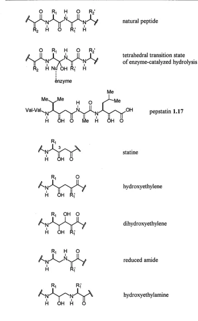
A classical approach in the design of enzyme inhibitors is the incorporation of hydrolytically stable dipeptide isosteres into peptide sequences. This approach was developed after the discovery of a variety of novel enzyme inhibitors from screening done on culture filtrates of microorganisms.37 The peptide natural product pepstatin 1.17 was discovered to be an inhibitor of aspartic proteases (pepsin, renin, cathepsin D, zymosin and later HIV protease) and contained a previously unknown y-amino acid statine. Statine mimics the tetrahedral intermediate formed by enzyme-catalyzed hydrolysis of an amide bond (figure 1.6). A ligand which more closely resembles a transition state or intermediate species of a reaction would be expected to bind better to the catalyzing enzyme, than one resembling the starting substrate. X-ray crystallographic studies of enzyme-inhibitor complexes have confirmed that the (3S)-hydroxyl group of statine hydrogen bonds to catalytically essential aspartate residues in the active site. 38 These clues on reaction mimetics provided by 1.17 lead to the idea of transplanting critical features of active natural products into other structural frameworks to develop selective and potent inhibitors of different protease enzymes.
Chapter One Introduction
0 R1 H 0 R21
y~~~-(l~~
I II
enzyme
natural peptide
tetrahedral transition state of enzyme-catalyzed hydrolysis
Me
Me~Me
H 0~Me
Vai-Vak.~~N0~~0H
pepstatin 1.17H OH 0 Me H OH 0
statine
hydroxyethylene
dihydroxyethylene
reduced amide
hydroxyethylamine
Figure 1.6: Hydrolytically stable dipeptide isosteres.
[image:28.595.85.480.78.715.2]Chapter One Introduction 29 The hydroxyethylene isostere was the first of such structures used, being incorporated into the amino acid sequence of the renin substrate to produce potent renin inhibitors. 39 Renin was the subject of much interest due to its implications in cardiovascular disease. Molecular modelling studies suggested that the related dihydroxyethylene isostere might interact via both hydroxyl groups with the catalytic aspartate residues, and so compound 1.18 was prepared40 and found to be a potent inhibitor of renin (ICso 0.35 nM).
o~~J)""'=
N 1 hN
OH~
H
0Boc-Phe-His-N
H
1.18
The hydroxyethylamine isostere has been incorporated into effective inhibitors of HIV protease41 that mimic the Phe-Pro cleavage site in the viral protein gag-pol. JG-365 1.19 was an initial potent inhibitor (IC50 0.24 nM) based on incorporation of the hydroxyethylamine isostere into the minimum substrate peptide sequence required for activity. Extensive structure-activity relationship studies lead to inhibitor 1.20 (Ro 31-8959, ICso 0.4 nM) with improved pharmacological properties. X-ray crystallographic studies of these HIV protease-inhibitor complexes has facilitated the development of new ligands.
Ph
Ac-Ser-Leu-Asn-~~Nx_
H OHO lle-Vai-OMe
1.19
Ph~
H~
QC-Asn-~~N
H OH0 1.20
Chapter One Introduction 30 isosteres were designed, based on this principle, and incorporated into potent inhibitors (figure 1.7).42 Interestingly, the configuration of the COH centre has little effect on the activity of these compounds.
Ph~i
~ :N~
N ...
H
HO OH: 0
I I
I
~
Ph .,...Ph
Cbz-Vai,NVN' Val-cbz
H
OHH
Phe-Pro scissile amide bond
pseudo-C2 symmetric inhibitor
Figure 1.7: Design of a pseudo-C2 symmetric HIV protease inhibitor.
Conformational Restriction
Conformational restriction43 is an important principle in peptidomimetics. Analogues which assume the bioactive conformation of a target peptide are potentially more selective and potent ligands for a receptor. Also, ligands which are 'preorganized' for receptor-binding have a lower relative entropy cost. Techniques of conformational restriction include a variety of short or long-range cyclizations and the introduction of other rigid elements which make the molecule less flexible and may induce a favourable bioactive conformation. An understanding of the conformational implications of such restrained analogues is important to further design and development.
Chapter One Introduction 31 Substrate-based cyclization techniques can link two side chains 1.2144, two backbone units 1.2245 or a side chain and a backbone unit 1.2346. Much research has been done on the synthetic problems encountered in developing such systems.
Many potent macrocyclic peptides are found in nature, such as somatostatin 1. 7 and cyclosporin A 1.10. The structure of cyclosporin A incorporates a number of peptidomimetic design techniques. It is cyclic, and possesses seven N-methylated amide bonds, several D-amino acids and some unusual C-alkylated substituents, which combined with intramolecular hydrogen bonding, all limit conformational freedom. In addition to their conformational restriction, macrocyclic peptides frequently possess more favourable pharmacological properties than linear peptides. Hydrophobic side chains of cyclic peptides provide a hydrophobic exocyclic surface that shelters cleavable amide bonds from degrading proteases and facilitates the penetration of cell membranes.
1.24 1.25
Synthetic macrocyclic peptidomimetics (for a recent review see Fairlie et al. 47)
have been developed based on X-ray crystal structures of HIV protease-inhibitor complexes. Macrocycles were incorporated into inhibitors 1.19 and 1.20, to link the P1 and P3 residues which are arranged closely in space in the active site, to give
Chapter One Introduction 32
Cis amide bond analogues
The partial double bond character of the amide bond leads to cis and trans isomers. In peptides the more stable trans configuration predominates. Exceptions include N-alkylated amino acids, cyclic peptides and turns (discussed below). Many cis amide bonds occur in the biologically active conformations of peptides. The biological activity of angiotensin II 1.5 is correlated with the isomerization of the His-Pro amide bond to the cis configuration. 49 The simulation of cis amide bonds which could occur in the biologically active conformation of a peptide is an important technique in designing peptidomimetics. Compounds 1.26, 50 1.27 and 1.2851 are examples of dipeptide analogues mimicking cis amide bonds. Each of these compounds incorporates a rigid ring structure as surrogate for the amide bond which restricts the relevant torsion angle to a value close to 0°. Incorporation of 1.27 into a cyclic hexapeptide analogue of somatostatin gave a compound· with some biological activity. 52
H N-N I II ,, Cbz-NY'N"N O
Ph/
Me~~
OMe1.26
M~
Boc-N
~
0
Ph OHChapter One Introduction 33
Imitation of secondary structure
0 ~ 0 Me 0
R30~¥
$
0~
oj:
~ ~
~so
R
~JYNy
N---i
~Pl
N--1
H R1
H'
M H
f3-turn 1.29 1.30
Figure 1.8: f3-turn mimietics.
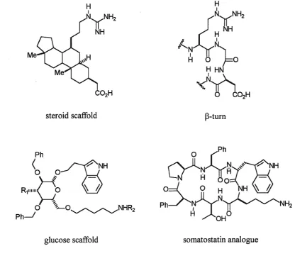
The retention of peptide secondary structure in mimetics is crucial to retention of biological activity. a.-Helix and f3-sheet structures, turns and loops are essential components of peptide and protein conformation. The f3-turn (figure 1.8) is a structure common to many biologically active cyclic peptides, and has been postulated in many cases for the active conformation of linear peptides. In many proteins, f3-turn structures are exposed and may be a part of ligand recognition sites. The secondary structures of peptides can interchange because of the inherent flexibility of peptides. Therefore, conformational restriction of bioactive secondary structures has been of great interest. The f3-turn is the most frequently imitated secondary structure. Examples of f3-turn mimetics include structures with still recognizable peptide chains 1.29,53 to those with completely non-peptidic components 1.30. Structure 1.30 has been used to produce two antiparallel peptide chains54 or, simply by changing the anchor groups, two parallel peptide chains (f3-sheet mimetics). 55
Chapter One Introduction
34
1.31
In addition, many of the techniques discussed above which restrict the conformational flexibility of a backbone and sidechains, can be applied to stabilize secondary structures. For example, the incorporation of cis amide surrogates into a backbone can stabilize the conformation of a ~-turn. The use of such constrained units has been reviewed.43
Scaffold peptidomimetics
Chapter One Introduction
steroid scaffold
glucose scaffold
35
B-turn
O (Ph
~Jl~~~,)"'~:sNH
LJ
-Hoo:::(_
-o:d_
0 H NH
~
hPh___)--...~)y.N~NH2
H
~OHO
[image:35.595.87.504.95.456.2]somatostatin analogue
Chapter One Introduction 36
Work Described in this Thesis
The research described in this thesis is divided into two related projects, which represent two distinct approaches towards the development of peptidomimetics as discussed above, rational design and the development of leads from natural products. Chapters two through four describe the design, synthesis and biological activity of a new, generally applicable cis-hydroxyethylamine dipeptide isostere, and its applications to the inhibition of HIV protease. The principles on which this rationally designed peptidomimetic is based are discussed above. It is hoped that the shift towards rational design will continue so that drugs may be designed de novo, based on a knowledge and understanding of receptor-ligand interactions.
37
Chapter Two
Design of a cis-Hydroxyethylamine Isostere
Introduction
A central underlying principle of peptidomimetics is conformational restriction (see chapter one for a discussion), where the flexibility of a system is restricted and bioactive conformations are promoted. The replacement of components of a target peptide with various structural modifications (isosteres) can be a probe of the conformational requirements for biological activity, by allowing different degrees of rotational freedom along the peptide backbone. Such modifications can also improve the pharmacological properties of the analogue by reducing its peptidic character, making it more biostable and hence more suitable as a therapeutic agent. The ultimate goal being to produce a non-peptidic ligand constrained to the appropriate bioactive conformation.
Cis-trans isomerization of amide bonds occurs as a result of the partial double
bond character of the CO-NH bond. The configurations of the amide bonds in a peptide is of crucial importance to the conformation of the molecule and therefore, to its ability to bind to a receptor. In many cases, binding to a receptor will only occur with a cis configuration at certain amide bonds. Cis amide bonds can also occur in N-alkylated amides, secondary structures such as turns, constrained cyclic peptides and bioactive conformations.
Chapter Two Design of a cis-Hydroxyethylamine Isostere 38 residues in protein X-ray crystal structures have been found with cis amide bonds. The NMR studies of morphiceptin (Tyr-Pro-Phe-Pro-NH2) have shown four different isomers generated from cis-trans isomerization of the Tyr-Pro and Phe-Pro amide bonds.60 N-Methylated amino acids are commonly found in naturally occurring peptide antibiotics. Conformational calculations and NMR studies on Sar-Sar dipeptide 2.1 (where Saris N-methylglycine) showed that the cis isomer is only 0.6 kcal/mol higher in energy than the trans (scheme 2.1).61
0
MeHN~~~OH
Me 0trans-2.1
Scheme 2.1: Cis-trans isomerization of2.1.
cis-2.1
There are many examples of the cis amide configuration in biologically active peptides and analogues. In a series of angiotensin II 1.5 analogues, trans to cis isomerization of the His-Pro bond was observed by NMR,62 when the compound was titrated in D20 from acidic to neutral pD. The amount of cis isomer present was correlated with biological activity for the angiotensin II receptor in rat uterine. This suggested that the cis isomer was the one bound to the receptor and responsible for the observed activity. It has also been proposed63 that the cis-trans isomerization of proline amide bonds might also play a role in transduction of membrane proteins. The isomerization of these membrane-bound proteins, and the resulting redirection of the protein chain, is proposed to provide the conformational change necessary for the reversible opening and closing of transport channels. Others suggested that cis-trans isomerization is also responsible for many of the slow kinetic events observed in enzyme kinetics and protein folding.64
Chapter Two Design of a cis-Hydroxyethylamine Isostere 39
Cyclosporin A 1.10, an immunosuppressant drug, is a conformationally constrained cyclic peptide containing seven N-methylated amide bonds. It is known that 1.10 inhibits a rotamase enzyme that catalyzes cis-trans isomerization of peptidyl-prolyl amide bonds. In non-polar solvents or in the solid state, it was observed that the hydrophobic peptide forms a twisted f3-sheet structure with the MeLeu-MeLeu amide bond (shown in box) fixed in the cis conformation. In more polar solvents, like methanol or water, several conformations were observed with isomerization occurring about that amide bond.
Somatostatin 1. 7 is a large cyclic peptide with an intramolecular disulfide bridge between two cysteine sidechains. Structure-activity relationship studies indicated that the Phe7-Trp8-Lys9-Thr10 sequence (the numbers indicate the position of the residue in the peptide sequence) was important for activity and that a f3-turn within this sequence was required for receptor recognition. Based on these studies, conformationally restricted analogues with smaller ring structures to stabilize the turn, were synthesized, 65 such as 2.2. Conformational analyses of these highly active analogues indicated that a cis amide bond between Phe-Pro was important for activity.
Chapter Two Design of a cis-Hydroxyethylamine Isostere
Ala-Giy-Cys-Lys-Asn-Phe-P he-Trp
1
I
HO-Cys-Ser-lhr-Phe-Thr-Lys
Pro-Phe-D-Trp
Phe-lhr-Lis
40
1.7
2.2
2.3
The dipeptide analogue 1.27, containing an ortho-substituted benzene ring as a
cis amide surrogate, was incorporated into the somatostatin analogue 2.2 as a replacement for the Phe-Pro sequence to give 2.3. The analogue 2.3 showed substantial bioactivity compared to the parent somatostatin. 52 A cis amide surrogate based on 1.27 has also been incorporated into analogues of the RGD peptide, leading to an active inhibitor of cell adhesion, 66 and also into inactivators of trypsin-like proteases. 67
1.27 1.28
Chapter Two Design of a cis-Hydroxyethylamine Isostere 41
to be easily extended in the C direction. By the correct choice of side chain groups, this surrogate can be applied specifically as a peptidomimetic.
H H
~
ArH
trans cis
Scheme 2.2: Isomerization of an olefin.
The trans olefinic group68 has been successfully used as a surrogate for a trans amide bond. However, the corresponding cis olefin is unsuitable as a cis amide surrogate due to the ease of cis to trans olefin isomerization (scheme 2.2). The CO-NH portion of the urethane bond of common N-terminal protecting groups such as Cbz and Boc has a high tendency to assume a cis configuration. 69a A synthetic scheme
was developed69b where a urethane bond was derived from the sidechain hydroxyl
group of tyrosine (figure 2.1 ). The incorporation of these urethane linkages into backbones promoted cyclization of the compounds and gave rise to a family of macrocyclic peptide analogues, with numerous possible applications.
/,_N
H
0
Figure 2.1: Tyrosine cis-urethane linkage (shown in box).
Chapter Two Design of a cis-Hydroxyethylamine Isostere 42 a novel completely non-peptidic scaffolding with attached sidechain groups (figure 2.2). Such compounds were shown to adopt the favorable pleated backbone conformations and antiperiplanar sidechain trajectories observed in some HIV protease inhibitors such as 1.19. Pyrrolinone-based compounds also have improved pharmacological properties than the corresponding peptide-based peptidomimetics.
Marshall et a!. 71 first proposed the tetrazole ring system as a cis amide bond surrogate to lock a dipeptide analogue in an equivalent configuration. The major concern in designing a surrogate is the amount of geometric and steric similarity to the
cis amide bond. Using a novel procedure50 for assessing conformational mimicry, it was shown that approximately 88% of the conformations accessible to the cis isomer of the dipeptide 2.4 are also accessible to the tetrazole analogue 2.5. This index of conformational mimicry was measured by the ability of 2.5 to orient the backbone and sidechains, on either side of the tetrazole ring, in a similar manner to that of 2.4. As the receptor-bound conformation of a target peptide is not always known, such statistical arguments for the suitability of analogues are useful. During these studies, the tetrazole 2.5 was found to have more conformational freedom than 2.4. This probably reflects the increased valence angle a.C-C=N of the tetrazole relative to the corresponding a.C-C=O angle of the dipeptide. Based on these studies, Smith et al. 72
concluded that the tetrazole analogue is an excellent conformational mimic of the cis
amide bond.
2.4
N-N
II ''
AcHN~N"N
Me = ~~ ~0
Me'~
NHMe
2.5
Tyr-Chapter Two Design of a cis-Hydroxyethylamine Isostere 43
Pro scissile amide bond. 74 Molecular modelling of the tetrazole analogue and the substrate showed good correlation between the two conformations. However the tetrazole analogue was found not to inhibit the HIV protease. This result may indicate that the increased steric bulk of the tetrazole ring in the active site may prevent effective binding of the analogue.
H N-N I // ''
Cbz-N~N"N
=
L/P
Ph/ Me~~\ OMe
1.26
(N-Me)Aia-Tyr-0-Trp
PLVai-L/.
2.6
The tetrazole surrogate has also been incorporated as a replacement for the Leu-Gly amide bond of deaminooxytocin75 2. 7 (see oxytocin 1.2 for comparison), to give 2.8 as a conformational probe of the importance of the cis configuration to this amide bond. Comparison of the bioactivity of 2.8 with that of a corresponding analogue incorporating a trans olefin surrogate suggested that a trans configuration was important to this amide bond. Replacement of the Pro-Pro and Ser-Pro amide bonds of bradykinin with tetrazole surrogates 76 gave analogues with greatly diminished activity relative to the parent peptides, suggesting either a cis configuration was not required or that the steric bulk of the tetrazole prevented key binding interactions.
S - - - ,
~
Tyr-lle-Gin-Asn-C s-Prn-Leu-Giy-NH,0
2.7
s
I
H N-NI ''
Tyr-lle-Gin-Asn-Cys-Pro-N0N"N
o
Me=
'--f
0Y.
NH2Chapter Two Design of a cis-Hydroxyethylamine Isostere
44
A classic strategy in the design of protease inhibitors is the incorporation of non-hydrolyzable dipeptide isosteres into the natural enzyme substrate at the P1-P1'
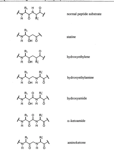
cleavage site. Isosteric replacement of the scissile amide bond CO-NH (with CH2NH in the case of the hydroxyethylamine isostere) increases the metabolic stability of the inhibitor.
A number of dipeptide isosteres have been developed. The application of the transition state mimic principle has considerably increased the activity of these inhibitors. In such compounds, an sp3 hydroxymethylene group is inserted in the centre of the isostere as a mimic of the tetrahedral transition state for hydrolysis of the P1-P1' amide bond. Enzymes bind more favourably to ligands resembling the transition
Chapter Two Design of a cis-Hydroxyethylamine Isostere 45
The hydroxyethylamine isostere was first used to yield potent renin inhibitors, 77 such as 2.9, by incorporation into the substrate sequence of angiotensinogen as a replacement for the scissile P1-P1' residues. However, due to its relatively large size and peptidic character, 2.9 possesses poor pharmacological properties and is not promising as an oral antihypertensive.
Me
Me~
Me MeBoc-Phe-His-';IY~X:IIe-Phe-OMe
H OH H 0
2.9
Chapter Two Design of a cis-Hydroxyethylamine Isostere 46
normal peptide substrate
statine
hydroxyethylene
hydroxyethylamine
hydroxyamide
a-ketoamide
[image:46.595.107.493.73.604.2]amino ketone
Figure 2.2: Non-hydrolyzable dipeptide isosteres.
Chapter Two Design of a cis-Hydroxyethylamine Isostere 47
(3R)-epimer of 2.10 possessing 400-fold greater activity than the (3S)-epimer. The removal of the hydroxyl group altogether resulted in extremely poor activity. The secondary amine of the hydroxyethylamine core was also shown to be important for activity when replaced by an ether function, resulting in a poorly active analogue. This suggested that the amino group acts as an important hydrogen bond acceptor-donor in the active site.
Ph~
., MePhCO~~~~Pro
H OH H 0
2.10
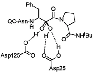
The hydroxyamide79 and the a-ketoamide80 dipeptide isosteres (figure 2.2) have been used successfully in potent HIV protease inhibitors (for a discussion see the following section on HIV protease inhibition). The extra amide bonds of these structures may make them less hydrolytically stable than the hydroxyethylamine structure, however they provide more possible hydrogen bonding interactions with a receptor. It was proposed that a-ketoamide based inhibitors of HIV protease are hydrated within the active site to form a stabilized hydrate which is a good transition state mimic (figure 2.3).
Ph~ ~
QC-Asn-~~N~
H <50,H'
H \
0 NHiBu
, 0 \
0, I \
: H
A
I IAsp125
o 6Yo
[image:47.595.227.389.555.680.2]Asp25
Chapter Two Design of a cis-Hydroxyethylamine Isostere 48
Related to the hydroxyethylamine isostere is the aminoketone structure (figure 2.2), resulting from conversion of the hydroxyl group of the hydroxyethylamine isostere to a ketone. The resulting ACE inhibitor 2.11 was synthesized78 to compare with 2.10 and was found to be more potent. The mode of action of 2.11 would be similar to an cx-ketoamide (figure 2.3), where nucleophilic attack on the ketone in the active site, would lead to a hemi-ketal transition state mimic. The ketomethylene (COCH2) component of the aminoketone isostere is found in the naturally occurring
aminopeptidase inhibitor arphamenine, and has also been incorporated into inhibitors of a number of other enzymes, including aminopeptidase81 and renin.82
Ph~
MPhCO~'ll~~Pro
H 0 H 0
2.11
Ph~
MPhCO~~~~Pro
H OH H 0
2.10
Chapter Two Design of a cis-Hydroxyethylamine Isostere
cis-
Hydroxyethylamine Isostere
cis amide bond surrogate
cis-hydroxyethylamine isostere 2.12
[image:49.595.96.503.127.451.2]hydroxyethylamine dipeptide isostere
Figure 2.4: Design of our cis-hydroxyethylamine isostere.
49
The subject of a significant part of the research undertaken in this thesis is a peptidomimetic structure combining the design principles of a cis amide bond surrogate and the hydrolytically stable hydroxyethylamine dipeptide isostere. This new cis-hydroxyethylamine isostere 2.12 incorporates a tetrazole ring in the centre of the dipeptide analogue to conformationally constrain the 1 and 5 substituents in a cis
configuration (figure 2.4). The isostere 2.12 is generally applicable in the design of peptidomimetics by selection of suitable natural or unnatural sidechains at R1 and R2,
and peptidic or non-peptidic extensions in theN and C directions.
Chapter Two Design of a cis-Hydroxyethylamine Isostere 50
2.13
One envisaged application for isosteres 2.12 and 2.13 is in the design of HIV protease inhibitors, based on the known binding conformation of JG365 1.19 (see following for a discussion).
HIV Protease Inhibition
The Human Immunodeficiency Virus (HIV) infects almost 20 million people worldwide and leads to the deadly disease AIDS. Enormous research is being directed toward developing therapeutic drugs to treat HIV. Until recently AIDS has been an inevitably fatal, incurable disease. However this year, exciting new clinical results have emerged. Powerful combinations of HIV protease and HIV reverse transcriptase inhibitors have been shown to reduce viral levels in the blood of infected patients to undetectable levels. These combinations of drugs include the more established HIV reverse transcriptase inhibitors, such as AZT and 3 TC, and three recently approved HIV protease inhibitors, including 1.20.
A second development is the discovery of chemokines, 83 compounds secreted by certain white blood cells that potently inhibit HIV replication. It has been shown that a chemokine receptor has a critical role in HIV' s ability to infect cells. Complete eradication of the virus from patients is now a possibility.
Chapter Two Design of a cis-Hydroxyethylamine Isostere 51
active site, resulted in non infectious virions and lead to the targeting of HIV protease inhibition as a viable strategy for treatment of the virus. The HIV protease belongs to the class of aspartic proteases (see chapter one) that includes renin and pepsin and which catalyzes the hydrolysis of amide bonds via aspartate residues in the active site. An enormous effort has been directed toward developing renin inhibitors as antihypertensive drugs, based on the mechanism of action of aspartic proteases. Many of the principles established in these efforts have now been applied to the design of HIV protease inhibitors.
Chapter Two Design of a cis-Hydroxyethylamine Isostere 52
[image:52.598.120.450.121.619.2]Chapter Two Design of a cis-Hydroxyethylamine Isostere 53
\
[image:53.595.102.496.76.714.2]Chapter Two Design of a cis-Hydroxyethylamine Isostere
Ph
ac.Asn-~YNx
H OHOIBu
0
2.14
0
Ph~ ~
~N0Asn-~~NX
~
H OHO NH!Bu
2.15
54
The hydroxyamide (norstatine) isostere (figure 2.2) was incorporated into potent HIV protease inhibitors, 79 such as 2.15, to compare with related hydroxyethylamine-based compounds, such as 2.14. The two types of inhibitors differ by one function, a carbonyl group in 2.15 which is a methylene group in 2.14. The extra amide bond of 2.15 may make it less hydrolytically stable than 2.14, however it provides more possible hydrogen bonding interactions in the active site. Compounds based on 2.15 were found to be 10-20 times more potent inhibitors of HIV protease than compounds based on 2.14.
Ph~ ~
Cbz-~~Nx
H 0
O OMe
2.16
Chapter Two Design of a cis-Hydroxyethylamine Isostere 55
Ph QC-Asn-N
H
2.17
Other isosteric Pt-Pt' replacements have been employed and exhibit potent activity. Benzamide-based inhibitor 2.17 was developed by Kaldor et al. 88 at the Lilly laboratories based on enzyme-inhibitor X-ray crystal structures. Molecular modelling calculations indicated that the benzamide carbonyl would be able to rotate out of the resonance plane of the adjacent benzene ring to enable a crucial hydrogen bonding interaction with a highly localized water molecule in the flap region. This prediction was later confirmed by the X-ray crystal structure of the IDV protease-2.17 complex.
A number of benzene-substituted derivatives with varying activities were also prepared, to investigate the dimensions of the benzamide binding pocket at S 1'.
Ph
MeO~H~ ~
HO~N
-N~OH
lJHOH
OMePh
2.18
In a novel approach, IDV protease inhibitors were designed which mimic the approximate C2 symmetry of the enzyme. Kempf et al. 42 imagined placing the C2
symmetry axis of the enzyme through the proposed tetrahedral intermediate for amide hydrolysis and performing the Cz operation on either the P1 or P1' regions of the
substrate (see figure 1. 7). Such structures may provides advantages over traditional substrate-based inhibitors, in terms of potency and selectivity for
mv
protease over other aspartic proteases. An X-ray crystal structure of themv
protease-2.18 complex showed an extended binding conformation, spanning the S2 to S2' subsites. 89 SeveralChapter Two Design of a cis-Hydroxyethylamine Isostere 56
A key feature of all the potent peptide-based inhibitor-HIV protease complexes is the hydrogen bonding from the two central carbonyls of the inhibitor to a water molecule which bridges to the Ile50 and Ile150 residues in the flaps of the enzyme.
Ph
Ac-Ser-Leu-Asn-~~2
H OH
O lle-Vai-OMe
1.19
Ph~ H,,,.
QC-Asn-~~N
H OH
1.20
Potent and selective HIV protease inhibition is possible by mimicking the Phe-Pro cleavage site unique to retroviral proteases. Since this cleavage site is rare in mammalian endopeptidases, such inhibitors have a selective advantage for HIV protease and are not as easily degraded by other enzymes. Incorporation of the hydroxyethylamine dipeptide isostere into the minimum substrate peptide sequence required for recognition gave 1.19.41'90 It is a potent inhibitor in vitro but failed to
Chapter Two Design of a cis-Hydroxyethylamine Isostere 57
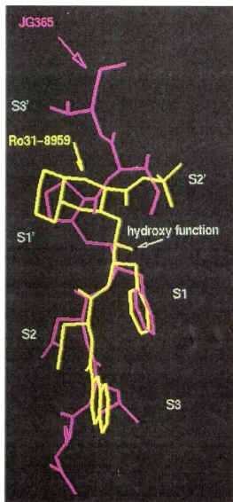
[image:57.603.169.432.112.678.2]Chapter Two Design of a cis-Hydroxyethylamine Isostere 58 Two distinct inhibitor binding modes91 are observed in the X-ray crystal structures of the HIV protease-inhibitor complexes of 1.1992 and 1.20 (figure 2.7). In 1.19 the He-Val (P2'-P3') residues occupy the S2'-S3' enzyme subsites as expected and force a favourable (S)-OH configuration on the transition state mimic. However, in 1.20 the C-terminal tert-butyl amide group fits well into the S2' subsite with the large decahydroisoquinoline (Diq) ring structure occupying the entire S1' subsite, with some S3' interactions. This binding mode shows a clear preference for the opposite (R)-OH
configuration at the transition state mimic. Extension of 1.20 to P3' showed a marked decrease in activity. Similarly, inhibitors based on 1.19, lacking a P3' ligand and with Pro at P1', show moderate potency and a slight preference for (R)-OH, indicating the second binding mode. The superimposition of the HIV protease-bound structures of 1.19 and 1.20 in figure 2.7, clearly shows the two distinct binding modes with differing backbone conformations between the hydroxyl groups and the prolyl or Diq rings. The backbone of 1.19 adopts a cis geometry about this region while 1.20 has a trans arrangement at the equivalent position from the hydroxyl group to the Diq ring.
Chapter Two Design of a cis-Hydroxyethylamine Isostere
Ph
Ac-Ser-Leu-Asn-~~~
H OH
0 lle-Vai-OMe
Ph~N
N-,,
3 NQC-Asn-~ ~\.f~"
,OH OH~~~'--f
Me~
NHtBu59
1.19
2.19
Figure 2.8: Relevant torsion angles of 1.19 and modelled 2.19, from the superimposition in figures 2.9 and 2.10.
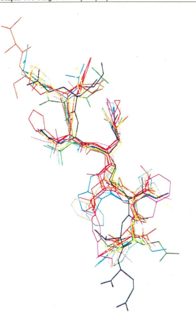
Chapter Two Design of a cis-Hydroxyethylamine Isostere 60
Figure 2.9: Front view of the superimposition of modelled 2.19 (in red) on the
enzyme-bound X-ray crystal structure of 1.19 (in blue) (only the P1-P1' regions are
shown, for clarity).
Figure 2.10 also shows that the two central carbonyl groups (pointing out to the left) are in a suitable position to form crucial hydrogen bonds with an active site-bound water molecule (not shown). The P1 phenylalanine side chain groups can also
be seen projecting into where the S 1 pocket of the enzyme would be. It is evident from the modelling studies that the P3-P1 ligands of 2.19 are able to occupy the same
enzyme subsites as 1.19. However, the steric bulk of the tetrazole ring may be a hindrance to binding if it is unable to project into the S 1' subsite occupied by the prolyl
and Diq ring structures of 1.19 and 1.20 (figure 2.7). Another important factor in enzyme binding is the choice of a P/ ligand which will completely fill the S1' binding pocket. The large Diq ring of 1.20 completely fills the S 1' subsite, as observed in the
[image:60.600.132.486.76.441.2]Chapter Two Design of a cis-Hydroxyethylamine Isostere 61
compared with the smaller prolyl ring of 1.19. On this basis, it may be favourable in
the future to alter the P 1' methyl side chain of our designed2.19 to a larger group.
Figure 2.10: Side view of the superimposition of modelled 2.19 (in red) on the
enzyme-bound X-ray crystal structure of1.19 (in blue).
The (JS)-epimer of 2.19, compound 2.20 is shown in figure 2.11 superimposed
on structure 1.19. Figure 2.11 clearly shows the C3-hydroxyl group of modelled 2.20
projecting in the opposite direction to the corresponding hydroxyl group of 1.19.
Comparison to figure 2.9 where the hydroxyl groups of modelled 2.19 and 1.19 are
superimposed, clearly suggests a preference for the (R)-configuration at C3, in order for our designed inhibitors, based on 2.12, to assume the known bioactive
[image:61.602.132.489.152.503.2]Chapter Two Design of a cis-Hydroxyethylamine Isostere
62
Figure 2.11: Superimposition of modelled 2.20 (in red), with a (S)-configuration at
C3 , on the enzyme-bound X-ray crystal structure of 1.19 (in blue) (only the P,-P/ regions are shown, for clarity).
Traditionally, the activity of dipeptide isosteres has been enhanced by the addition of amino acid residues to both the N and C terminals, to improve recognition in the active site. Although this approach has been used to develop potent enzyme inhibitors, they generally exhibit poor pharmacological properties due to their peptidic character. Extensive research93 is being done to develop non-peptidic ligands for the traditional P3-P2' positions of potent HIV protease inhibitors, which will show potent
[image:62.600.128.507.66.437.2]Chapter Two Design of a cis-Hydroxyethylamine Isostere
Table 2.1: HIV protease activity of 1.19 based inhibitors.
X y C3 IC5o (nM) Ref
1.19 Ac-Ser-Leu-Asn Ile-Val-OMe
s
0.24 412.21 Ac-Ser-Leu-Asn Ile-Val-OMe R 20 41
2.22 Cbz-Asn OtBu
s
300 412.23 Cbz-Asn OtBu R 140 41
2.14 QC-Asn OtBu R 23 41
2.24 Cbz-Asn NHtBu R 210 41
2.25 Cbz OtBu R 6500 41
2.26 Ac-Leu-Asn Ile-Val-OMe RS Ki21 nM 79
2.27 Ac-Leu-Asn Ile-OMe RS Ki4520nM 79
Ph'l
H~.,
QC..Asn-~~N
H OH1.20 ICso 0.23 nM 2.17 ICso 1.5 nM
Ph'l
w
Cbz-~~Nx
H 0O OMe
2.16
rc
50 405 nM0
QC=
oc:iY
Chapter Two Design of a cis-Hydroxyethylamine Isostere 64 In order to make comparisons to the activity of known potent inhibitors ofHIV protease (table 2.1), suitable P3-P3' ligands were chosen for our designed isostere 2.12 (table 2.2). The phenylalanine side chain at P1 and asparagine residue at P2 are
favoured in all the inhibitors. The 2-quinolinylcarbonyl (QC) ligand at P3 seems
favoured over Cbz by comparison of the activities of 2.14 and 2.23 (table 2.1). Inhibitors 1.20 and 2.17 also favour QC-Asn at P3-P2 . Ideally, to promote the binding
mode of 1.19 (which our mode