Experimental Intramammary Infection with a Strain of
Escherichia coli Isolated from a Cow with Persistent
E. coli Mastitis
Stephen P. Oliver1, Susan I. Headrick1, Mark J. Lewis2, Barbara E. Gillespie1, David L. Johnson3, Raul A. Almeida1*
1
Department of Animal Science, The University of Tennessee, Knoxville, USA 2
East Tennessee Research and Education Center, Little River Animal and Environmental Unit, The University of Tennessee, Knoxville, USA
3
Middle Tennessee Research & Education Center, The University of Tennessee, Spring Hill, USA Email: *ralmeida@utk.edu
Received August 30,2012; revised October 11, 2012; accepted October 21, 2012
ABSTRACT
Transient E. coli intramammary infections (IMI) are usually associated with rapid onset of clinical signs including mammary gland swelling and abnormal milk with rapid clearance of bacteria from milk. Conversely, reports have de-scribed strains of E. coli showing very different clinical trends. Persistent E. coli IMI are associated with mild clinical symptoms that disappear shortly after the onset of infection, possibly flaring-up intermittently during lactation. In the present study, we evaluated a strain of E. coli isolated from a cow with persistent mastitis to determine if the experi-mental infection model mimics naturally occurring persistent E. coli IMI. Uninfected mammary quarters of 7 Holstein heifers were infused within 10 days of calving with 50 colony-forming units of a persistent E. coli strain. Six of 7 heif-ers developed mild clinical mastitis with elevated rectal temperatures within 9 to 36 h after infusion. The challenge strain was isolated intermittently in milk from all infected mammary quarters during the first two weeks after infusion and 3 animals continued to shed E. coli periodically during the sampling period. One animal shed E. coli intermittently in milk for 172 d after challenge and developed clinical mastitis four times during this period. The isolated strain had an identical pulsed-field gel electrophoresis profile as the E. coli strain used to infuse mammary glands. The experimental IMI model described here mimics very closely naturally occurring persistent E. coli IMI, thus providing an excellent in vivo model to better understand pathogenesis and to facilitate development of control strategies for this important masti-tis pathogen.
Keywords:Escherichia coli;Intramammary Experimental Infection; Persistent Mastitis; Dairy Cows
1. Introduction
Current mastitis control programs devised in the 1960’s are based on hygiene including teat disinfection, antibi- otic therapy, and culling of persistently infected cows. Acceptance and application of these measures has led to considerable progress in controlling contagious mastitis pathogens. However, these same procedures are less ef- fective against environmental pathogens such as E. coli
because of the low susceptibility of E. coli to common mastitis treatments [1], lack of efficacy of teat disinfec- tion for the prevention of new E. coli intramammary in- fections (IMI), and low efficacy of vaccination programs [2]. Therefore, it is not surprising that E. coli mastitis has become a major problem in many well-managed dairy farms that have successfully controlled contagious patho-
gens.
Transient E. coli mastitis is associated with rapid onset of clinical symptoms including mammary gland swelling and abnormal milk with rapid elimination of bacteria from milk. In some cases, severe systemic involvement occurs and when not properly diagnosed and treated, it could result in the death of the affected animal. Reports have also described strains of E. coli showing very dif- ferent clinical trends. For example, there are reports on E. coli strains associated with persistent IMI which often times start with mild clinical symptoms that disappear soon after the onset of infection only to flare-up again during lactation, usually resulting in mild clinical masti- tis [3-5]. Escherichia coli associated with persistent mas- titis have been isolated in milk for long periods in spite of a high number of somatic cells in milk [3]. Published data suggests that adhesion to and internalization into
mammary epithelial cells and subsequent intracellular survival might be important virulence attributes of strains of E. coli associated with persistent mastitis [3,5]. It was shown that internalization of persistent E. coli strains occurred by an endocytic mechanism that avoids bacte- rial uptake into acidified lysosomal compartments al- lowing bacteria to remain in the endosome evading host immune responses [6]. Studies conducted in our lab showed that transient E. coli strains internalize into bo- vine mammary epithelial cells preferentially exploit- ing receptor-mediated endocytosis (clathrin mediated), whereas persistent strains of E. coli appear to exploit caveloae-mediated endocytosis (CME). Exploitation of CME would allow persistent strains to circumvent host cell intracellular bactericidal/bacteriostatic mechanisms such as endosome acidification and endosome-lysosome fusion, which is consistent with characteristics of persis- tent IMI that they cause under natural conditions [7]. Experimental infection models have been an important tool for studying the pathogenesis of infections, man- agement of infectious diseases, and development and evaluation of therapeutic and prevention strategies for disease control. In the present study, we evaluated a strain of E. coli isolated from a cow with persistent mas- titis to determine if experimental intramammary infection mimics naturally occurring persistent E. coli mastitis in dairy cows.
2. Materials and Methods
2.1. Bacterial Strain and Growth Conditions
The E. coli strain ECC-1470 [3,7] isolated originally in milk from of a cow with persistent mastitis was used. The challenge inoculum was a frozen stock that was thawed, grown in Luria Bertani broth (Becton Dickinson Company, Sparks, MD, USA) overnight at 37˚C and di-luted serially to a concentration of approximately 50 colony-forming units (CFU) in 5 ml of sterile phosphate buffered saline (PBS, pH 7.4).
2.2. Experimental Animals
Seven healthy Holstein heifers free of mastitis and any other infectious disease were used. For the experimental infection protocol, animals were grouped together, milked last and included in normal herd practices.
2.3. Experimental Infection Protocol
One uninfected mammary gland of 7 Holstein heifers was infused with E. coli strain ECC-1470. Following disinfection of the teat end with ethanol, a bacterial sus-pension with a concentration of approximately 50 CFU in 5 ml of sterile PBS was infused into the teat cistern using sterile disposable syringes fitted with sterile dis-
posable teat canulas. Heifers were challenged within 10 days of calving; one heifer was challenged 2 d before calving, one at calving, two at 1 d after calving, one at 2 d after calving, one at 6 d after calving, and one at 8 d after calving. Post-calving infusions with the bacterial challenge suspension were administered within 20 min after milking. The protocol used for experimental infec-tion of heifers was reviewed and approved by The Uni-versity of Tennessee Institutional Animal Care and Use Committee.
2.4. Animal Inspection and Clinical Evaluation
Heifers and mammary glands were monitored exten- sively during the first week after experimental challenge. Animal observations including appetite, restlessness, mobility, and responsiveness were evaluated. Rectal temperatures were taken using a digital thermometer and recorded. Milk samples for microbiological evaluation and enumeration of somatic cell counts (SCC) were ob- tained twice daily for one week after challenge and twice weekly thereafter for a minimum of 56 days, or longer if
E. coli was still isolated from milk. Pulsed-field gel elec- trophoresis (PFGE) patterns of E. coli isolated at inter- vals following challenge were evaluated. Milk and mammary condition were evaluated using the following scoring system: Milk Score: 1 = normal milk, 2 = flakes,
3 = small slugs, 4 = large slugs/clots, 5 = stringy/watery.
Mammary Gland Score: 1 = normal; the udder was
pliable when totally milked out. Heat, pain, redness, and/or swelling were not detectable; the animal exhibited no signs of discomfort; 2 = slight swelling; the udder was less pliable with some firmness as if not totally milked out. Additional milking or stripping did not return the gland to normal. Redness, heat and pain were generally not detectable and animals generally did not exhibit signs of discomfort; 3 = moderate swelling; the udder was definitely firm, reddened and warm to the touch. The udder did not return to normal size when milked out. The animal generally exhibited signs of discomfort (irritable, performs a stepping motion with feet and/or kicks) dur- ing prepping and milking procedures; 4 = severe swelling; the udder was very hard, red, hot and noticeably larger than other mammary quarters before milking with little or no change in size following milking. The animal was extremely uncomfortable and very irritable.
2.5. Milk Sample Collection and Processing for Bacteriological Evaluation
ends were cleaned with swabs containing 70% isopropyl alcohol. Milk samples for microbiological analysis were collected into sterile screw-cap tubes and keep at –20˚C until evaluation. Samples for SCC were collected into snap cap 50 ml tubes containing milk preservative pills and kept at 4˚C until processing. Bacteriological evalua- tion of milk samples was conducted at the Tennessee Milk Quality Laboratory. Samples (10 and 100 l) were plated onto blood and MacConkey agar, and one ml was placed on a Petrifilm Coliform Count Plate (3M Com- pany, St. Paul, MN, USA). Somatic cell counts were conducted at the Tennessee Dairy Herd Improvement Association laboratory following standard procedures.
2.6. Pulsed-Field Gel Electrophoresis
Pulsed-field gel electrophoresis (PFGE) patterns of E. coli isolated at intervals following challenge were ana- lyzed as described [8]. Genomic DNA was digested with
XbaI (Sigma Chemical Co. St. Louis, MO, USA) fol- lowed by PFGE. Gel DNA band patterns were analyzed using Molecular Analyst software version 1.6 (Bio-Rad Laboratories, Hercules, CA, USA) to determine strain relatedness. Band position tolerance of 2.5% was used for comparison of DNA patterns.
3. Results
3.1. Clinical Findings
Six of 7 heifers became infected following intramam- mary challenge. These heifers developed mild cases of clinical mastitis characterized by abnormal milk for a few days after challenge and slight to moderate swelling of the challenged mammary gland for up to a week after challenge. Initial clinical manifestations as evidenced by the presence of flakes and small slugs in milk and slight swelling and redness of the infused mammary gland were noticed at 18 h after inoculation reaching a peak at ap- proximately CH + 72 h. At this inspection point, inocu- lated mammary glands showed swelling, firmness, red- ness, and were warm to the touch. Milk from inoculated mammary glands had moderate to large slugs. After 72 h, clinical symptoms declined, becoming moderate with slight swelling of the infused mammary gland and pres- ence of small slugs in milk. Elevated rectal temperatures were detected approximately at CH + 9 h, reaching a peak approximately at CH + 12 h and declining to nor-mal values 96 h after challenge (Figure 1).
3.2. Microbiological Findings
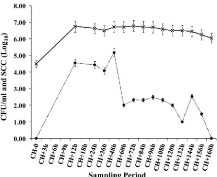
[image:3.595.309.537.86.256.2]Escherichia coli were isolated in milk from all infected mammary glands frequently during the first week after challenge (Figure 2). Three animals continued to shed E. coli intermittently during the two-month sampling period.
Figure 1. Milk scores, mammary scores, and rectal tem- peratures of heifers following intramammary challenge (CH) with a strain of E. coli (ECC-1470) isolated from a cow with persistent mastitis.
Figure 2. Milk somatic cell counts (*; log10 CFU/ml) and colony forming units per ml (X; log10 SCC/ml) following intramammary challenge (CH) with a strain of E. coli (ECC-1470) isolated from a cow with persistent mastitis.
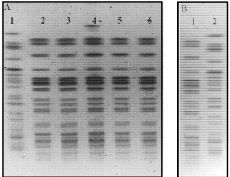
One animal shed E. coli in milk intermittently for 172 d after challenge (Figure 3), and developed clinical masti- tis 4 times from which the challenge strain was isolated, as evaluated by PFGE typing (Figure 4). From all the infected animals (positive isolation of the challenge strain in milk), two animals cleared the infection within one week while the remaining animals cleared E. coli
from milk between 11 and 172 days after infusion. For all infected animals, the range of the infection was 168 days, with a median of 40 days.
3.3. Somatic Cell Counts
[image:3.595.312.535.317.499.2]reaching values of ~7.0 log10 at CH + 12 h. These SCC
values were maintained throughout the entire sampling period and appeared to be unaffected by fluctuations in CFU/ml of E. coli in milk (Figures 2 and 3).
4. Discussion
Several studies have shown the increasing importance of
[image:4.595.59.285.216.393.2]E. coli IMI and indicated that these IMIhave become a major problem in many well-managed dairy farms since
Figure 3. Milk somatic cell counts per ml (◊; log10 SCC/ml) and colony forming units per ml (・; log10 CFU/ml) of cow 7489 for 172 d following intramammary challenge (CH) with a strain of E. coli (ECC-1470) isolated from a cow with persistent mastitis. Clinical mastitis (arrows) was observed at 1, 60, 91 and 116 days after challenge.
Figure 4. PFGE patterns of scherichia coli isolated from
use significant milk loss and frequent culling
increased preva-le
, E. coli was also shown to be th
cally, E. coli mastitis is associated with acute on- se
ation using a strain of E. coli isolated fr
d that clinical manifestations of any in
E
cow 7489. Panel A; lane 1: PFGE control G5244, lane 2: E. coli strain isolated from IMI at CH + 56 - 63, lane 3: from IMI at CH + 91, from IMI at CH + 116, and from milk of the infused mammary gland at CH+172. Panel B; lane 1: challenge strain (E. coli ECC-1470), lane 2: PFGE control G5244.
they ca
and/or death of infected cows [9-12]. A study from Wisconsin reported an
nce of E. coli from 17.7% to 24.9% of the total mastitis pathogens isolated [13].
In the United Kingdom
e most important pathogen in well-managed dairies [14]. Collectively, these studies demonstrate that E. coli
is an increasingly important cause of mastitis in dairy cows.
Typi
t of clinical signs including mammary gland swelling and abnormal milk with rapid elimination of bacteria from milk. In some cases, severe systemic involvement occurs and when not properly diagnosed and treated, it could result in death of the animal. However, an increas- ing number of reports have described strains of E. coli
showing chronic and persistent IMI [3-5]. These persis- tent IMI usually start with mild clinical symptoms that disappear soon after the onset of infection only to flare- up again during lactation, generally resulting in mild clinical mastitis.
In this investig
om a cow with persistent mastitis, we were able to in-duce IMI with positive isolation of the challenge strain of
E. coli in milk and clinical manifestations resembling persistent mastitis from which the strain was isolated initially [3,4]. Results from this investigation also showed that the strain used for infusing mammary glands was repeatedly isolated during the sampling period and into lactation. One animal shed E. coli in milk intermittently for 172 d after challenge (Figures 3 and 4) and devel-oped clinical mastitis four times (Figure 3) which was caused by the challenge strain as evaluated by PFGE typing (Figure 4).
It is well accepte
fectious disease result from the interplay between host defenses and virulence of the infectious agent. Results of the present study showed that experimental infection of heifer mammary glands during the periparturient period with a strain of E. coli isolated originally from a cow with persistent IMI caused mild clinical mastitis initially. In some animals, the challenge strain persisted for long periods of time in spite of a high number of somatic cells in milk similar to what was observed in cows with natu-rally occurring persistent E. coli mastitis [3,4]. Thus, based on experimental and natural infection data, E. coli
[image:4.595.56.289.476.655.2]ed an experimental E. co
5. Acknowledgements
the Tennessee Agricultural
REFERENCES
[1] S. Pyorala, L. d V. Rainio,
“Effi-mastitis and the strain persists in milk for very long pe- riods in spite of a high number of somatic cells in milk. Previous results showed that persistent E. coli strains possess enhanced adherence and internalization capabili- ties as compared with acute E. coli strains [3,4]. Fur- thermore, work from our lab and other laboratories de- monstrated that persistent strains of E. coli were capa- ble of circumventing intracellular bactericidal mecha- nisms such as endosome acidification and endosome- lysosome fusion thus allowing intracellular persistence of these strains [6,7]. Such avoidance mechanisms are very likely the cellular basis to explain persistent IMI caused by these E. coli strains. From a practical standpoint, these results indicate that E. coli strains with these characteris- tics are able to persist for long periods in the affected mammary glands thus creating reservoirs of persistent E. coli strains in the herd. Such reservoirs are very difficult to control since these strains are resistant to host surveil- lance systems, and specialized in gaining access to mi- croenvironments where host defenses or antimicrobials present in milk are not effective.
In conclusion, we have develop
li IMI model that mimics clinical symptoms observed in natural persistent E. coli mastitis. Strains of E. coli
associated with persistent IMI appear to behave quite differently from strains of E. coli causing transient masti- tis. The in vivo experimental infection model developed in the present study could be a useful and valuable tool to better understand pathogenesis and to facilitate develop- ment of control strategies for this important mastitis pa- thogen.
This research was funded by
Experiment Station, The University of Tennessee Col-lege of Veterinary Medicine Center of Excellence Re-search Program in Livestock Diseases and Human Health, and Epitopix, LLC, Wilmar, MN.
Kaartinen, H. Käck an
cacy of Two Therapy Regimens for Treatment of Ex- perimentally Induced Escherichia coli Mastitis in Cows,”
Journal of Dairy Science Vol. 77, No. 2, 1994, pp. 453- 461. doi:/jds.S0022-0302(94)76973-3
[2] D. J.Wilson, Y. T. Grohn, G. J. Bennett, R. N. Gonzales, Y. H. Schukken and J. Spatz,“Comparison of J5 Vacci- nates and Controls for Incidence, Etiologic Agent, Clini- cal Severity, and Survival in the Herd Following Natu- rally Occurring Cases of Clinical Mastitis,” Journal of Dairy Science, Vol. 90, No. 9, 2007, pp. 4282-4288. doi:/jds.2007-0160
[3] B. Dogan, S. Klaessig, M. Rishniw, R. A. Almeida, S. P. Oliver, K. Simpson and Y. H. Schukken, “Adherent and
Invasive Escherichia coli are Associated with Persistent Bovine Mastitis,” Veterinary Microbiology, Vol. 116, No. 4, 2006, pp. 270-282. doi:10.1016/j.vetmic.2006.04.023 [4] D. Döpfer, H. W. Barkema,T. J. G. M. Lam, Y. H.
Schukken and W. Gaastra, “Recurrent Clinical Mastitis Caused by Escherichia coli in Dairy Cows,” Journal of Dairy Science, Vol. 82, No. 2, 1999, pp. 80-85.
doi:/jds.S0022-0302(99)75211-2
[5] D. Dopfer, R. A. Almeida, T. J. G. M. Lam, H. Neder-
5(00)00191-7
bragt, S. P. Oliver and W. Gaastra, “Adhesion and Inva- sion of Escherichia coli from Recurrent Clinical Cases of Bovine Mastitis,” Veterinary Microbiology, Vol. 74, No. 4, 2000, pp. 331-343.
doi:10.1016/S0378-113
, “Escherichia coli
[6] S. Passey, A. Bradley and H. Mellor
Isolated from Bovine Mastitis Invade Mammary Cells by a Modified Endocytic Pathway,” Veterinary Microbiology, Vol. 130, No. 1-2, 2008, pp. 151-164.
doi:10.1016/j.vetmic.2008.01.003
[7] R. A. Almeida, B. Dogan, S. Klaessing, Y. H. Schukken and S. P. Oliver, “Intracellular Fate of Strains of Es- cherichia coli Isolated from Dairy Cows with Acute or Chronic Mastitis,” Veterinary Research Communications, Vol. 35, No. 2, 2011, pp. 89-101.
doi:10.1007/s11259-010-9455-5
[8] S. E. Murinda, S. D. Batson, L. T. Nguyen, B. E. Gilles- pie and S. P. Oliver, “Phenotypic and Genetic Markers for Serotype-Specific Detection of Shiga Toxin-Producing
Escherichia coli O26 Strains from North America,” Food- borne Pathogen and Disease, Vol. 1, No. 2, 2004, pp. 125- 135. doi:10.1089/153531404323143657
[9] S. P. Oliver and B. A. Mitchell, “Prevalence of Mastitis Pathogens in Herds Participating in a Mastitis Control Program,” Journal of Dairy Science, Vol. 67, No. 10, 1984, pp. 2436-2440. doi:/jds.S0022-0302(84)81592-1 [10] D. A. Todhunter, K. L. Smith and J. S. Hogan,
”Envi-Hertl, H. ronmental Streptococcal Intramammary Infections of the Bovine Mammary Gland,” Journal of Dairy Science, Vol. 78, No. 11, 1995, pp. 2366-2374.
[11] Y. T. Gröhn, D. J. Wilson, R. N. González, J. A.
Schulte, G. Bennett and Y. H. Schukken, “Effect of Pathogen-Specific Clinical Mastitis on Milk Yield in Dairy Cows,” Journal Dairy Science, Vol. 87, No. 10, 2004, pp. 3358-3374. doi:/jds.S0022-0302(04)73472-4 [12] Y. T. Gröhn, R. N. González, D. J. Wilson, J. A. Hertl, H.
Schulte, G. Bennett and Y. H. Schukken, “Effect of Pathogen-Specific Clinical Mastitis on Herd Life in two New York State Dairy Herds,” Preventive Veterinary Medicine, Vol. 71, No. 1-2, 2005, pp. 105-125.
doi:10.1016/j.prevetmed.2005.06.002
[13] J. A. Makovec and P. L. Ruegg, “Results of Milk Sam- ples Submitted for Microbiological Examination in Wis- consin from 1994 to 2001,” Journal of Dairy Science, Vol. 86, No. 11, 2003, pp. 3466-3472.
doi:/jds.S0022-0302(03)73951-4