http://www.scirp.org/journal/apd ISSN Online: 2169-9720
ISSN Print: 2169-9712
DOI: 10.4236/apd.2018.71002 Feb. 26, 2018 7 Advances in Parkinson’s Disease
Frequency of Low Vitamin D3 Levels
in Subjects with Parkinson’s Disease.
A Study Conducted at PMCH,
a Tertiary Care Hospital, Nawabshah
Anwar Ali Jamali
1*, Ghulam Mustafa Jamali
1, Bhojo Mal Tanwani
2, Niaz Hussain Jamali
2,
Moti Ram Bhatia
31Department of Medicine, Peoples Medical University of Medical and Health Sciences, Nawabshah, Sindh, Pakistan 2Department of Physiology, Peoples Medical University of Medical and Health Sciences, Nawabshah, Sindh, Pakistan 3Department of Psychiatry, Peoples Medical University of Medical and Health Sciences, Nawabshah, Sindh, Pakistan
Abstract
Background: Lack of serum vitamin D3 is related to PD (Parkinson’s disease). Currently a valid place for vitamin D3 deficiency in Parkinson disease (PD) has been anticipated. The aim of present research was to evaluate insufficiency of D3 (vitamin) in subjects with PD (Parkinson’s disease). Many of physio-logical functions connected with higher risk of illness are maintained by vita-min D, which also plays significant task in pathogenesis of calcium homeosta-sis and skeletal ailments. It forecasts hazard of perhomeosta-sistent ailments like malig-nancy, CVS conditions, and T2DM. Continuous insufficiency of this vitamin may lead to PD. Method: This was a cross sectional study. Conducted at People’s Medical College Hospital, Nawabshah during period of Jan. 2014-Dec. 2016, the sample size of 243 subjects clinically diagnosed as PD was enlisted. Inclusion criteria were all male and female subjects aged >50 years, clinically diagnosed Parkinson’s disease enlisted in research. Results: In 151 (62.1%) subjects, vitamin D3 levels were <30 ng/ml while in 92 (37.9%) subjects, vitamin D3 values were normal (30 - 150 ng/ml) (p = 0.000). Conclusion: Considerably low levels of vitamin D3 were seen in Parkinson’s disease. Our information sustains a legitimate part of vitamin D insufficiency in PD.
Keywords
Parkinson’s Disease, Vitamin D Deficiency, Nawabshah How to cite this paper: Jamali, A.A.,
Ja-mali, G.M., Tanwani, B.M., JaJa-mali, N.H. and Bhatia, M.R. (2018) Frequency of Low Vitamin D3 Levels in Subjects with Parkin-son’s Disease. A Study Conducted at PMCH, a Tertiary Care Hospital, Nawabshah. Ad-vances in Parkinson’s Disease, 7, 7-18. https://doi.org/10.4236/apd.2018.71002
Received: January 24, 2018 Accepted: February 23, 2018 Published: February 26, 2018
Copyright © 2018 by authors and Scientific Research Publishing Inc. This work is licensed under the Creative Commons Attribution International License (CC BY 4.0).
DOI: 10.4236/apd.2018.71002 8 Advances in Parkinson’s Disease
1. Introduction
Vitamin D, a fat-soluble vitamin, has many biological consequences. It increases absorption of calcium, phosphate and magnesium in gut. Vitamin D3 and D2 (Cholecalciferol, Ergocalciferol) are essential compounds in our body [1]. Par-kinson’s Disease is widespread neurological ailment of old age with unidentified cause. PD has great financial burden and social status of people universally. Un-even prevalence and incidence rates may be affected by ecological or hereditary components, approaches for case determination, diagnostic criterion, or age disseminations of investigation populaces may affect outcomes. Equivalence of existing researches is restricted [2]. Vitamin D3 assumes a critical part in patho-genesis of skeletal ailments and calcium homeostasis [3]. Vitamin D insufficien-cy likewise predicts expanded danger of other perpetual ailments, malignaninsufficien-cy,
[4], cardiovascular sicknesses [5] and DM (type 2) [6]. Constantly deficient vi-tamin D values promote a constant loss of dopaminergic neurons and propose to assume a major part in the pathogenesis of PD [7]. The epidemiological confir-mation of a relationship among vitamin D and PD is, however, constrained to cross-sectional researches [8] [9] [10].
Studies had declared that in America (North), hundreds of populations who suffer from PD are vitamin D deficient. There is connection among PD and D3. These results have a strong correlation in old age peoples and fall risks and inti-mates additional search into the method essential for this connection [11]. Sub-jects with PD have decreased vitamin D concentrations in relation to controls. PD is a noteworthy reason for incapacity in older people. Biological credibility and epidemiological information show that vitamin D inadequacy may add to PD progression [7]. The recent research explored whether D3 level predicts Par-kinson disease occurrence in populace of Pakistan where solar exposure is high from different zones of world. Vitamin D inadequacy had turned into universal issue in the older, kids and grown-ups [12] [13]. Lack of vitamin D3 can occur from decreased solar contact [14]. Altered bone mineralization and bony injury are associated with insufficiency leading to softening bony ailments (osteomala-cia and rickets) [15] [16]. This study will help in future to manage PD patients properly by reducing risk, treating and avoiding complications by giving addi-tional supplements of vitamin D as a primary step by adding the basic element to diet and as drug.
2. Methods
2.1. Subjects and Setting
life-DOI: 10.4236/apd.2018.71002 9 Advances in Parkinson’s Disease style or nutritional status excluded. A questionnaire based interview and com-plete clinical examination performed in subjects. All aspects of research updated to subjects and signed consent obtained. Educational situation, routine daylight contact, cigarettes, alcohol, head injury, pesticide exposure and medical history taken by direct questions. This study conducted after authorization of Peoples Medical University Hospital ethical committee.
2.2. Diagnoses of PD and Vitamin D3 Deficiency
Parkinson’s disease was diagnosed through UK PD Society Brain Bank clinical diagnostic criteria, [17] clinical history and relevant signs on examination of pa-tients. Vitamin D3 deficiency diagnosed through laboratory analysis of blood samples of PD sufferers. Serum 25(OH)D concentrations > 30 ng/ml normal, >20 and <30 ng/ml insufficiency and deficiency < 20 ng/ml [18].
A well-versed printed consent dully signed by subjects with diagnosis of PD obtained and gratifying inclusion criterion attending Medical Departments PMC Hospital Nawabshah. The venous blood drawn, sent to laboratory for analysis of serum Vitamin D3 by Mini Vidas Biomerieux Global Company France. Levels < 30 ng/ml were labeled as Hypovitaminosis D. After collection of investigations, serum Vitamin D3 levels in PD subjects were determined and proforma filled accordingly.
2.3. Statistical Analysis
The important outcome of study was assessment of vitamin D levels in subjects of PD. All gathered figures analyzed by Statistical Package for Social Science (SPSS) software, edition 20.0. Frequency & percentages computed for categorical variables like gender, and Vitamin D levels. Mean and standard deviation consi-dered for variables (quantitative) as age and vitamin D3 levels. Significance of se-rum Vitamin D was seen with age, gender, duration of PD to see the impact of these on outcomes. P value < 0.05 was considered statistically significant. Variables (Clinical) communicated as mean ± standard deviation (SD) or percentage as suit-able. Chi-square test utilized to review distinctions in ratios. Affiliation among serum vitamin D3, PD and its duration were investigated by bivarate correlation analysis by changing for the covariates (age, sex, BMI, smoking, liquor utilize, pesticide history, BMI and vitamin D). The relationships between serum 25(OH)D and length of PD were examined by bivarate correlation investigation.
3. Results
3.1. Analyses of Age and Vitamin D
DOI: 10.4236/apd.2018.71002 10 Advances in Parkinson’s Disease
3.2. Analyses of Demographic Data
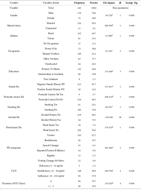
Most participants were males 170 (70%) and 73 (30%) females, 232 (95.5%) married and 11 (4.5%) were unmarried (p = 0.000). A large number of subjects 162 (66.7%) were from rural and 81(33.3%) from urban community (p = 0.000). By occupation 27 (11.1%) have no any occupation, 73 (30%) house-wives, 100 (41.2%) manual workers and 43 (17.7%) were office workers (p = 0.000). Re-garding educational status 64 (26.3%) uneducated, 123 (50.6%) primary to ma-triculation, 48 (19.8%) intermediate to graduation and only 08 (3.3%) were postgraduates (p = 0.000). A large number of study subjects 213 (87.7%) had no any evidence of PD in family, where as positive family history was observed in 30 (12.3%) subjects (p = 0.000). Evaluating risk factors for PD, history of pesti-cide contact in 09 (3.7%), smoking 61 (25.1%), alcohol abuse 24 (9.9%) and his-tory of head injury observed in 18 (7.4%) subjects (p = 0.000). BMI normal in 143 (58.8%), over weight in 91 (37.4%) and 09 (3.7%) were obese subjects (p = 0.000). Baseline characteristics of participants as tremors, bradykinesia, speech changes, impaired posture and balance, rigidity and writing changes were ob-served in 67.1%, 10.7%, 6.2%, 5.8%, 5.3% and 4.9% respectively (p = 0.000). Re-garding duration of PD 82 (33.7%) were <01 year, 40 (16.5%) 1 - 2 years, 41 (16.9%) 2 - 5 years, 40 (16.5%) 5 - 10 years and 40 (16.5%) had duration > 10 years (p = 0.000). We identified 151 (62.1%) subjects with vitamin 25(OH)D < 30 ng/ml, remaining 92 (37.9%) have normal vitamin D3 values (30 - 150 ng/ml) (p = 0.000). Insufficiency (10 - 30 ng/ml) found in 148 (60.9%) and deficiency (<10 ng/ml) was seen in 03 (1.2%) subjects (p = 0.000). Rest of chi-square values and df were shown in Table 1.
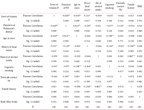
3.3. Analyses of Different Correlations
The correlation of different variables assessed as shown in Table 2. We found that vitamin D3 levels (p = 0.000) were strongly correlated with age (p = 0.000), duration of PD (p = 0.000) these were statistically significant, and the analysis of other risk factors of PD were also assessed there was not significant correlation of various risk factors of PD and vitamin D deficiency like head injury (p = 0.017) alcohol (p = 0.358), smoking (p = 0.566), pesticide contact (p = 0.512), family history (p = 0.840) and BMI (p = 0.572) as shown in Table 2. The p-value of less than 0.05 was considered statistically significant.
4. Discussion
DOI: 10.4236/apd.2018.71002 11 Advances in Parkinson’s Disease
Table 1. Frequency, percentage and chi-square values of study variables.
Variable Variables details Frequency Percent Chi-Square df Asymp. Sig.
Variable Total 243 100.0 Non-parametric
Gender Male 170 70.0 38.720b 1 0.000
Female 73 30.0
Marital status Married 232 95.5 200.992b 1 0.000
Unmarried 11 4.5
Address Rural 162 66.7 27.000b 1 0.000
Urban 81 33.3
Occupation
No Occupation 27 11.1
51.765c 3 0.000
House Wife 73 30.0
Manual Workers 100 41.2
Office Workers 43 17.7
Education
Uneducated 64 26.3
112.440c 3 0.000
Primary To Matric 123 50.6
Intermediate to Graduate 48 19.8
Post Graduate 8 3.3
Family Hx Negative Family History PD 213 87.7 137.815a 1 0.000
Positive Family History PD 30 12.3
Pesticide contact Hx Pesticide Contact Hx Yes 9 3.7 208.333b 1 0.000
Pesticide Contact Hx No 234 96.3
Smoking Hx Smoking Yes 61 25.1 60.251b 1 0.000
Smoking No 182 74.9
Alcohol Hx Alcohol History No 219 90.1 136.585 50 0.000
Alcohol History Yes 24 9.9
Head injury Hx Head Injury Yes 18 7.4 176.333b 1 0.000
Head Injury No 225 92.6
PD symptoms
Tremor 163 67.1
447.840e 5 0.000
Bradykinesia 26 10.7
Speech Changes 15 6.2
Impaired Posture & Balance 14 5.8
Rigidity 13 5.3
Writing Changes & Others 12 4.9
Vit D
Deficiency: 0 - 10 ng/ml 3 1.2
209.704c 2 0.000
Insufficiency: 10 - 30 ng/ml 148 60.9
Sufficiency: 30 - 150 ng/ml 92 37.9
Duration of PD (Years) <1 82 33.7 132.025d 4 0.000
DOI: 10.4236/apd.2018.71002 12 Advances in Parkinson’s Disease Continued
>2 - 5 41 16.9
>5 - 10 40 16.5
>10 40 16.5
Vit D Level Normal 92 37.9 14.325b 1 0.000
Hypovitaminosis D 151 62.1
BMI
Normal 143 58.8
112.691a 2 0.000
Over Weight 91 37.4
[image:6.595.58.540.268.637.2]Obese 9 3.7
Table 2. Correlation of different variables in Parkinson’s disease subjects. Correlations of low levels of Vitamin D levels with other
variables of study.
Level of
vitamin D Duration of PD Age in years Hx of
head injury
Hx of alcohol
intake
Cigarette smoking
Pesticide contact
Hx
Family history BMI
Level of vitamin D
Pearson Correlation 1 −0.649** −0.656** 0.153* −0.059 −0.037 −0.042 0.013 0.036
Sig. (2-tailed) 0.000 0.000 0.017 0.358 0.566 0.512 0.840 0.572
Duration of Parkinson’s disease
Pearson Correlation −0.649** 1 0.814** −0.129* 0.021 −0.077 0.130* −0.014 0.013
Sig. (2-tailed) 0.000 0.000 0.045 0.742 0.232 0.043 0.832 0.840
Age in years Pearson Correlation −0.656** 0.814** 1 −0.065 −0.030 −0.198** 0.019 −0.098 0.004
Sig. (2-tailed) 0.000 0.000 0.314 0.646 0.002 0.771 0.128 0.953
History of head injury
Pearson Correlation 0.153* −0.129* −0.065 1 −0.064 −0.164* −0.055 −0.180** 0.002
Sig. (2-tailed) 0.017 0.045 0.314 0.318 0.011 0.389 0.005 0.975
History of alcohol intake
Pearson Correlation −0.059 0.021 −0.030 −0.064 1 0.001 0.065 0.882** 0.030
Sig. (2-tailed) 0.358 0.742 0.646 0.318 0.990 0.314 0.000 0.641
Cigarette smoking
Pearson Correlation −0.037 −0.077 −0.198** −0.164* 0.001 1 −0.114 0.044 0.006
Sig. (2-tailed) 0.566 0.232 0.002 0.011 0.990 0.077 0.493 0.925
Pesticide contact hx
Pearson Correlation −0.042 0.130* 0.019 −0.055 0.065 −0.114 1 0.074 0.001
Sig. (2-tailed) 0.512 0.043 0.771 0.389 0.314 0.077 0.253 0.982
Family history Pearson Correlation 0.013 −0.014 −0.098 −0.180** 0.882** 0.044 0.074 1 0.078
Sig. (2-tailed) 0.840 0.832 0.128 0.005 0.000 0.493 0.253 0.225
Body Mass Index
Pearson Correlation 0.036 0.013 0.004 0.002 0.030 0.006 0.001 0.078 1
Sig. (2-tailed) 0.572 0.840 0.953 0.975 0.641 0.925 0.982 0.225
N 243 243 243 243 243 243 243 243 243
*Significant Correlation at 0.05 levels (2-tailed). **Significant Correlation at 0.01 levels (2-tailed).
DOI: 10.4236/apd.2018.71002 13 Advances in Parkinson’s Disease described role of vitamin D3 lack in PD, vitamin D3 insufficiency leads or progresses to Parkinson’s disease. These issues were focused in different studies in rest of world. Findings of present study are match-able with previous studies as discussed below.
PD (Parkinson’s disease) is a neurodegenerative disease in a particular zone of the cerebrum called substantia nigra, [19] characterized by inflexible nature, tremors and dyskinesia along with postural insecurity and dementia. Vitamin D had critical effect on neurological illnesses as PD and Dementia. In cerebrum, hippocampus and substantia nigra neurons show high convergences of VDRs in their core and 1-OHase in their cytosol. In present study mean ± SD values of age were 67.64 ± 6.67 years (age range 56 - 85 years), males 170 (70%) and 73 (30%) were females, our findings are supported in a study by Moghaddasi M et al. in which mean age of the patients were 56.57 ± 11.71 years (age range 24 - 79 years); 3 (75.9%) males and 20 (24.1%) were females. Mean age of symptoms onset was 50.71 ± 12.10 years (range 20 - 77 years) [20]. With insufficiency of vitamin D, there is hazard of developing PD; this hazard increases to twofold when there is deficiency of vitamin D [21]. Vitamin D levels were low in subjects with PD and AD in comparison to the normal controls, [8] current study also determined insufficiency of vitamin D3 in 61.1% of PD subjects. 25(OH)D3 emphatically connected with intellectual execution, especially with measuring its role in elderly populace [22]. Many of studies (Cross Sectional) had shown the relationship of decreased levels of vitamin D with incident of PD also predicted increased hazard of PD [23]. Vitamin D levels in high-risk group and matched controls (age, sex) did not differ, and it was suggested that there is no deficiency of vitamin D before diagnosis of PD [24]. A majority of researches in established PD had shown lower vitamin D values as compared to fit controls [9]. Serum values of vitamin D decrease as severity of disease increases [25] [26] as low le-vels of vitamin D3 were observed in subjects of PD in this study.
Nitric Oxide (free radical) can damage to cells, its synthesis is inhibited by vi-tamin D, and vivi-tamin D3 also causes formation of glutathione (antioxidant) thus plays a neuro-protective role [27]. It is assumed that vitamin D3 is involved in initiating the synthesis of N G F (nerve growth factor), Glial cell line derived factor and NT3 (Neurotrophin) and in this way is considered as Neurotrophic Factor [28] [29] [30]. Peterson et al. in their research found strong relationship of automatic postural responses with serum vitamin D concentrations [31]. Dai-ly vitamin D3 supplements (1200 IU) for one year showed mild progression of disease and worsening of disease observed in those who did not receive incre-ments. [32] As there were presence of more than one symptom of PD, VDD was more common as age of patient increases with more than one symptom. Patients with PD had limited outdoor activities so solar exposure is decreased this may contributes to decreasing levels of vitamin D even with incremental intake [33].
DOI: 10.4236/apd.2018.71002 14 Advances in Parkinson’s Disease oral supplements of vitamin D. There study indicated that low values of this vi-tamin with reduced sun exposure are associated with increase hazard of PD [34], above associations of VDD and PD are evident in present study. Yoon JH et al. in their study subjects with early PD, observed the relation among serum vita-min D values and endothelial cell dysfunction [35]. Probable racial variations in passageway for consumed vitamin D may be dilemma crossways to panel sug-gestions meant for D3 values as in Inuit. Decreased synthesis of this vitamin is balanced in Inuit through transforming lot of vitamin-D towards its chiefly ac-tive type [36]. A Toronto research on Canadians (young) from various origins had average vitamin D3 values that were fundamentally elevated from autho-rized proposals [37]. 22% European, 78% and 77% of Asian (East, South) herit-age had vitamin D3 level < 40 nmol/l (15 ng/ml), compared with previous stu-dies. Toronto study in Asians (East) observed decreased vitamin D3 in contrast with White community [38].
Rural men around Delhi had average 44 nmol/L of D3, in current study 162 (66.7%) subjects from rural and 81 (33.3%) were urban (p = 0.000), with mean vitamin D3 levels 27.68 ± 21.72 ng/ml, these findings are matching with results of study by Rajasree S et al. Normal Indians have decreased vitamin D3, not much unusual than Canadian Asians (South). South Indians with IHD had tre-mendously increased (>222.5 nmol/l) D3 values [39]. In present study with ref-erences to above studies we found that 151 (62.1%) subjects have vitamin D3 <30 ng/ml and remaining 92 (37.9%) have sufficient D3 values (30 - 150 ng/ml) (p value 0.000). Insufficiency (10 - 30 ng/ml) in 148 (60.9%) and deficiency (<10 ng/ml) observed in 03 (1.2%) subjects (p = 0.000). Melanin substance demon-strated opposite association with serum 25(OH)D [37]. Uniformly deficient 25(OH)D values seen in Indians (living in India and China). Noteworthy Here-ditary minority of French Canadians didn’t buildup consumed vitamin D3. Vi-tamin D3 protein binding polymorphisms had a significant part of variety in se-rum D3 as totaled intake of vitamin D3 [40] [41]. Different methods controlling metabolism with limited extent of vitamin D values in which vascular capacity is streamlined were associated with increased mortality [42], abnormal functioning and premature aging [43].
Worldwide prevalence of vitamin D deficiency/insufficiency accounts for 1 billion people [13] where south Asians are uniformly affected despite abundant sunshine [44]. Pakistan a rising nation of Asia (South) with an area spreading over scope 24˚35' North and longitude 61˚ East to 78˚ East, seriously facing D3 in-sufficiency in pregnant ladies, neonates, babies, youngsters, teenagers, grown-ups, and elderly individuals regardless of plentiful daylight [45]. 70% fit volunteers in Pakistan, 84% pregnant ladies in India are distressed by VDD. Sri Lanka and Bangladesh are no exception where 26% boys and 8% girls are victims of VDD
DOI: 10.4236/apd.2018.71002 15 Advances in Parkinson’s Disease evaluation of population groups [47]. Mansoor et al. elucidated that 56.9% men and 43.1% normal women had Vitamin D3 < 20 ng/ml [48]. Sheikh et al. ob-served in 84.3% of tested healthy subjects (38 - 55 years) in Karachi had 25(OH)D levels < 30 ng/ml suggesting extensive VDD prevalence throughout Pakistan declaring Pakistani population a vitamin D deficient [49].
Findings of above studies were considerably in contest with present study where we identified 151 (62.1%) subjects have vitamin D3 < 30 ng/ml. A nar-rowed danger of death in old age observed with high Vitamin D3 levels while others didn’t benefit [50]. Taking supplements are valuable or not still unclear
[51]. Increased danger of vitamin D deficiency observed in Blacks comparison to White populace [52]. Further studies needed to find out reasons for these dif-ferences and clarify probable part of vitamin D in pathogenesis and clinical path of PD.
5. Conclusion
Vitamin D deficiency is commonly associated in patients suffering from Parkin-son’s disease. As concluded in present research that as the age advances, risk of Parkinson’s disease increases with simultaneous decrease in vitamin D level. As concluded in our study, 62.1% subjects of Parkinson’s disease were vitamin D deficient.
References
[1] Holick, M.F. (2006) High Prevalence of Vitamin D Inadequacy and Implications for Health. Mayo Clinic Proceedings, 81, 353-373. https://doi.org/10.4065/81.3.353 [2] Von Campenhausen, S., Bornschein, B., Wick, R., Bötzel, K., et al. (2005) Prevalence
and Incidence of Parkinson’s Disease in Europe. European Neuropsychopharma-cology, 15, 473-490. https://doi.org/10.1016/j.euroneuro.2005.04.007
[3] Grant, W.B. (2006) Epidemiology of Disease Risks in Relation to Vitamin D Insuffi-ciency. Progress in Biophysics and Molecular Biology, 92, 65-79.
https://doi.org/10.1016/j.pbiomolbio.2006.02.013
[4] Kilkkinen, A., Knekt, P., Heliovaara, M., et al. (2008) Vitamin D Status and the Risk of Lung Cancer: A Cohort Study in Finland. Cancer Epidemiology, Biomarkers & Prevention, 17, 3274-3278. https://doi.org/10.1158/1055-9965.EPI-08-0199
[5] Giovannucci, E., Liu, Y., Hollis, B.W. and Rimm, E.B. (2008) 25-Hydroxyvitamin D and Risk of Myocardial Infarction in Men: A Prospective Study. Arch Intern Med, 168, 1174-1180. https://doi.org/10.1001/archinte.168.11.1174
[6] Knekt, P., Laaksonen, M., Mattila, C., et al. (2008) Serum Vitamin D and Subse-quent Occurrence of Type 2 Diabetes. Epidemiology, 19, 666-671.
https://doi.org/10.1097/EDE.0b013e318176b8ad
[7] Newmark, H.L. and Newmark, J. (2007) Vitamin D and Parkinson’s Disease—A Hypothesis. Movement Disorders, 22, 461-468. https://doi.org/10.1002/mds.21317 [8] Evatt, M.L., Delong, M.R., Khazai, N., et al. (2008) Prevalence of Vitamin D
Insuffi-ciency in Patients with Parkinson Disease and Alzheimer Disease. Arch Neurol, 65, 1348-1352. https://doi.org/10.1001/archneur.65.10.1348
DOI: 10.4236/apd.2018.71002 16 Advances in Parkinson’s Disease
https://doi.org/10.1002/mds.20658
[10] Sato, Y., Kikuyama, M. and Oizumi, K. (1997) High Prevalence of Vitamin D Defi-ciency and Reduced Bone Mass in Parkinson’s Disease. Neurology, 49, 1273-1278. https://doi.org/10.1212/WNL.49.5.1273
[11] Hongliu, D., Kaltra, D., Kaitlin, C., et al. (2013) Unrecognized Vitamin D3 Defi-ciency Is Common in Parkinson Disease: Harvard Biomarker Study. American Academy of Neurology, 81, 1531-1537.
[12] Eriksen, E.F. and Glerup, H. (2002) Vitamin D Deficiency and Aging: Implications for General Health and Osteoporosis. Biogerontology, 3, 73-77.
https://doi.org/10.1023/A:1015263514765
[13] Holick, M.F. (2007) Vitamin D deficiency. The New England Journal of Medicine, 357, 266-281.
[14] Schoenmakers, I., Goldberg, G.R. and Prentice, A. (2008) Abundant Sunshine and Vitamin D Deficiency. The British Journal of Nutrition, 99, 1171-1173.
https://doi.org/10.1017/S0007114508898662
[15] Grant, W.B. and Holick, M.F. (2005) Benefits and Requirements of Vitamin D for Optimal Health: A Review. Alternative Medicine Review, 10, 94-111.
[16] Brown, J.E., Isaacs, J., et al. (2013) Nutrition through the Life Cycle. Cengage Learning, Boston, MA.http://www.revolvy.com/main/index.php?s
[17] Hughes, A.J., Daniel, S.E., Kilford, L. and Lees, A.J. (1992) Accuracy of Clinical Di-agnosis of Idiopathic Parkinson’s Disease: A Clinico-Pathological Study of 100 Cas-es. Journal of Neurology, Neurosurgery & Psychiatry, 55, 181-184.
https://doi.org/10.1136/jnnp.55.3.181
[18] Shen, L. and Ji, H.F. (2015) Associations between Vitamin D Status, Supplementa-tion, Outdoor Work and Risk of Parkinson’s Disease: A Meta-Analysis Assessment. Nutrients, 7, 4817-4827.https://doi.org/10.3390/nu7064817
[19] Evatt, M.L., DeLong, M.R., Kumari, M., et al. (2011) High Prevalence of Hypovita-minosis D Status in Patients with Early Parkinson Disease. Archives of Neurology, 68, 314-319. https://doi.org/10.1001/archneurol.2011.30
[20] Moghaddasi, M., Mamarabadi, M. and Aghaii, M. (2013) Serum 25-Hydroxyvitamin D3 Concentration in Iranian Patients with Parkinson’s Disease. Iranian Journal of Neurology, 12, 56-59.
[21] Lv, Z., Qi, H., Wang, L., et al. (2014) Vitamin D Status and Parkinson’s Disease: A Systematic Review and Meta-Analysis. Neurological Sciences, 35, 1723-1730. https://doi.org/10.1007/s10072-014-1821-6
[22] Jennifer, S.B., Tammy, M.S., Bess, D.H., et al. (2009) The Journals of Gerontology: Series A, Vitamin D Is Associated with Cognitive Function in Elders Receiving Home Health Services. The Journals of Gerontology. Series A, Biological Sciences and Medical Sciences, 64, 888-895.
[23] Knekt, P., Kilkkinen, A., Rissanen, H., et al. (2010) Serum Vitamin D and the Risk of Parkinson Disease. Archives of Neurology, 67, 808-811.
https://doi.org/10.1001/archneurol.2010.120
[24] Fullard, M.E., Xie, S.X., Marek, K., et al. (2017) Vitamin D in the Parkinson Asso-ciated Risk Syndrome (PARS) Study. MovementDisorders, 32, 1636-1640. https://doi.org/10.1002/mds.27127
DOI: 10.4236/apd.2018.71002 17 Advances in Parkinson’s Disease [26] Ding, H., Dhima, K., Lockhart, K., et al. (2013) Unrecognized Vitamin D3 Defi-ciency Is Common in Parkinson Disease: Harvard Biomarker Study. Neurology, 81, 1531-1537. https://doi.org/10.1212/WNL.0b013e3182a95818
[27] Garcion, E., Wion-Barbot, N., Montero-Menei, C.N., et al. (2002) New Clues about Vitamin D Functions in the Nervous System. Trends in Endocrinology Metabolism, 13, 100-105. https://doi.org/10.1016/S1043-2760(01)00547-1
[28] Naveilhan, P., Neveu, I., Wion, D. and Brachet, P. (1996) 1,25-Dihydroxyvitamin D3, an Inducer of Glial Cell Line-Derived Neurotrophic Factor. Neuroreport, 7, 2171-2175. https://doi.org/10.1097/00001756-199609020-00023
[29] Musiol, I.M. and Feldman, D. (1997) 1,25-Dihydroxyvitamin D3 Induction of Nerve Growth Factor in L929 Mouse Fibroblasts: Effect of Vitamin D Receptor Regulation and Potency of Vitamin D3 Analogs. Endocrinology, 138, 12-18.
https://doi.org/10.1210/endo.138.1.4858
[30] Neveu, I., Naveilhan, P., Baudet, C., Brachet, P. and Metsis, M. (1994) 1, 25-Dihydroxyvitamin D3 Regulates NT-3, NT-4 but Not BDNF mRNA in Astro-cytes. Neuroreport, 6, 124-126. https://doi.org/10.1097/00001756-199412300-00032 [31] Peterson, A.L., Mancini, M. and Horak, F.B. (2013) The Relationship between
Bal-ance Control and Vitamin D in Parkinson’s Disease—A Pilot Study. Movement Disorders, 28, 1133-1137. https://doi.org/10.1002/mds.25405
[32] Suzuki, M., Yoshioka, M., Hashimoto, M., et al. (2013) Randomized, Double-Blind, Placebo-Controlled Trial of Vitamin D Supplementation in Parkinson Disease. The American Journal of Clinical Nutrition, 97, 1004-1013.
https://doi.org/10.3945/ajcn.112.051664
[33] Miyake, Y., Tanaka, K., Fukushima, W., et al. (2011) Lack of Association of Dairy Food, Calcium, and Vitamin D Intake with the Risk of Parkinson’s Disease: A Case Control study in Japan. Parkinsonism & Related Disorders, 17, 112-116.
https://doi.org/10.1016/j.parkreldis.2010.11.018
[34] Juan, W., Deyu, Y., Yu, Y., et al. (2016) Vitamin D and Sunlight Exposure in New-ly-Diagnosed Parkinson’s Disease. Nutrients, 8, 142.
https://doi.org/10.3390/nu8030142
[35] Yoon, J.H., Park, D.K., Yong, S.W. and Hong, J.M. (2015) Vitamin D Deficiency and Its Relationship with Endothelial Dysfunction in Patients with Early Parkin-son’s Disease. Journal of Neural Transmission (Vienna), 122, 1685-1691.
https://doi.org/10.1007/s00702-015-1452-y
[36] Rejnmark, L., Jørgensen, M.E., Pedersen, M.B., et al. (2004) Vitamin D Insufficiency in Greenlanders on a Westernized Fare: Ethnic Differences in Calcitropic Hormones between Greenlanders and Danes. Calcified Tissue International, 74, 255-263. https://doi.org/10.1007/s00223-003-0110-9
[37] Gozdzik, A., Barta, J.L., Wu, H., et al. (2008) Low Wintertime Vitamin D Levels in a Sample of Healthy Young Adults of Diverse Ancestry Living in the Toronto Area: Associations with Vitamin D Intake and Skin Pigmentation. BMC Public Health, 8, 336. https://doi.org/10.1186/1471-2458-8-336
[38] Abnet, C.C., Chen, W., Dawsey, S.M., et al. (2007) Serum 25(OH)-Vitamin D Con-centration and Risk of Esophageal Squamous Dysplasia. Cancer Epidemiology, Biomarkers & Prevention, 16, 1889-1893.
https://doi.org/10.1158/1055-9965.EPI-07-0461
DOI: 10.4236/apd.2018.71002 18 Advances in Parkinson’s Disease
https://doi.org/10.1023/A:1014559600042
[40] Sinotte, M., Diorio, C., Bérubé, S., Pollak, M. and Brisson, J. (2009) Genetic Poly-morphisms of the Vitamin D Binding Protein and Plasma Concentrations of 25-Hydroxyvitamin D in Premenopausal Women. The American Journal of Clinical Nutrition, 89, 634-640. https://doi.org/10.3945/ajcn.2008.26445
[41] Labuda, M., Labuda, D., Korab-Laskowska, M., et al. (1996) Linkage Disequilibrium Analysis in Young Populations: Pseudo-Vitamin D-Deficiency Rickets and the Founder Effect in French Canadians. American Journal of Human Genetics, 59, 633-643.
[42] Hsu, J.J., Tintut, Y. and Demer, L.L. (2008) Vitamin D and Osteogenic Differentia-tion in the Artery Wall. Clinical Journal of the American Society of Nephrology, 3, 1542-1547. https://doi.org/10.2215/CJN.01220308
[43] Tuohimaa, P. (2009) Vitamin D and Aging. The Journal of Steroid Biochemistry and Molecular Biology, 114, 78-84. https://doi.org/10.1016/j.jsbmb.2008.12.020 [44] Harinarayan, C.V., et al. (2009) Vitamin D Status in India—Its Implications and
Remedial Measures. A Review of over 50 Studies of 25(OH)D. Journal of the Asso-ciation of Physicians of India, 57, 40-48. http://www.japi.org/jan_2009/R-1.html [45] Akhtar, S. (2016) Prevalence and Correlates of Vitamin D Deficiency-Perspectives
from Pakistan. MINI REVIEW. Department of Food Science and Technology, Ba-hauddin Zakariya University, Multan, Pakistan. Pakistan Journal of Pharmaceutical Sciences, 29, 1325-1330.
[46] Akhtar, S. (2016) Vitamin D Status of South Asian Populations—Risks and Oppor-tunities. Critical Reviews in Food Science and Nutrition, 56, 1925-1940.
https://doi.org/10.1080/10408398.2013.807419
[47] Anjum, P., Safder, N., Khalid, M. and Mehboob, I. (2013) Vitamin D Deficiency in Pakistani Population. Journal of Pakistan Orthopaedic Association, 25, 18-19. [48] Mansoor, S., Habib, A., Ghani, F., et al. (2010) Prevalence and Significance of
Vita-min D Deficiency and Insufficiency among Apparently Healthy Adults. Clinical Bi-ochemistry, 43, 1431-1435.https://doi.org/10.1016/j.clinbiochem.2010.09.022 [49] Sheikh, A., Saeed, Z., Jafri, S.A.D., et al. (2012) Vitamin D Levels in Asymptomatic
Adults—A Population Survey in Karachi, Pakistan. PLoSONE, 7, e33452. https://doi.org/10.1371/journal.pone.0033452
[50] Bjelakovic, G., Gluud, L.L., Nikolova, D., et al. (2014) Vitamin D Supplementation for Prevention of Mortality in Adults. The Cochrane Database of Systematic Re-views (Systematic Review), 1, CD007470.
https://doi.org/10.1002/14651858.CD007470.pub3
[51] Bolland, M.J., Grey, A., Gamble, G.D. and Reid, I.R. (2014) The Effect of Vitamin D Supplementation on Skeletal, Vascular, or Cancer Outcomes: A Trial Sequential Meta-Analysis. The Lancet Diabetes & Endocrinology (Meta-Analysis), 2, 307-320. https://doi.org/10.1016/S2213-8587(13)70212-2