ORIGINAL RESEARCH
PEDIATRICS
Pseudo-Leptomeningeal Contrast Enhancement at 3T in
Pediatric Patients Sedated by Propofol
XA.M. McKinney,X A. Chacko Achanaril,XB. Knoll,XD.R. Nascene, andXR.S. Gawande
ABSTRACT
BACKGROUND AND PURPOSE: Propofol is a cerebral vasoconstrictor that modulates cerebral perfusion by decreasing the metabolic rate of oxygen. Because younger children often undergo intravenous sedation for MR imaging, this study set out to evaluate the degree of leptomeningeal contrast enhancement on 3T postcontrast brain MR imaging and to determine whether this phenomenon relates to sequence, sedation dosage, or patient age or weight.
MATERIALS AND METHODS: During a 2-year period, of 152 children 1–5 years of age who underwent MR imaging, 43 were included for MRI review. Of these, 37 underwent postcontrast imaging with either solely gradient-echo T1WI (n⫽20) or spin-echo T1WI (n⫽17); notably, 6 patients underwent both sequences. Three neuroradiologists separately graded the degree of leptomeningeal contrast enhancement (grades 0 –3) that was correlated with various factors and calculated the interobserver reliability.
RESULTS:For the 43 patients, the mean patient age was 3.1⫾1.4 years. The leptomeningeal contrast-enhancement grade was significantly greater (P⬍.0001) on spin-echo T1WI (1.9 –2.1) versus gradient-echo TIWI (1.2–1.4). Patient weight (r⫽ ⫺0.366 to⫺.418,P⫽.003–.01) and age (r⫽ ⫺0.315 to⫺0.418, P⫽.004 –.032) moderately and inversely correlated with the leptomeningeal contrast-enhancement grade, while the propofol dosage, sedation duration, and time to T1WI post-contrast administration did not (each,P⬎.05). The interobserverwas strong regarding the leptomeningeal contrast-enhancement grade on both spin-echo T1WI (⫽0.609 – 0.693,P⬍.0001) and gradient-echo TIWI (⫽0.567– 0.698,P⬍.0001).
CONCLUSIONS: Leptomeningeal contrast enhancement (or “pseudo”-leptomeningeal contrast enhancement) occurs with a greater frequency and degree on 3T postcontrast spin-echo T1WI relative to gradient-echo TIWI in younger children sedated with propofol and should not be mistaken for disease. This phenomenon may be more prominent with lower age or size and may arise from propofol-induced vascular smooth-muscle dilation.
ABBREVIATIONS:GE⫽gradient-echo; LMCE⫽leptomeningeal contrast enhancement; SE⫽spin-echo; TTI⫽time to postcontrast T1-weighted imaging
E
xtra-axial enhancement in the CNS can be either leptomenin-geal, occurring along the surface of the brain and subarach-noid space, or pachymeningeal, comprising the dura and its re-flections.1The vessels within the pachymeninges do not have a blood-brain barrier, which causes the typical appearance of thin, linear, and smooth enhancement on postcontrast T1WI following the intravenous administration of gadolinium-based contrast agents; in contrast, the main mechanism of leptomeningealcon-trast enhancement (LMCE) (ie, pial enhancement) is disruption of the blood-brain barrier.1,2
Often, children younger than 8 years of age require sedation to undergo a high-quality MR imaging examination. Anesthetic agents have been shown to cause changes in cerebral homeostasis and vascular reactivity.3-5Such agents can cause a global decrease in cerebral metabolism, with resultant decreases in both CBF and CBV.3-5Prior studies have also demonstrated that 2,6 diisopropyl phenol (propofol) can modulate CBF by decreasing the metabolic rate of oxygen; in addition, speculation based on animal studies suggests that propofol can dilate vascular smooth muscle in other regions of the body.6
The basis of this study is that the authors had noted prominent LMCE on brain MR imaging in some sedated children, but based on clinical notes, they neither were acutely ill nor exhibited men-ingeal signs. Thus, this study was initiated to determine whether
Received February 13, 2018; accepted after revision June 5.
From the Department of Radiology (A.M.M., A.C.A., D.R.N.), Neuroradiology Divi-sion, University of Minnesota, Minneapolis, Minnesota; Department of Radiology (B.K.), Hennepin County Medical Center, Minneapolis, Minnesota; and Department of Radiology (R.S.G.), Johns Hopkins University, Baltimore, Maryland.
Please address correspondence to Alexander M. McKinney, MD, University of Min-nesota, Department of Radiology, 420 Delaware St SE, MMC 292, Minneapolis, MN 55455; e-mail: mckinrad@umn.edu
the degree of this phenomenon of apparent LMCE (termed here “pseudo”-LMCE) relates to the type of T1WI sequence, time to acquiring postcontrast T1WI, propofol dosage, or various patient demographics.
MATERIALS AND METHODS
This retrospective study was performed after Hennepin County Medical Center, Minneapolis, Minnesota, review board approval. Review of the imaging data base and electronic clinical records yielded 152 healthy pediatric patients between the ages of 1 and 5 years who underwent 3T brain MR imaging and were sedated with intravenous propofol between November 2011 and November 2013. Inclusion criteria were the following: 1) ages were between 1 and 5 years; 2) either axial gradient-echo (GE) T1WI or spin-echo (SE) T1WI was performed; 3) the child received gadolinium-based intravenous contrast; 4) intravenous propofol was used for sedation; 5) the patient had either normal examination findings or only mild, nonacute, and noncongenital abnormalities (eg,⬍5 white matter foci); and 6) the patient had not had meningitis or other clinical diseases that could cause LMCE. Exclusion criteria consisted of the following: studies performed on a 1.5T magnet, an incomplete MR imaging examination, moderate-to-severe structural abnormalities, or clinical signs of meningitis (Fig 1). Anesthesia was induced via intravenous administration of propo-fol by a pediatric intensivist, without the use of inhalational anes-thetics, akin to sedation methods described previously.7
MR Imaging Technique
All studies were performed on a single 3T MR unit (Intera; Philips Healthcare, Best, the Netherlands), with sedation performed by a pediatric intensivist. The imaging parameters for GE TIWI were a volumetric acquisition of 9.8 ms/4.6 ms/8°/15–20 cm/1 (TR/TE/flip angle/FOV/NEX), a 169⫻169 to 240⫻240 matrix, 1-mm section thickness (0-mm gap), and an acquisition time of approximately 5 minutes; these scans were reconstructed in the axial plane at a section thickness of 3 mm. For SE T1WI, the parameters were 353–734 ms/10 ms/14 –20 cm/1 (TR/TE/FOV/NEX), with a 168⫻132 to 265⫻205 matrix, axial 3-mm thickness (0.3–1.0 mm gap), and an acquisition time of about 5 minutes. We attempted to approximate and coregister the GE TIWI and SE TIWI to each other at the same thickness and level. Axial spin-echo T2WI, FLAIR, and DWI were also performed in each patient; the axial spin-echo T2WI and DWI acquisitions were performed after the intravenous administration of gadolinium-based contrast but prior to the postcontrast GE TIWI or SE TIWI acquisitions, to ensure a minimum delay of several minutes before the T1WI acquisitions were performed. The standard weight-based intravenous dose of gadolinium-weight-based contrast was 0.1 mL/kg of body weight (0.1 mmol/kg) of gadobutrol (Gadavist; Bayer Scher-ing Pharma, Berlin, Germany).
Imaging Interpretation
Two staff neuroradiologists (A.M.M.,B.K., each with⬎10 years of imaging experience) and 1 neuroradiology fellow (R.S.G., with
[image:2.594.55.531.46.388.2]2 years of dedicated neuroradiology experience) independently graded the degree of LMCE as follows: grade 0, minimal thin vascular structures barely visible within the sulci; grade 1, thin vascular structures extending into the depths of the sulci; grade 2, smooth and slightly thickened LMCE; and grade 3, almost nodu-lar, diffusely thickened LMCE or apparent involvement of adja-cent parenchyma. The time between the commencement of the administration of intravenous contrast and the start of the post-contrast T1WI sequence was also recorded and was termed “time to imaging” (TTI).
Statistical Analysis
The interobserver variability was calculated regarding LMCE grades using the Cohen. The LMCE grade was correlated with the propofol dosage, duration of sedation, patient age, weight, and TTI using the Spearman correlation. A Mann-WhitneyUtest was used to compare the grades of LMCE within the group (n⫽6) who underwent both GE TIWI and SE TIWI. The significance threshold was set toP⬍.05.
RESULTS
Of 152 pediatric patients (1–5 years of age) sedated by propofol for 3T MR imaging, 109 were excluded due to the lack of postcon-trast T1WI (n⫽ 96), the MR imaging being at 1.5T (n⫽3), moderate-severe brain injury or congenital abnormalities, or sev-eral other factors, as listed under “Excluded Patients” within the organization chart ofFig 1. A total of 43 patients were ultimately included for MRI review; of these, 37 underwent postcontrast imaging with either solely gradient-echo T1WI (n⫽20/43) or spin-echo T1WI (n⫽17/43); notably, 6 patients underwent both sequences (n⫽6/43).Table 1lists the mean patient age, weight, propofol dosage, sedation duration, and TTI for both sequences. While the postcontrast TTI range was similar between sequences, it was slightly greater on SE TIWI than on GE TIWI (mean, 12.6 versus 11.0 minutes), being significantly different (P⫽.01).
As shown inTable 1, the range of LMCE grades of the review-ers was greater on SE TIWI (1.9 –2.1) vreview-ersus GE TIWI (1.2–1.4) and was significantly different (P⬍.0001). Interobserver be-tween reviewers was strong for both GE TIWI (⫽0.567– 0.698, P ⬍ .0001) and SE TIWI (⫽0.609 – 0.693, P⬍ .0001). No patients had grade 0 LMCE on SE TIWI. Examples of the LMCE grades are provided inFigs 2–5.
Regarding the 6 patients who underwent both T1WI se-quences, the mean LMCE grade on SE TIWI (1.83) was greater than that on GE TIWI (1.33) but was not significantly different (P⫽.546). Examples of LMCE on both sequences in the same patient are shown inFigs 3and5.
When we attempted to correlate various factors with the LMCE grade, there were significant, inverse, moderate correla-tions between patient weight and LMCE grade, as well as age and LMCE grade (each,P⬍.05;Table 2). Neither the propofol dose nor the sedation duration significantly correlated with the LMCE grade. The TTI did not correlate significantly with the grade of LMCE on GE TIWI, while on SE TIWI, there was a significant, moderate correlation between the LMCE grade and TTI with only 1 of the 3 observers (a staff neuroradiologist), but not the other 2 (Table 2).
DISCUSSION
[image:3.594.303.532.48.175.2]Because MR imaging is noninvasive and does not use ionizing radiation, it is often a technique of choice for pediatric patients requiring neuroimaging. However, its potentially long imaging
Table 1: Demographics, TTI, dosages, and LMCE grades of the study patients
Parameter Range Mean SD
Age (yr) 1.3–5.0 3.1 1.4
Weight (kg) 5.7–24.6 15.3 4.5
Propofol dose (mcg/kg/min) 73–303 192 52
Sedation duration (min) 43–110 66.6 13.8
TTI GE T1WI (min) 8.0–17.0 12.6 2.2
TTI SE T1WI (min) 8.0–17.0 11.0 2.1
LMCE score on SE T1WI 1.9–2.1 2.0 0.8
LMCE score on GE T1WI 1.2–1.4 1.2 0.8
FIG 2. Grade 0 pseudo-LMCE in a 2-year-old girl post-trauma. Axial (A) and coronal (B) GE TIWI shows only minimal vasculature within the sulci. This grade of enhancement was present only on GE TIWI in 13%–17%, while no patients were graded as 0 on SE TIWI.
[image:3.594.54.285.64.158.2] [image:3.594.301.533.232.490.2]time often requires sedation in pediatric patients younger than 8 years of age. Thus, propofol is a lipid emulsion agent that is com-monly the preferred anesthetic for children younger than 1 year of age who require intravenous sedation, due to its rapid onset, short duration, and infrequent side effects.7-9Because the presence of truly abnormal LMCE would be of concern in children, this study set out to determine whether pseudo-LMCE is a sequence-depen-dent (SE TIWI versus GE TIWI), TTI-depensequence-depen-dent (time to post-contrast T1WI), propofol dosage– dependent phenomenon, or whether it is related to demographics such as age and weight. Ultimately, it was found that overall, the degree of apparent LMCE is significantly greater on SE TIWI compared with GE TIWI and that the only factors that correlated (inversely) with the degree of LMCE were patient weight and age. Hence, the type of T1WI sequence and patient size may be factors to consider when a pattern of apparent LMCE (so-called pseudo-LMCE) is identi-fied, to distinguish this phenomenon from true meningeal abnor-malities. Because this study focused solely on children between 1 and 5 years of age, future studies would be necessary to evaluate whether this phenomenon also occurs to some degree in older children and juveniles.
The mechanism of how this pattern of pseudo-LMCE occurs is not yet known, but there are several plausible explanations. The various determinants of cerebral blood flow are the patient’s age, cerebral metabolic rate for oxygen, cerebral perfusion pressure, arterial oxygen, and carbon dioxide tensions. First, children un-der propofol sedation breathe spontaneously, but propofol causes a decrease in the tidal volume with a maintained respiratory rate
and mild reduction in the partial pressure of oxygen in the blood, as well as a mild reduction in the fraction of inspired oxygen. By this phenomenon, one likely mechanism of pseudo-LMCE may be cerebral vasodilation secondary to an increase in the partial pressure of carbon dioxide (due to smooth-muscle relaxation); this increase in the partial pressure of carbon dioxide is likely due to the lack of “breathing off” CO2.
9,10Another factor could be that
the leptomeninges may be more reactive or sensitive (in a sense “immature”) in children compared with adults, an effect perhaps amplified by intravenous sedation. Additionally, because propo-fol sedation may affect respiration, studies have shown that the end-tidal volume of CO2has an inverse relationship with the de-gree of venous contrast, which could also contribute to LMCE.11 Hence, on the basis of reviewing the images within this study as well as the authors’ experience, the authors opine that pseudo-LMCE on cerebral postcontrast T1WI often represents prominent venous vasculature of the subarachnoid space in younger chil-dren, being anecdotally described previously as less common in older children and adults; because older ages were not included in this study, this should be proved by a prospective study compar-ing age groups.12However, the finding in the current study of a significant, inverse correlation between patient weight and the grade of pseudo-LMCE suggests that smaller and younger pa-tients have more vasoreactivity, perhaps because their vasculature is not as mature. This theory may be supported by a study by
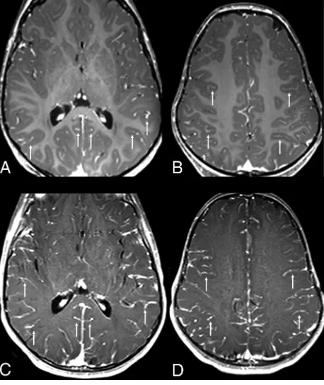
FIG 4. Examples of grade 2 pseudo-LMCE, demonstrated on both GE TIWI and SE TIWI in 2 different patients. In a 3-year-old girl with weakness, grade 2 pseudo-LMCE appears as smooth and slightly thickened enhancement (arrows) throughout the depths of the sulci on axial (A) and coronal (B) GE TIWI. In a 5-year-old boy with head-aches, there is mildly thickened vasculature diffusely throughout the sulci (arrows) on axial (C) and coronal (D) SE TIWI. Note that grade 2 enhancement was slightly more frequent on SE TIWI (35%– 45%) than on GE TIWI (22%–35%).
[image:4.594.53.286.46.288.2] [image:4.594.303.532.48.320.2]Harreld et al,4which noted that in propofol-sedated children, the usual age-related decreases in CBF were reversed and increases in CBF and CBV were weight-dependent.
Unfortunately, exuberant pseudo-LMCE in children may sim-ulate serious disorders that have implications for diagnosis ther-apy, such as leading to an unnecessary lumbar puncture to ex-clude meningitis. A radiologist should use other available imaging sequences to exclude true leptomeningeal abnormalities before the patient leaves the MR imaging scanner. The authors com-monly experience the scenario in which a child with an unrelated diagnosis (eg, developmental delay, autism, and so forth) is im-aged during sedation with propofol, in which the presence of pseudo-LMCE is spurious and varies with the postcontrast T1WI sequence used and the findings of other tests such as a resultant lumbar puncture, serum culture, and so forth are negative. This phenomenon being more common and greater in degree on SE TIWI versus GE TIWI is thought to be related to GE TIWI having a longer TR and lower contrast-to-noise ratio than SE TIWI; these features have been confirmed by studies noting that GE TIWI has a lower lesion detectability and visibility of contrast enhancement for a similar slice thickness.13-15While there was a small difference in slice gap between the 2 sequences in this study, the slice thick-ness and acquisition plane were coregistered between the 2 se-quences, so this small gap is unlikely to account for the difference in the degree of LMCE.
Intravenous contrast is not required in most pediatric brain MR imaging examinations, and gadolinium-based contrast should be avoided when unnecessary due to the possibility of deposition within particular brain structures, especially with re-peat administrations in children.16-20While this study did use a macrocyclic agent (the class of agents least likely to result in brain deposition), the use of most gadolinium-based agents is off-label for most gadolinium based intravenous contrast agents in the infantile population but is considered a standard of care in various clinical scenarios.16-18 For example, particular known or sus-pected pathologies that may require either gadolinium-based contrast for diagnosis or follow-up or to exclude related pathol-ogy including infectious disorders (eg, abscess, empyema, or me-ningoencephalitis), neoplasms, syndromic disorders (eg, phako-matoses), vascular malformations, or vasculitis, to name a few. Hence, while stewardship is critical to lessen the use of gadolini-um-based contrast, there will continue to be subsets of patients that necessitate such contrast in the foreseeable future, and an awareness of this appearance of pseudo-LMCE may help prevent a misdiagnosis of leptomeningeal disease in children.
This study has several limitations, including its retrospective nature and the relatively small sample size of groups that under-went both T1WI sequences. The role of supplemental oxygen dur-ing sedation was not accounted for, which may also affect cerebral
hemodynamics and alter subarachnoid signal intensity on other sequences, such as previously noted on FLAIR.21In this regard, the authors found it difficult to obtain an accurate tabulation of the exact fractionation of oxygen and the length of time admin-istered while the patient was under sedation, although the elec-tronic record did note that there was titration of the supplemental oxygen in some patients. Thus, it is recommended that future studies prospectively tabulate the oxygen fraction accurately. An-other potential limitation is that there was a small but significant difference between the TTI of both SE TIWI (12.6 minutes) and GE TIWI (11.0 minutes), which might create a bias toward having a greater LMCE score on SE TIWI; however, because no signifi-cant association was noted between the degree of LMCE and TTI, such bias (if present) was unlikely to affect the LMCE grade be-tween sequences. Another limitation was that several factors such as CSF protein, fraction of inspired oxygen, end-tidal CO2, and leakage of propofol across the BBB were not evaluated in this study. These factors, previously implicated on T2WI and FLAIR imaging, could be assessed with respect to T1WI in a future study.21,22
CONCLUSIONS
The phenomenon of apparent LMCE, termed pseudo-LMCE herein, is relatively common on postcontrast T1-weighted MR imaging of younger children sedated by intravenous propofol and should not be mistaken for disease. This effect occurs more com-monly and to a greater degree on SE TIWI compared with GE TIWI and inversely correlates with age and weight. The presence of this finding may relate to the immaturity of younger children’s vasculature but needs to be studied further.
Disclosures: Alexander M. McKinney—UNRELATED:Board Membership: VEEV Inc Informatics,Comments: owner, Informatics Solutions.
REFERENCES
1. Smirniotopoulos JG, Murphy FM, Rushing EJ, et al.Patterns of
con-trast enhancement in the brain and meninges.Radiographics2007;
27:525–51CrossRef Medline
2. McKinstry CS, Worthington BS, Niendorf HP, et al.Demonstration of meningeal contrast enhancement on magnetic resonance imag-ing.Acta Radiol Suppl1986;369:564 – 67Medline
3. Kaisti KK, Långsjo¨ JW, Aalto S, et al.Effects of sevoflurane, propofol, and adjunct nitrous oxide on regional cerebral blood flow, oxygen
consumption, and blood volume in humans.Anesthesiology2003;
99:603–13CrossRef Medline
4. Harreld JH, Helton KJ, Kaddoum RN, et al.The effects of propofol
on cerebral perfusion MRI in children.Neuroradiology 2013;55:
1049 –56CrossRef Medline
5. Klein KU, Fukui K, Schramm P, et al.Human cerebral microcircu-lation and oxygen saturation during propofol-induced reduction of bispectral index.Br J Anaesth2011;107:735– 41CrossRef Medline 6. Gragasin FS, Davidge ST.The effects of propofol on vascular func-Table 2: Correlation coefficients andPvalues for LMCE versus other factorsa
Correlation
TTI Overall
(SE and GE T1WI) TTI SE T1WI Only TTI GE T1WI Only Weight (kg) Age (yr)
Dose/Weight (mg/kg)
Duration of Sedation (min) LMCE () ⫺0.232 to⫺0.302 ⫺0.358 to⫺.475 0.016–0.190 ⫺0.366 to⫺0.418 ⫺0.315 to⫺0.418 0.103–0.210 0.023–0.147
Pvalue .051–.130 .036b–.122c .371–.940 .003–.011b .004–.032b .151–.484 .318–.875
a
Ranges provided are per the 3 reviewers.
b
Pvalues⬍.05.
c
[image:5.594.52.534.58.103.2]tion in mesenteric arteries of the aging rat.Am J Physiol Heart Circ Physiol2009;297:H466 –74CrossRef Medline
7. Machata AM, Willschke H, Kabon B, et al.Propofol-based sedation regimen for infants and children undergoing ambulatory magnetic resonance imaging.Br J Anaesth2008;101:239 – 43CrossRef Medline 8. Martin LD, Pasternak LR, Pudimat MA.Total intravenous
anesthe-sia with propofol in pediatric patients outside the operating room.
Anesth Analg1992;74:609 –12Medline
9. Szabo´ EZ, Luginbuehl I, Bissonnette B.Impact of anesthetic agents
on cerebrovascular physiology in children.Pediatr Anesth2009;19:
108 –18CrossRef Medline
10. Remsen LG, Pagel MA, McCormick CI, et al.The influence of anes-thetic choice, PaCO2, and other factors on osmotic blood-brain
barrier disruption in rats with brain tumor xenografts.Anesth
Analg1999;88:559 – 67Medline
11. Kwong KK, Wanke I, Donahue KM, et al.EPI imaging of global increase of brain MR signal with hold preceded by
breath-ing O2.Magn Reson Med1995;33:448 –52CrossRef Medline
12. McKinney AM.Atlas of Normal Imaging Variations of the Brain, Skull, and Craniocervical Vasculature. New York: Springer-Verlag; 2017: 413–26; chap 18
13. Komada T, Naganawa S, Ogawa H, et al.Contrast-enhanced MR imaging of metastatic brain tumor at 3 Tesla: utility of T(1)-weighted SPACE compared with 2D spin echo and 3D gradient echo
sequence.Magn Reson Med Sci2008;7:13–21CrossRef Medline
14. Chappell PM, Pelc NJ, Foo TK, et al.Comparison of lesion
enhance-ment on spin-echo and gradient-echo images.AJNR Am J
Neurora-diol1994;15:37– 44Medline
15. Mugler JP 3rd, Brookeman JR.Theoretical analysis of gadopentetate
dimeglumine enhancement in T1-weighted imaging of the brain: comparison of two-dimensional spin-echo and three-dimensional
gradient-echo sequences. J Magn Reson Imaging 1993;3:761– 69
CrossRef Medline
16. Saunders DE, Thompson C, Gunny R, et al.Magnetic resonance
imaging protocols for paediatric neuroradiology. Pediatr Radiol
2007;37:789 –97CrossRef Medline
17. American College of Radiology. ACR Appropriateness Criteria. http://www.acr.org/Quality-Safety/Appropriateness-Criteria. Ac-cessed May 1, 2018
18. Soares BP, Lequin MH, Huisman TA.Safety of contrast material use
in children. Magn Reson Imaging Clin N Am 2017;25:779 – 85
CrossRef Medline
19. Roberts DR, Chatterjee AR, Yazdani M, et al.Pediatric patients dem-onstrate progressive T1-weighted hyperintensity in the dentate nu-cleus following multiple doses of gadolinium-based contrast agent.
AJNR Am J Neuroradiol2016;37:2340 – 47CrossRef Medline 20. Ryu YJ, Choi YH, Cheon JE, et al.Pediatric brain: gadolinium
depo-sition in dentate nucleus and globus pallidus on unenhanced
T1-weighted images is dependent on the type of contrast agent.Invest
Radiol2018;53:246 –55CrossRef Medline
21. Frigon C, Shaw DW, Heckbert SR, et al.Supplemental oxygen causes increased signal intensity in subarachnoid cerebrospinal fluid on brain FLAIR MR images obtained in children during general
anes-thesia.Radiology2004;233:51–55CrossRef Medline
22. Filippi CG, Ulug AM, Lin D, et al.Hyperintense signal abnormality in subarachnoid spaces and basal cisterns on MR images of children anesthetized with propofol: new fluid-attenuated inversion