warwick.ac.uk/lib-publications
A Thesis Submitted for the Degree of PhD at the University of Warwick
Permanent WRAP URL:
http://wrap.warwick.ac.uk/102341
Copyright and reuse:
This thesis is made available online and is protected by original copyright.
Please scroll down to view the document itself.
Please refer to the repository record for this item for information to help you to cite it.
Our policy information is available from the repository home page.
BACILLUS SUBTILIS AN D BACILLUS LICHENIFORMIS
AS HOSTS FOR GENETIC MANIPULATION
By
DAVID A. BARSTOW, B.Sc. (Warwick)
This thesis is presented for the degree of Doctor of Philosophy
in the Department of Biological Sciences, University of Warwick
SUMMARY
Because o f the potential use of the Bacilli for genetic manipulation,
experiments were undertaken to investigate the usefulness of Bacillus subtilis
and Bacillus licheniformis strain L02, as hosts.
Attem pts at shotgun-cloning directly in B. subtilis met with repeated
failure. However, subsequently the 03T-thyP3 gene, pC19^ chloramphenicol
acetyl transferase gene, B. licheniformis 9/C penP gene and the E. coli lacZ
gene were expressed in B. subtilis when cloned into the plasmid vector pAB224
or one of its derivatives. Consequently, such plasmids are useful vectors for
genetic manipulation in B. subtilis.
The properties of the thyP3-containing hybrids w ere investigated. Of
particular interest was the finding that monomeric plasmid DNA, containing the
thyP3 gene, was active in the transformation of competent B. subtilis cells.
Transformation resulted in integration of the thyP 3-containing region of the
plasmid into the host chromosome.
The secretion of fusion proteins by B. subtilis was investigated employing
the B. licheniformis 7**9/C penicillinase protein signal-peptide. This resulted in
the secretion of the E. coli S-galactosidase enzyme from the B. subtilis cell.
The thermotolerant Bacillus licheniformis strain L02 was investigated as
a possible host for genetic manipulation. A series of mutant strains were
isolated but the induction of competence in two such strains could not be
achieved. Additionally, transformation of protoplasts of strain L02 could not
be demonstrated. Thus contrary to previous hopes, at present this strain does
CONTENTS
Page
SUMMARY 1
CONTENTS 11
ACKNOWLEDGEMENTS vl
DECLARATION v il
LIST OF FIGURES v l 11
LIST OF TABLES xl
ABBREVIATIONS X111
CO N TENTS
2.2.3 Transformation o f B. subtilis
protoplasts 57
2.2.4 Transformation o f competent
B. licheniformis cells 57
2.2.5 Transformation o f B. licheniformis
protoplasts 58
2.2.6 Transformation o f competent E. coli
cells 58
2.2.7 Large scale isolation of plasmid DNA
from Bacillus strains 59
2.2.8 Small scale isolation of plasmid DNA
from Bacillus strains 60
2.2.9 Large scale isolation of plasmid DNA
from E. coli 61
2.2.10 Isopycnic centrifugation 61
2.2.11 Isolation of chromosomal DNA from
Bacillus strains 62
2.2.12 Precipitation o f DNA 63
2.2.13 Phenol extraction o f DNA 63
2.2.14 Treatm ent of D N A with enzymes 63
2.2.14.1 Restriction endonucleases 63
2.2.14.2 T4 polynucleotide ligase
2.2.14.3 DNA polymerase I, large
64
fragment (Klenow) 64
2.2.14.4 Nick-translation 64
2.2.15 A garose gel electrophoresis 65
2.2.16 R ecovery of DNA from agarose gels 67
2.2.17 Southern transfer 67
2.2.18 D N A -D N A hybridisation and
autoradiography 68
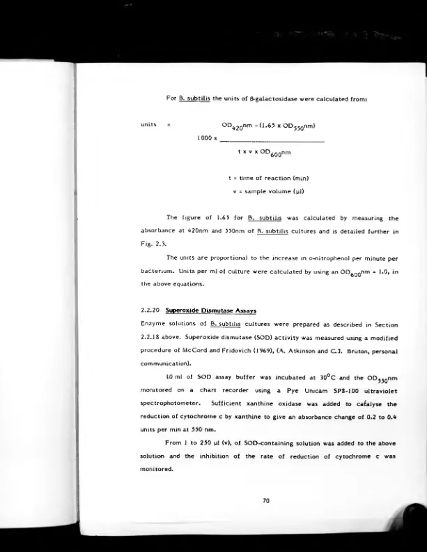
2.2.19 0-galactosidase assays 68
2.2.20 Superoxide dismutase assays 70
2.2.21 Chloramphenicol acetyl transferase
assays 72
2.2.22 B. licheniformis mutagenesis 73
2.2.23 B. licheniformis Diate UV
mutagenesis
CONTENTS
Chapter III
Chapter IV
2.2.24 Determination of reversion frequency
of B. licheniformis mutants 74
2.2.25 Analysis o f plasmid stability 75
SHOTGUN-CLONING IN B. subtUis 76
3.1 Introduction 77
3.2 Results 80
3.2.1 Attem pted shotgun-cloning o f the
B. subtilis trpC gene 80
3.2.2 Attem pted shotgun-cloning o f the
B. licheniformis 749/C penP gene 84
3.2.2.1 Employing competent
B. subtilis cells 85
3.2.2.2 Employing B. subtilis 87
protoplasts
3.3 Discussion 91
ISOLATION AND CHARACTERISATION OF THY
P3-CONTAINING PLASMIDS IN B.subtilis 94
4.1 Introduction 95
4.2 Results 97
4.2.1 Construction and characterisation
of thy P3-containing plasmids 98
4.2.2 Transformation of competent cells
with the hybrid pTT plasmids 110
4.2.3 E ffects ot restriction endonuclease
digestion on plasmid transformation 113
4.2.4 Transformation with ccc monomeric
plasmid DNA 116
4.2.5 Construction and characterisation of
CAT-containing plasmids 118
4.2.6 Transformation o f competent cells
with pTTC l 126
4.2.7 Hybridisation analysis of
antibiotic-sensitive, Thy* transformants 128
CONTENTS
Chapter V
Chapter VI
Chapter VII
Page
4.2.8 Stability of hybrid plasmids 136
4.3 Discussion 138
PENICILLINASE-0-GALATOSIDASE FUSIONS IN
E. coli AND B. subtilis 147
5.1 Introduction 148
5.1.1 Protein secretion 148
5.1.2 B-galactosidase fusions 153
5.2 Results 154
5.2.1 Construction and characterisation
of penP-lacZ fusions in E. coli 154
5.2.2 Construction and characterisation
of pTAHl 169
5.2.3 Construction and characterisation
of penP-lacZ fusions in B. subtilis 170
5.2.4 Construction and characterisation of
bifunctional plasmids containing
a penP-lacZ fusion 184
5.3 Discussion 197
B. lichen if ormis STRAIN L02 AS A HOST FOR
GENETIC MANIPULATION 201
6.1 Introduction 202
6.2 Results 204
6.2.1 Conventional UV mutagenesis of
strain L02 204
6.2.2 Plate UV mutagenesis of strain L02 206
6.2.3 Attempted induction of competence of
strain L02 mutants 208
6.2.4 Attempted transformation of protoplasts
of strain L02 211
6.3 Discussion 215
ACK NO W LED GEM ENTS
I should like to thank my supervisors Professor A. Atkinson and D r. S.B. Primrose
for enthusiastic help and constructive criticism throughout th e course of this
work. I am also indebted to my colleagues at CAMR for their frequent advice and
assistance. Many thanks to Miss Karen Miller for typing the manuscript.
Special thanks to my family for continued support and encouragement.
This project was funded by the Health and Safety Executive.
»!*•
«2
A. r . % ■ ;*5JDrThe work contained in this thesis was the result of original research conducted by
myself under the supervision of Professor A. Atkinson and Dr. S.B. Primrose. A ll
sources of information have been specifically acknowledged by means of
reference.
None of the work contained in this thesis has been used in any previous application
FIGURES
Figure 1.1: Transformation o f competent B. subtilis cells
by multimeric plasmid DNA 18
Figure 1.2: Transformation o f competent B. subtilis cells by plasmid DNA which is partially homologous with
the recipient chromosome 19
Figure 1.3: Plasmid-rescue transformation by competent
B. subtilis cells 21
Page
Figure 1.**: Properties of a generalised transcriptional unit 31
Figure 1.5: Major properties o f a generalised E. coli promoter 32
Figure 2.1: Growth of a culture of B. subtilis strain IG20
to competence 56
Figure 2.2: Standard curve for the determination o f DNA
fragment sizes 66
Figure 2.3: Absorbance o f B. subtilis cultures at 550nm and
420nm 71
Figure 3.1: Restriction endonuclease cleavage maps of the
plasmid pAB124 and the deletion derivative pAB224 79
Figure 4. 1 : Photograph showing EcoRI-digested plasmid DNAs
after agarose gel electrophoresis 99
Figure <».2: Restriction endonuclease cleavage maps of p T T l, pTT2 and pTT3 and the parental plasmids used in
their construction 103
Figure 4.3: Photograph of an autoradiograph showing
hybridisation of a pTTl probe to restriction
endonuclease-digested plasmid DNAs 109
Figure 4.4: Photograph showing restriction
endonuclease-digested plasmid DNAs after agarose gel e le ctro
phoresis 123
Figure 4.5: Restriction endonuclease cleavage maps of pTTC l
and the parental plasmids used in its construction 125
Figure 4.6: Photograph o f an autoradiograph showing hybridi sation of a 1.5Md Bglll fragment of pCDl to
restriction endonuclease-digested chromosomal DNAs 131
Figure 4.7: Photograph of an autoradiograph showing hybridi sation of a pC194 probe to restriction
endonuclease-digested chromosomal DNAs 134
Figure 4.S: Photograph of an autoradiograph showing hybridi sation of a pAB224 probe to restriction
endonuclease-digested chromosomal DNAs 135
FIGURES
Figure <*.9: Schematic diagram showing the processing of (a) monomeric and (b) muitimeric plasmid DNAs
by competent B. subtilis cells 140
Page
Figure 5.1: Amino acid sequence of some prokaryotic
signal-peptides 150
Figure 5.2: DNA sequence of the promotor-proximal
region of the B. licheniformis 749/C
penP gene 152
Figure 5.3: Predicted DNA sequence o f a penP -lacZ
fusion 156
Figure 5.4: Restriction endonuclease cleavage map of
p.MC1396 and the DNA sequence at the
5'-end o f the lacZ gene 157
Figure 5.5: Restriction endonuclease cleavage map of
pUB1660 and the DNA sequence of the
Bglll region 158
Figure 5.6: Restriction endonuclease cleavage map of
pMC1871 and the DNA sequences flanking
the lacZ gene 160
Figure 5.7: Photograph showing pEClacl, pEClac2 and
pECIac3 after agarose gel electrophoresis 164
Figure 5.8: Restriction endonuclease cleavage maps of
pEClac2 and the parental plasmids used in
its construction 166
Figure 5.9: Photograph showing restriction
endonuclease-digested pTAHl plasmid DNA 172
Figure 5.10: Restriction endonuclease cleavage maps of pTAH l and the parental plasmids used in its
construction 173
Figure 5.11: Restriction endonuclease cleavage maps of the predicted structure of the pTAHlacZ plasmids and the parental plasmids used in
their construction 175
Figure 5.12: Photograph showing the different plasmid
species present in a pTAHlacZlOl preparation 177
Figure 5.13: Photograph showing restriction endonuclease-
digested pEBlacl plasmid D N A after agarose
FIGURES
Page
Figure 5.1*»: Restriction endonuclease cleavage maps of pEBlacl and the parental plasmids used in its
construction 190
Figure 5.15: Photograph showing restriction endonuclease-
digested pEBlac3 plasmid DNA after agarose
gel electrophoresis 192
Figure 5.16: Restriction endonuclease cleavage maps of
pEBlac3 and the parental plasmids used in
its construction 193
Figure 6.1: Survival of B. licheniformis strain L02
after UV irradiation 205
Figure 6.2: Transformation of B. licheniformis strain L89 212
✓
T v » . , . “ (r • I n
---
1
TABLES
PaRe
Table 1.1: Some natural plasmids isolated from species of
Bacillus 9
Table 1.2: Some natural S. aureus plasmids introduced
into B. subtilis 10
Table 1.3: Some hybrid plasmids for use in B. subtilis 12
Table 1.4: Some E. coli-B. subtilis bifunctional plasmids 13
Table 2.1: Bacterial strains 50
Table 2.2: Plasmids 55
Table 3.1: Regeneration and transformation o f B. subtilis
IG20 protoplasts 90
Table 0.1: DNA fragment sizes of pTTl, pTT2, pTT3 and pCDl
obtained after restriction endonuclease digestions 101
Table <».2: Transformation of competent B. subtilis QB903
cells 111
Table <*.3: Transformation of competent B. subtilis QB903
cells 110
Table 0.0: Transformation of competent B. subtilis BD393
cells by monomeric plasmid DNAs 117
Table <>.5: Properties of the plasmid pBD60 120
Table ¡*.6: Construction of pTTl-pBD60 hybrid plasmids 121
Table <*.7: DNA fragment sizes of pTTCl obtained after
restriction endonuclease digestion 122
Table <».8: Transformation of competent B. subtilis BD393
cells 127
Table 0.9: Stability analysis of p T T l, pTT2, pTT3 and pTTCl 137
Table 3.1: DNA fragment sizes of pMC1871 and pUB1660
obtained after restriction endonuclease digestions 159
Table 3.2: Transformation of E. coli MC1061 cells 162
Table 5.3: DNA fragment sizes of pEClac2 obtained after
restriction endonuclease digestions 165
Table 5.0: B-Kalactosidase activity of E. coli cultures 167
Table 5.5: DNA fragment sizes of pTAHl obtained after
restriction endonuclease digestions 171
TABLES
Table 5.6: 8-galactosidase activity of B. subtilis cultures
Table 5.7: Enzyme activities o f B. subtilis IG20-pTAHlacZ101
cultures
Table 5.8: Stability analysis of B. subtilis harbouring pTAH l and pTAHlacZlOl
Table 5.9: DNA fragment sizes o f pMC1396 obtained after
restriction endonuclease digestions
Table 5.10: Transformation of E. coli MC1061 and B. subtilis IG20 competent cells with pEBlacl, pEBlac2 and pEBlac3
Table 5.11: DNA fragment sizes of pEBlacl obtained after restriction endonuclease digestions
Table 5.12: DNA fragment sizes of pEBlac3 obtained after restriction endonuclease digestions
Table 5.13: Enzyme activities of B. subtilis IG20-pEBlac3 cultures
Table 5.10: Stability analysis of pEBlac3 and the lacZ~ derivatives pEBlac3 w l-0
Table 6.1: Plate UV mutagenesis of B. licheniformis L02
Table 6.2: B. licheniformis strain L02 mutants obtained
after plate UV mutagenesis
Table 6.3: Spontaneous reversion frequency of
B. licheniformis strain L02 mutants
Tabld 6.0: Transformation of B. licheniformis strain L02
ABBREVIATIONS
ccc covalently closed circular
Md megadaltons
s
sensitive
r
resistant
p.s.i* pounds per square inch
s
Svedberg unitsAG free energy of interaction
Kcal Kilo calories
w/v weight to volume ratio
rpm revolutions per minute
hrs hours
oz ounce
OD optical density
UV ultra violet
mRNA messenger RNA
rRNA ribosomal RNA
Tc tetracycline
Pc penicillin
Ap ampicillin
Km kanamycin
Fus fusaric acid
Sm streptomycin
Cm chloramphenicol
Neo neomycin
Cad cadmium
D T T DL-dithiothreitol
O N P C o-nitrophenyl-6-D-thiogalactopyranoside
B C I G 5-bromo-ti-chloro-3-indolyl-6-D-thiogalactopyranoside
D T N B 5-i'-dithiobis(2-nitrobenzoic acid)
I P T G isopropyl ß-D-thiogalactopyranoside
E D T A ethylene diamine tetraacetic acid
SDS sodium dodecyl sulphate
U uracil
A adenine
G guanine
T thymine
C cytosine
C A T chloramphenicol acetyl transferase
S O D superoxide dismutase
The following are used in restriction endonuclease cleavage maps:
E EcoRl
a B^lll
Ba BamHI
Bs BstEII
H Hindlll
S Sail
Sm Smal
Other symbols and units used are as detailed in the Biochemical Journal (1981) 193:
1
-21
.CHAPTER I
GENERAL INTRODUCTION
Page
1.1 GENETIC MANIPULATION IN E. coli USING
PLASMID VECTORS 2
1.2 ALTERNATIVE HOST-VECTOR SYSTEMS 4
1.3 WHY BACILLI? 5
1.4 PLASMIDS IN BACILLI 7
1.5 TRANSFORMATION AND TRANSFECTION
OF COMPETENT CELLS OF BACILLI 11
1.6 TRANSFORMATION OF PROTOPLASTS
OF BACILLI 20
1.7 RESTRICTION AND MODIFICATION
IN BACILLI 22
1.8 CLONING IN BACILLI 23
1.9 EXPRESION OF CLONED GENES IN BACILLI 29
1.10 PROTEIN SECRETION IN BACILLI 37
1.1 GENETIC MANIPULATION IN E- coli USING PLASMID VECTORS
The term plasmid, originally used by Lederberg (1952) to describe all
extrachromosomal hereditary elements of bacteria, is now restricted to the
extrachromosomal, autonomously replicating, genetic elements (Broda, 1979).
Plasmids are found in a wide variety of Gram-positive and Gram-negative
bacteria where they exist within the cell as covalently closed circular (ccc) DNA
molecules in the form of supercoils (Clewell and Helinski, 1969; Blair et al., 1972).
They range in size from 1 to greater than 200 Md (Broda, 1979) and determine a
wide variety of traits.
The use of plasmids for genetic manipulation stems from two major
properties which are common to most plasmids. Firstly, they can often be
purified easily from a bacterial strain and subsequently introduced into either the
same strain or into a different strain, species or genus and secondly, many but not
all plasmids have easily selectable genetic markers, the most important being
antibiotic resistance. Such markers greatly facilitate the selection of
transformed clones.
With the discovery of site-specific restriction endonucleases (see Roberts,
1981) particularly type II enzymes (Smith and Wilcox, 1970), it became possible to
controllably and specifically manipulate DNA molecules in vitro. Type II enzymes
cleave duplex DNA to produce either fully double-stranded (flush or blunt-ended)
termini or fragments with single-stranded, self-complementary (cohesive or
sticky-ended) termini. The restriction endonuclease EcoRI was shown to produce
DNA molecules with cohesive termini after cleavage (M ertz and Davis, 1972)
which could be reannealed by the use of E. coli polynucleotide ligase (Dugaiczyk
et al.. 1975) and introduced into bacterial cells. Blunt-ended DNA fragments also
can be joined by the use of T9 polynucleotide ligase (Heyneker et al., 1976).
Other methods for linking together DNA molecules have subsequently been
developed. The enzyme terminal-deoxynucleotidyl transferase (Chang and
Bollum, 1971) can be used to add homopolynucleotide tails to DNA fragments to
be joined. A fter mixing and annealing such DNA molecules then can be used
directly to transform E. coli where repair of single-stranded gaps occurs in vivo
(e.g. Hutchison and Halvorson, 1980). Synthetic DNA fragments (linkers) also can
be used for linking together DNA molecules (e.g. Maniatis et al., 1978).
The choice of a vector in which to clone foreign DNA is principally
determined by four factors. Firstly, the vector DNA must be able to infect a
suitable host organism and replicate within that host. Secondly, the vector must
possess at least one but preferably two selectable markers such as those
conferring resistance to antibiotics. These markers enable identification of host
cells which have taken up the required DNA molecule. Thirdly, the vector must
possess suitable restriction endonuclease sites for the insertion of foreign DNA;
ideally a unique site which lies within'an antibiotic-resistance gene. By cloning
foreign DNA into a site which lies within a second marker, it is possible to screen
transformants for those which harbour recombinants. The undamaged marker is
used to select transformants which then can be screened for loss of the second
marker. Fourthly, insertion of foreign DNA must not impair essential functions
such as control of plasmid replication.
To date most work involving the manipulation o f DNA molecules has made
use of E. coli K12 and its associated plasmids and bacteriophages. The major
reason for this is that our genetic and biochemical knowledge of this organism is
fa r greater than that of any other. This bacterium provided the basis for the
classical genetic studies of Jacob and Monod and much o f our understanding of
gene structure and gene regulation stems from work carried out using E. coli.
Furthermore, simple methods for introducing DNA into E. coli cells have been
developed (e.g. Mandel and Higa, 1970; Cohen et al.. 1972). A large variety of
plasmid and bacteriophage vectors are available for molecular cloning in this host
and its derivatives such as pAT153 (Twigg and Sherratt, 1980) whereas
bacteriophage X (for review see Brammar, 1979) is the most widely used
bacteriophage vector.
In summary, the discovery of restriction endonucleases and other DNA
modifying enzymes has made the rearrangement of DNA molecules in vitro a
relatively simple procedure. Fragments of DNA from any source can be
covalently joined to a suitable vector molecule and introduced into a bacterial
cell. Cells containing a particular DNA fragment then can be isolated and further
investigated as required. To date most of the developments in genetic
manipulation have been made in E. coli. However, more recently other organisms
have been investigated with the aim of developing genetic manipulation systems
similar to those developed for use in E. coli.
1.2 ALTERNATIVE HOST-VECTOR SYSTEMS
The host-vector systems of E. coli have a wide range of useful features but are
unlikely to be ideally suited for every purpose. Furthermore, E. coli may not
prove to be the most suitable organism for the production of cloned gene products
on an industrial scale since it has several unwanted attributes.
There has always been much controversy about the use of E. coli as a host
for genetic manipulation (e.g. Crobstein, 1977) since it is a normal inhabitant of
the alimentary tract of man and domestic animals. Most strains of E. coli
produce a lipopolysaccharide endotoxin which makes its use as a host to produce
pharmaceutical products, such as interferon, possibly undesirable. The
development of crippled strains such as X 1776 (Curtiss et al„ cited in Brammar,
1979) reduces the risk of infection and proliferation in the gut but does not avoid
the problem associated with endotoxin production. The development of genetic
manipulation techniques which are applicable to other organisms greatly
facilitates the study of both the genetics and biochemistry of that organism and in
addition, problems with E. coli may not be present in other systems.
Plasmids, bacteriophages and viruses have been isolated from, and genetic
exchange systems have been developed for a large number o f both prokaryotic and
eukaryotic species; these are the two basic attributes that are required for the
development of a useful genetic manipulation system. Among the prokaryotes, in
addition to the Bacilli, systems have been developed for organisms such as
Streptococcus (Stassi et al., 1981), Staphylococcus (Wilson and Baldwin, 1978),
Streptomyces (Bibb et__ al., 1980), Methylophilus (Hennam et__ al., 1982),
Haemophilus (Setlow et al., 1981), Salmonella (Lederberg and Cohen, 197*0 and
Pseudomonas (Sakaguchi, 1981). Among the eukaryotes systems have been
developed for the yeast Saccharomyces (e.g. Gerbaud et al., 1979), the fungus
Neurosporra (Vapnek and Case, 1981), animal ce lls (e.g. Mulligan and Berg, 1980)
and plant cells (for review see Maheshwari et a l., 1980). With further advances,
cloning systems in organisms other than E. coli will undoubtedly assume a much
more important role in the future.
1.3 WHY BACILLI?
B. subtilis is a Gram-positive, aerobic, soil bacterium and other than E. c o li is the
most widely studied prokaryotic organism (for reviews see Priest, 1977; Henner
and Hoch, 1980; Young, 1980). Studies have focussed on many aspects of this
organism including its biochemistry, physiology and genetics and a large number
of mutants have been isolated and characterised. Also, transformation and
transduction studies have led to the construction of a linked genetic map
(Lepesant-Kejzlarova et al.. 1975; Henner and Hoch, 1980). Several o f these
advances have laid the groundwork for the development of B. subtilis as a suitable
host for the cloning and expression of foreign genes.
Recently many studies on the regulation of gene expression during
sporulation have made use of genetic manipulation techniques (Jayaraman et al..
specific sporulation genes will give an understanding of the nature and function of
their products.
The Bacilli already are widely used for the industrial production of a wide
range of products such as antibiotics, insecticides, and enzymes and the large
scale fermentation requirements of Bacilli are well known. Genetic manipulation
now offers a new approach for the improvement of com m ercially important
strains in addition to the construction of strains producing novel products. The
ability of Bacilli to secrete proteins into the culture medium is of industrial
importance also. Currently greater than U0 extracellular enzymes are produced
commercially from Bacilli (Priest, 1977) and it is hoped to tailor strains to secrete
cloned gene-products such as interferon.
The Bacilli have several advantages over E. coli in terms of safety.
Unlike Gram-negative bacteria the Bacilli have a simple cell surface composed of
teichoic acid and peptidoglycan neither o f which are pyrogens. Although
B. subtilis can exist on human skin, it is a non-pathogenic organism and there are
no reports to date o f it causing disease in hosts that are not compromised by pre
existing disease processes (Ehrlich, 1978b). Also, unlike E. coli which can readily
transfer genetic material to many other species of Enterobacteriacae, B. subtilis
does not readily transfer genetic material to other species of Bacillus. Some
thermophilic species of Bacillus have an added safety feature in that they are
incapable of growth at temperatures below i»0°C and hence cannot grow at human
body temperature (A . Atkinson, personal communication). Human infections with
such species have never been reported.
The wide use of B. subtilis on an industrial scale results in a massive
release of bacteria into the environment without any adverse e ffe c ts having been
observed. Indeed, because of the bright orange colour of B. globigii colonies, and
the resistance of their spores, this organism is used as a biological tracer to
monitor air and water currents (A. Atkinson, personal communication). Also,
B. subtilis var natto is consumed in vast quantities in the orient in the form of
natto, a vegetable cheese produced by the fermentation o f boiled soya beans.
High levels of oral ingestion of this m aterial by humans have been reported not to
cause any ill e ffe c ts (Ehrlich, 1978b).
A disadvantage of using the Bacilli as hosts is that they can persist in the
environment for long periods of time in the form of spores (Roberts and Hitchins,
1969). If bacteria containing recombinant DNA which renders the host a
biohazard w ere to escape into the environment, this could present a problem due
to the persistence of spores. Asporogenic mutants of B. subtilis, which autolyse
when the cells reach the stationary phase of growth, have been isolated (Brown
and Young, 1970) and multiply auxotrophic strains have been constructed (Young,
1980). It is hoped that the use of such strains will overcom e the problems of
persistence in the environment of accidentally released organisms.
In addition to B. subtilis several other species of Bacillus may be useful
for the cloning and expression of foreign genes. Transformation, by plasmid DNA,
has been reported for B. megaterium (Vorobjeva et at., 1980; Brown and Carlton,
1980) ; B. thuringiensis (Alikhanian et al„ 1981; Martin et al.. 1981; Miteva et al„
1981) ; B. licheniformis (Imanaka et al., 1981b) and B. stearothermophilus (Imanaka
et al., 1982). In addition plasmids have been transferred to B. megaterium.
B. licheniformis and B. polymyxa by fusion of protoplasts o f these species to
B. subtilis protoplasts harbouring the S. aureus plasmid pC221 (Dancer, 1980). The
ability to transfer plasmid DNA into the above mentioned strains will allow
studies on the expression of cloned genes within these hosts. The commercial
usefulness of these strains then may be improved by genetic manipulation
techniques.
0.1 PLASMIDS IN BACILLI
Since the initial discovery of plasmid DNA in B. megaterium (Carlton and
Helinski, 1969) many plasmids indigenous to the Bacilli have subsequently been
Halvorson, 1980) to a ISOMd plasmid isolated from B. thuringienis (Lereclus et
al., 1982). Most of the Bacillus plasmids isolated appear to be cryptic in that they
confer no known phenotype on their host (e.g. Bernhard, et al., 1978) but
phenotypic traits such as bacteriocin production (Bernhard et al., 1978) have been
assigned to some plasmids. More recently antibiotic-resistance plasmids have
been isolated from several species of Bacillus and been shown to replicate and
express antibiotic-resistance in B. subtilis (e.g. Bingham et al., 1979). Table 1.1
lists some o f the antibiotic-resistance plasmids isolated from species of Bacillus.
Although several of these indigenous Bacillus plasmids have been further
developed as vectors for molecular cloning (e.g. K re ft et al., 1978), the use of
antibiotic-resistance plasmids from the Gram-positive bacterium Staphylococcus
aureus has proved more successful. Several small antibiotic-resistance plasmids
from S. aureus have been shown to transform B. subtilis (e.g. Ehrlich, 1977),
undergo autonomous replication and express 'their antibiotic-resistance markers.
Table 1.2 summarises the properties of some such plasmids.
Not all the S. aureus plasmids transformed into B. subtilis have behaved in
a similar manner. Gryczan et al. (1978) reported that pUBlOl (Penr, Cadr, Fusr in
S. aureus), pK595 (K m r in S. aureus) and pSH2 (Km r in S. aureus) could repeatedly
transform competent B. subtilis cells to Fusr, Km r and Kmr respectively.
However, extrachromosomal plasmid DNA could not be isolated from such
transformants. It was suggested by these workers that chromosomal integration
may have occurred in each case. In contrast Erlich (1977) reported that pK5<*5
and pSl77 (Smr in S. aureus) failed to produce Kmr and Smr B. subtilis
transformants respectively. Also the plasmid pTP2 (T cr, Pcr in S. aureus)
fragmented when introduced into B. subtilis to give either Tcr or P cr
transformants (Kono et al., 1978).
Several of these natural plasmids isolated from S. aureus have proved to
be useful vectors for molecular cloning directly in B. subtilis. particularly pUBllO
(Keggins et al., 1978; McDonald and Burke, 1982) and pC194 (Michel et al.. 1980;
[image:24.682.30.646.54.871.2]Plasmid
Size (Md)
Unique restriction endonuclease sites
Plasmid-borne
markers Reference pPL576 30.0 Involved in
sporulation
Lovett, 1973
pPLI «.7 Cryptic Lovett & Bramucci, 1975 pPL2 «6.3 Cryptic
pMBl 7.0 Cryptic
pV1B2 3.6 Cryptic pABl ISA ». 9 BamHI» Sail» Kpnl,
Xbal. Xmal
Cryptic Bingham et al., 1979 pABllSB 3.0 BamHI, Kpnl. Xbal Cryptic
pABl 2*1 2.9 Xbal. Hpal, Caull Tc-resistance pAB128 2.5 Bgll, Hpal, Caull Cryptic
pBC16 2.8 BamHI Tc-resistance Bernhard et al.» 1978 pIM 1 3 1.3 Hhal, Sacl Em-resistance Mahler and Halvorson, 1980 pTBl 1-pTBlS Cryptic Imanaka et al.» 1981a pTB19 17.2 BamHI Tc, Km-resistance
pJP3623 3.3 Tc-resistance Polak & Novick, 1982 pJP3633 3.1 Tc resistance
Size Unique restriction Plasmid-borne
Plasmid (Md) endonuclease sites markers Reference pCI94 1.8 Hindlll, Haelll,
Hpall, Hhal, BrIII, Mspl
Cm-resis»ance Ehrlich, 1977;
Wilson & Baldwin, 1978; Horinouchi & Weisblum, 1982a pC221 3.0 Hindlll. BstEII,
EcoRI
Cm-resistance pC223 3.0 Hindlll Cm-resistance pUBl 12 3.0 Hindlll Cm-resistance pT127 2.0 Tc-resistance pUBllO 3.0 BamHI. Bglll, EcoRI,
Xbal. A val. Pvull
Neo-resistance Gryczan et al., 1978 Lofdahl et al., 1978 pSA2IOO k . 7 EcoRI. Haelll. Xbal.
Hindlll
Cm, Sm-resistance pSAOJOl 2.8 EcoRI. Hindll,
Hindlll. Xbal
Sm-resistance pEI9<t 2.« Hpal. Pstl, Bell.
Xbal. Haelll
Em-resistance Horinouchi & Weisblum, 1982b pTP2 2.8 EcoRI Pc, Tc-resistance Kono et al.. 1978
pTP9 2.8 Hindlll Cm-resistance
TABLE 1.2s Some natural S. aureus plasmids introduced into B. subtilis
Rutberg et al., 1981). However, more versatile cloning vectors have been
developed from naturally isolated plasmids by recombination in vitro. Table 1.4
lists some of those hybrid vectors that can replicate in both B. subtilis and E. coli
and Table 1.3 lists some of those which cannot replicate in E. c o li. Examples of
the uses of some of these vectors will be given later, (section 18)
1.5 TRANSFORMATION AND TRANSFECTION OF COMPETENT c p i rLS
OF BACILLI
Unlike E. coli for which competence is an a rtificially induced state, several
species of Bacillus develop a natural physiological state in which they are capable
of absorbing DNA from the surrounding medium (Spizizen, 1958). Our current
understanding of the mechanism of transformation of competent Bacillus cells
stems from work with B. subtilis using mainly chromosomal DNA as donor
molecules. However, transfecting DNA and plasmid DNA, although intracellularly
processed in a different manner, probably share at least part of the same binding,
uptake and processing machinery.
Chromosomal DNA Transformation
The following is a summary of our understanding of chromosomal DNA
transformation of competent B. subtilis cells (for review see Dubnau, 1976). Upon
addition of double-stranded chromosomal DNA to competent cells the DNA
becomes bound to the competent cell surface at a few points along the length of
each DNA molecule (Dubnau and Cingliano, 1972b). The DNA undergoes
fragmentation at the cell surface to yield double-stranded molecules o f 10 to
20 Md (Dubnau and Cirigiiano, 1972a; Arwert and Venema, 1973) and a single
strand o f chromosomal DNA is taken up. There is concomitant hydroylsis of the
homologous strand to a low molecular weight form and its release into the culture
medium (Piechowska and Fox, 1971; Dubnau and Cirigiiano, 1972a; D avidoff-
Plasmid Size (Md) Unique restriction endonuclease sites Plasmid-borne markers Reference pBD6 3.1 BainHI. Tael, Bell.
Hindlll
Km, Srn-resistance Cryczan et ah. 1978, 1 pBD8 6.0 EcoRI. Hindlll.
Bglll. BainHI. Xbal
Km, Sm, Crn-resistance pBU9 3.6 EcoRI. BamHI. Tael,
Belli. Bell, Hpal. Pstl
Km, Em-resistance
pBDIO 6.« Belli. BamHI. Xbal. Hpal. Bell
Km, Cm, Em-resistance p B D Il 4.0 Xbal. BamHI. Belli.
Hpal. Bell
Km, Em-resistance pBOI2 4.3 EcoRI. Xbal, BamHI,
Tael. Belli. Hindlll
Km, Cin-resistance pBD6<* 3.2 EcoRI. Xbal. Tael,
BamHI. Bell
Km, Cm-resistance
p B C I6-l 1.8 EcoRI Tc-resistance K reft et al.. 1978 pBSlbl-1 2.3 EcoRI. Pstl. Hindlll Tc-rcsistance
pAB224 2.0 EcoRI. BstEII, Caull. Hpal. Hpall. Thai Hhal
Tc-resistance Bingham et al.. 1980
pAB324 2.3 Hpal. Hpall. Caull. BstEII
Tc-resistance pTL 12 6.4 EcoRI, Belli. BamHI.
Sinai. Xmal
leu A, B Tp- resistance
Tanaka & Kawano, 1980 pTB33 11.2 BainHI. Hindlll Kin, Tc-resistance Imanaka et al.. 1981a pKOlOl 6.3 BaniHI. Xbal. Belli Neo, SI-resistance McDonald & Burke, 1982 pBO90 4.6 Hindlll Cm, Em-resistance Docherty et al.. 1981
Plasmid-•borne antibiotic-Size Unique restriction resistance markers
Plasmid (Md) endonucleasc sites E. coli B. subtilis Reference pHV12 7.9 Cm, Ap Cm Ehrlich, 1978a pHVI6, 1) «.6 A vai. Panini. Sali, Cm, Ap Cm
EcoRI. P ili, l’ vull
pHVl6 9.7 Cm, Ap Cm pH V I8 Crn, Ap, Cm
Km
pJK3 3.1 Sali, BarnHl Tc, Ap Tc K re lt et al.. 1978 pJK20l 2.6 BamHl, Hindlll, Te, Cm Tc Goebel et al., 1979
Sali. EcoKl
pJK501 7.9 IM I. Sali, Pallili! Tc Tc P-JK302 6.3 Illudili. Sali. IM I, Tc Tc
BamHl
pHV23 6.1 Avai, Bamlll, EcoKl, Ap, Cm, Tc, Cin Michel et al.r 1980 Hpal. Kpnl. Pst,l, Tc
Pvull, Sali, Xbal
pTA13U2 3.3 Illudili. EcoKl Cm, Ap Cm Sakaguchi et al.. 1981 pHV33 6.6 P ili, EcoKl. Bamlll,
Sali
Tc, Cm, Ap
Cm Primrose 4 Ehrlich, 1981
pJK 3.1 P iti. Sali, Illudili. BamHl. EcoKl
Tc Tc K re lt 4 Hughes, 1981 p JK 302 Illudili. BamHl. Sali,
P ili,
Tc, Ap Tc POG2I63 3.0 Bulli. Illudili. Piti,
S ili
Cm, Ap Cm, Ap Gray 4 Chang, 1981
Strauss, 1970; Rudner et al., 1968) but some of the ends of the DNA molecule are
lost or otherwise made unavailable for subsequent recombination events (Guild et
al., 1968).
The single-stranded donor DNA rapidly interacts with a homologous
segment of recipient DNA to form an unstable complex (Davidoff-Abelson and
Dubnau, 1973) which then becomes stabilised by base-pairing (Buitenwerf and
Venema, 1977; 1978). A fter form ation of the nascent donor-recipient complex the
donor DNA becomes entirely base-paired to the homologous recipient D N A and
eventually becomes covalently attached, replacing the equivalent recipient
segment (Arwert and Venema, 1973; Davidoff-Abelson and Dubnau 1973). Finally
expression of the donor genetic material results following segregation o f the
heteroduplex.
The overall process of chromosomal DNA transformation requires a high
degree of donor-recipient DNA sequence homology. Transformation of B. subtilis
with chromosomal DNA from closely related species occurs, but at a much lower
frequency for most genetic markers (Wilson and Young, 1972). A functional recE4
gene-product, which is involved in recombination events, is also essential (Dubnau
et al., 1973; Prozorov et al., 1982).
Investigations into the mechanism of transformation of competent cells
by chromosomal DNA have exclusively involved B. subtilis. However, competence
transformation systems have been developed for other species of Bacillus, namely
B. amyloliquefaciens (Coukoulis and Campbell, 1971); B. licheniformis (Gwinn and
Thorne, 1964; Thorne and Stull, 1966); B. thuringiensis (cited in Martin e t al.,
1981) and B. caldotenax (M. Munster, personal communication) where similar
mechanisms of transformation may operate.
Bacteriophage DNA Transfection
Transfection is the process whereby cells are infected by naked nucleic acid from
a virus resulting in the production of a complete virus. The mechanism of
transfection has been widely investigated using several B. subtilis bacteriophages
and has proved to be a useful system for studying the uptake and processing of
D N A by competent B. subtilis cells (for review see Trautner and Spatz, 1973).
Competence for both chromosomal D N A transformation and transfection
develop with a similar time course (Trautner and Spatz, 1973) and, as with
chromosomal D NA, transfecting DNA is processed to single-stranded
intermediates during uptake (Loveday and Fox, 1978; Dishman, 1972, cited in
Contente and Dubnau, 1979a). Following uptake, repair of damaged transfecting
D N A occurs by a recombination event (Trautner and Spatz, 1973) and depending
on the bacteriophage DNA used, from 2 to 9 molecules are required to produce a
successful infection. Therefore transfection exhibits a 2nd to 9th order dose-
response curve. Unlike chromosomal DNA transformation, transfection is a very
inefficient process which could result from heavily damaged DNA molecules being
unable to participate in a successful repair event (Trautner and Spatz, 1973;
Contente and Dubnau, 1979a).
Plasmid DNA Transformation
Investigations into the uptake and processing of DNA molecules by competent
B. subtilis cells using chromosomal DNA have been hindered by the size and
sequence heterogeneity of the transforming DNA and also the large size of the
recipient chromosome. Therefore transformation using plasmid DNA has been
investigated both as a model of chromosomal DNA transformation and also
because an understanding of plasmid DNA transformation is advantageous for
genetic manipulation in the Bacilli.
All plasmid DNA molecules are probably taken up into competent cells by
the same mechanism. However, subsequent processing events may d iffer
depending on the nature o f the transforming plasmid and also on whether the
Firstly consider the case where the recipient does not contain a
homologous resident plasmid and the transforming plasmid has no homology with
the recipient chromosome. The follow ing observations have been made of plasmid
DNA transformation of competent B. subtilis cells. Plasmid DNA transformation,
like chromosomal DNA transformation is a first order process (Contente and
Dubnau, 1979a) suggesting that a single plasmid molecule is sufficient for a
successful transformation event or that the interaction o f a cell with a single
plasmid molecule is rate limiting. However, in contrast to chromosomal DNA
transformation, plasmid DNA transformation does not require the recEfr gene-
product (Gryczan et al., 1978) and is a very inefficient process (Contente and
Dubnau, 1979a). The addition of 10^ to 10^ molecules per transformant is
required with an average uptake of 10^ to 10** molecules per competent cell.
Linear and nicked plasmid DNAs, in addition to ccc monomeric plasmid DNA, are
not active in the transformation of competent cells (Ehrlich, 1977; Canosi et al.,
1978; Contente and Dubnau, 1979a), multimeric forms of plasmid DNA alone are
responsible for all the transforming a ctivity of a plasmid preparation.
Although the exact mechanism of transformation o f competent B. subtilis
cells by plasmid D N A is not known, the following model has been put forward (de
Vos et al., 1981). The model proposes that the entry of plasmid DNA is analogous
to the entry of chromosomal DNA. A double-stranded scission of the plasmid
DNA occurs and one strand is taken up whereas the other strand is degraded to
acid soluble products. There is extensive intracellular breakdown of entered DNA
resulting in the release of acid-soluble material. The degree o f polymerisation of
the transforming plasmid affects subsequent processing steps. Whereas
transformation w ith monomeric species of plasmid DNA does not lead to the
production of a transformed cell, transformation with multimeric plasmid DNA
does. It is assumed that complementary single strands produced by
transformation w ith a multimeric plasmid molecule, form partially double-
converted to fully double-stranded monomer-length plasmid molecules by an ill-
defined process. A model of plasmid DNA transformation of B. subtilis competent
cells is summarised in Figure 1.1.
Secondly, consider the case where the transforming plasmid contains a
piece of D N A which has homology with a region of the recipient chromsome or
even a resident prophage; such DNA can be processed by a d ifferen t mechanism to
that described above. However, multimers of such plasmids may be processed as
described in Figure 1.1. Monomeric plasmid molecules of this type are active in
the transformation of B. subtilis competent cells but only if the host cell is recEfr-
proficient (Bensi et al., 1981; Canosi et al., 1981). It has been suggested that
repair of damaged plasmid DNA can occur by a recEfr-dependant recombination
event involving base-pairing of homologous regions of the transforming plasmid
and host chromosome. This occurs providing that the damage is within the
homologous region of the plasmid. Figure 1.2 is a model summarising this event.
If additional DNA fragments are inserted into the homologous region of such
plasmids then plasmid rearrangements may occur upon transformation of recEfr-
proficient competent cells (Iglesias et al., 1981). "C orrected" plasmids are
produced which have the configuration of the plasmid into which a given
alteration was initially introduced.
Thirdly, if the recipient harbours a plasmid which has homology with the
transforming plasmid, then plasmid DNA is processed by a differen t mechanism to
those described above. Contente and Dubnau (1979b) observed a ten-fold increase
in plasmid transformation frequency when the recipient contained a homologous
resident plasmid compared with strains which were plasmid-free or contained a
non-hornologous plasmid. Also, markers on linear plasmid molecules could be
"rescued" providing that the linearising cut was within the homologous region of
the transforming plasmid and from 0.2 to 0.5 Md of the junction between the
homologous and non-homologous regions of the plasmid. This process was found to
Figure 1.1: Transformation of competent B. s u fatilis c e l l s by
multimeric plasmid DNA (adanted from Canosi et a l . , 1981) 1 Plasmid DNA binds to the comnetent c e l l surfa ce .
2. 3 Uptake o f s in g le -stra n d ed plasmid DNA.
4 Base pairinn o f complementary s in g le -stra n d ed plasmid DNA strands.
5, G C irc u la r is a tio n t o form f u l l y double-stranded , monomer- length plasmid DNA.
[image:34.690.24.667.74.867.2]Figure 1 . 2 : Transformation o f competent B. s u b t i l l s c e l l s by plasmid DNA which is p a r t i a l l y homologous w it h the r e c ip ie n t chromosome (adapted from Canosi e t a l ■. 1981) 1 Plasmid DNA binds to the competent c e l l su r fa ce . 2. 3 Uptake o f sing le -stra n d ed plasmid ONA.
4 Base pairinq o f plasmid DNA with complementary region o f the r e c ip ie n t chromosome.
[image:35.683.28.661.75.872.2]The process of plasmid-rescue transformation has been modified to permit
shotgun-cloning of heterologous chromosomal genes in B. subtilis (Gryczan et al..
1980a). This process relies on repair of damaged plasmid molecules by
recombination between a homologous resident plasmid and the transforming
hybrid plasmid. Figure 1.3 is a model showing how plasmid-rescue transformation
probably occurs.
1.6 TRANSFORMATION OF PROTOPLASTS OF BACILLI
Following work on the regeneration and fusion o f Bacillus protoplasts (e.g.
Landman et al., 1968; Fodor and A lfoldi, 1976; Schaeffer et al.. 1976), Chang and
Cohen (1979) have developed a PEG-induced protoplast transformation system for
B. subtilis. These workers reported that up to 80% of a population of protoplasts
could be transformed with plasmid DNA with an efficien cy of greater than 107
transformants per pg of DNA. Although Chang and Cohen (1979) could not detect
transformation of B. subtilis protoplasts with chromosomal D N A, low frequency
transformation with chromosomal DNA has subsequently been reported (L
evi-Meyrueis et__ al., 1980). Also transformation of B. subtilis L-forms by
bacteriophage DNA has been reported (White et al., 1981).
In contrast to the B. subtilis competence plasmid transformation
procedure, the PEG-induced protoplast transformation procedure does not have a
requirement for multimeric plasmid DNA; ccc, open circular and linear
monomeric plasmid molecules also are active in transformation although open
circular and linear molecules are one to three orders of magnitude less effic ie n t
in transformation (Chang and Cohen, 1979). de Vos and Venema (1981)
demonstrated that plasmid DNA entered protoplasts in a double-stranded form
and that the entered DNA was present predominantly as ccc DNA molecules. The
efficien cy of plasmid DNA transformation was found to be close to one indicating
that each entered plasmid molecule could give rise to a transformed cell. This
supported the observations made by these workers that little, if any, damage was
Plasmid
A Homologous _______
res id ent --- D t A
plasmid
c
1
c
Figure 1 . 3 : Plasmid-rescue transformation by competent B. s u b t i l i s c e l l s (adapted from Contente and Dubnau, 1979b and Gryczan e t a l . . 1980a). 1 Plasmid ONA binds to the competent c e l l surface.
2. 3 Uptake o f s in g le -st ra n d ed plasmid DNA.
4 I n t e r a c t io n o f r e s id e n t plasmid with homologous reg io n o f the transforming plasmid.
[image:37.687.28.660.85.859.2]done to plasmid DNA upon entry into protoplasts. Thus, PEG-induced protoplast
transformation has two major differences to simple competence plasmid
transformation. Firstly, protoplast transforamtion is much more efficient and
secondly, it does not have a requirement for multimeric plasmid DNA.
In summary, although naturally induced competence has been widely used
to introduce plasmid DNA into B. subtilis cells, this method is not generally
applicable to other species of Bacillus. Plasmid DNA transformation of
competent cells has been reported for only one other species of Bacillus,
B. licheniformis (Docherty et__al., 1981). However, plasmid DNA has been
introduced into protoplasts of several species of Bacillus including B. megaterium
(Brown and Carlton, 1980), B. licheniformis (Bingham, 1980; Imanaka et al.,
1981b), B. thuringiensis (Alikhanian et al., 1981; Martin et al., 1981; Miteva et al.,
1981), and B. stearothermophilus (Imanaka et al.. 1982) and therefore may be
applicable to many other species of Bacillus.
1.7 RESTRICTION AND MODIFICATION IN BACILLI
Restriction endonucleases have been isolated from (see Roberts, 1981) and
restriction and modification systems probably operate in many species of Bacillus.
Bron et al., (1980a) and Trautner et al. (1979) have shown that in transformation
with chromosomal DNA and in transfection o f lysogenic recipients, restriction of
donor DNA does not occur providing that it is homologous with the recipient DNA.
Conversely, restriction of bacteriophage DNA does occur upon transfection o f a
non-lysogenic strain (Trautner et al.. 1979; Bron et al., 1975; 1980a, 1980b).
Plasmid DNA is restricted upon transformation of competent B. subtilis cells
(Prozorov et al., 1980; Tanaka, 1979b) but if the transforming plasmid has
homology with a resident plasmid in the recipient, restriction is not observed
(Canosi et al., 1981). Also if a chromosomal DNA fragment is inserted into a
plasmid, this fragment is not sensitive to restriction whereas the vector region of
the plasmid is (Canosi et al., 1981).
Bron et__ al. (19S0a) have postulated that in chromosomal DNA
transformation and in systems where there is homology between the donor and
recipient DNAs, pairing of the donor moiety with a modified recipient D N A strand
occurs. This results in the formation of a restriction-resistant heteroduplex. In
the absence of DNA homology single-stranded donor molecules can anneal upon
themselves to yield restriction-sensitive homoduplexes. The above can be used to
explain the observations made also with transfecting and plasmid DNAs, i.e.
restriction is not observed if the transforming DNA has homology with host DNA.
Restriction of plasmid DNA upon transformation of Bacillus protoplasts
has been reported for some species (Bingham, 1980; Vorobjeva et_aj., 1980;
lmanaka et al., 1982) but not others (Chang and Cohen, 1979; Alikhanian et al.,
1981). However, if restriction of plasmid DNA does prove to be a problem, it may
be possible to isolate restriction-deficient mutants such as B. subtilis strain IG20
(trpC2, hsm", hsr”).
1.8 CLONING IN BACILLI
For several years the Bacilli, particularly B. subtilis, have been used as hosts for
gene cloning and although problems have been encountered, many successful
cloning events have been reported in the literature (e.g. Gryczan and Dubnau,
1978). Suitable cloning vehicles are a prerequisite for DNA cloning in any
organism and therefore many early reports detailed the construction o f hybrid
plasmids for use in B. subtilis (see Table 1.3). Plasmid cloning vehicles have been
developed along similar lines to those developed for use in E. co li; mainly by the
in vitro linkage of 2 or more plasmid molecules. A large number o f hybrids have
been constructed (e.g. Gryczan and Dubnau, 1978; Gryczan et al., 1980b) and some
have been shown to possess several unique restriction endonuclease sites which
can be used to clone foreign DNA. In addition, in some cases insertional
inactivation of plasmid-borne markers has been demonstrated (e.g. Gryczan et al.,
Although the construction of hybrid plasmids has been a relatively simple
process, shotgun-cloning of chromosomal genes has proved to be far more
difficu lt. Using the naturally occuring S. aureus plasmid pU BllO, Keggins et al.
(1978) reported the first successful shotgun-cloning of chromosomal genes in
B. subtilis. However, following this initial report, many workers have had great
difficulty in shotgun-cloning chromosomal genes directly in B. subtilis employing
competence transformation procedures (Gryczan et al., 1980a; A.J.P. Docherty
and A.H.A. Bingham, personal communications). The concensus of opinion now is
that the failure to readily shotgun-clone chromosomal genes directly in B. subtilis
is due to the inherent properties of the competence transformation process of this
species. Gryczan et al. (1980a) reported that when foreign DNA was ligated to a
plasmid vector, the yield of transformants was markedly reduced and that the
extent of the reduction was greater than that expected solely from competition at
the level of DNA uptake. This probably occurred because as the ratio of insert
DNA to plasmid DNA increases, the number of vector oligomers decreases on
subsequent ligation. Therefore, attem pting to increase the number of hybrids
decreases the number of plasmid oligom ers produced. As reported by Canosi et
al. (1978), only plasmid oligomers are a ctive in the transformation o f competent
cells and hence, increasing the frequency of hybrid plasmids decreases the
transformation frequency of a plasmid-chromosomal DNA mix.
Although it appeared that shotgun-cloning utilising competent B. subtilis
cells was extremely difficult, recently several reports describing the successful
shotgun-cloning of various chromosomal genes have appeared. Jayaraman et al.
(1981) cloned the B. subtilis spoOB and pheA genes using pUBllO. However, the
spoOB and gheA genes, which were shown to reside on the same BamHI fragment,
were partially purified by agarose gel electrophoresis prior to cloning. Selection
was made for pheA * transformants, which were screened for a spoOB'*' phenotype.
McDonald and Burke (1982) have cloned a B. subtilis chromosomal sulfanilamide-
resistance gene and Palva (1982) has cloned the B. amyloliquefaciens a-amylase
gene using pU BllO. In the latter case no direct selection for a-amylase a ctivity
was applied, instead Neor transformants w e r e screened for a-amylase production.
A systematic study of random-segment cloning in B. subtilis using
competent cells was made by Michel e t al. (1980). Random fragments of
B. amvloliquefaciens chromosomal DNA w e re cloned in pHV33 and pC19U. A fter
selection for Cmr transformants, clones were screened for the presence of
chromosomal DNA fragments. Y ields of 16% and 18% of transformants
containing inserts were obtained respectively. The average size of the cloned
DNA fragments was 1.1 and 0.9 Md respectively with sizes ranging from 0A to
2.0 Md. This compared to an average size of the starting insert DNA of 3 Md with
sizes ranging from less than 0.*» Md to greater than 12 Md. Also, M ichel et al.
(1980) used the insertional inactivation vectors p H V ll and pHV23 to clone
S. cerevisiae chromosomal DNA fragm ents. A fter initial selection for Cm-
resistance the transformants were screened for Tc-resistance. 11.0% and 12.5%
of the p H V ll and pHV23 Cmr transformants respectively were found to be Tcs.
Two Cmr , T c s clones examined were found to contain an insert indicating that
these insertional inactivation vectors can be used for random-segment cloning. In
summary, although a high frequency o f insertion was found, the average size of
the inserts was small compared to the size of the starting m aterial. These
workers suggested that the small size o f the cloned segments may explain why
other workers have had difficulties in isolating active genes by random-segment
cloning in B. subtilis.
Although Michel et al. (1980) did not determine how and why small
fragments of DNA were preferentially cloned, they suggested several reasons why
this may occur. The hybrids detected may result from transformation by plasmid
molecules having internal repeats of the cloning vector. Intracellular processing
may then occur yielding hybrids w ith small inserts. Alternatively, fragments
which do not contain recognition sites for the hosts restriction system may be
of com petent cells is a ffected by the restriction system of the host. If restriction
does prove to be a problem then restriction deficient hosts are available to
circum vent this problem. A third explanation for the cloning of small DNA
fragments is that large DNA fragments may be unstable and subject to deletion.
If this is the case then it is difficult to imagine how such a problem could be
overcom e.
It is evident from the above that much more work needs to be done to
determine the exact mechanism of, and the parameters that govern the cloning of
random D N A fragments in B. subtilis before further progress can be made.
Because o f the difficulties encountered with cloning directly in B. subtilis
employing competent cells, other approaches have been investigated and are
detailed below.
A successful system for the shotgun-cloning of heterologous DNA
fragments has been developed by Gryczan et al., (1980a). This stratagem was
based on the findings of Contente and Dubnau (1979b) who showed that
transformation of competent B. subtilis cells by linear, or nicked plasmid DNA
occurred providing that the host carried a homologous resident plasmid and was
recE*»-proficient. Gryczan et al., (1980a) suggested that a damaged vector DNA-
chrornosomal DNA hybrid plasmid molecule could be rescued by recombination
with a homologous resident plasmid if the damage to the transforming plasmid
was within the homologous sequences. The method was demonstrated by cloning
several B. licheniformis chromosomal DNA segments which complemented
B. subtilis trp and his mutations. However, complementation of several other
B. subtilis auxotrophic markers was not detected. This could have been because
the equivalent B. licheniformis genes w ere unable to complement the B. subtilis
mutations tested or the B. licheniformis genes may have been inactivated by the
enzyme used to try and clone them. A ltern atively, as was suggested by Michel et
al. (1980) for the direct cloning system in B. subtilis, small fragments may be
preferentially cloned by this method and markers not detected may have resided
on large DNA fragments. The sizes of the cloned fragments of Gryczan et al.
(1980a) ranged from 0.6 to U.7 Md, however the size of their starting material was
not given.
Other workers also have successfully used the plasmid-rescue shotgun
cloning system to clone B. licheniformis chromosomal DNA genes in B. subtilis
(Docherty et a l., 1981; Dubnau et al., 1981). However, a major drawback of this
system is that since the process is re cEU-dependent, it cannot be used to clone
homologous (i.e. B. subtilis) chromosomal DNA genes. The development of a
similar system in other species of Bacillus would, however, allow the cloning of
B. subtilis chromosomal genes.
The B. subtilis protoplast transformation system described by Chang and
Cohen (1979) appears to be ideally suited to shotgun-cloning. Yields of 80%
transformants with an efficiency of 4 x 10^ transformants per ug of supercoiled
DNA were obtained by these workers. The transformation procedure does not
require oligom eric plasmid DNA and is recE4-independent. Chang and Cohen
(1979) showed that hybrid plasmids constructed in vitro were active in
transformation although at a frequency of one to three orders of magnitude lower
than the frequency observed for ccc plasmid DNA.
The regeneration of B. subtilis protoplasts to the bacillary form requires a
complex medium and hence a direct selection for complementation of auxotrophic
markers cannot be applied. However, subsequently a minimal medium for the
regeneration o f B. subtilis protoplasts has been developed (Sanchez-Rivas, 1982).
Therefore, using this regeneration medium it may be possible to apply direct
selection for complementation of auxotrophic markers.
Although Goebel et al. (1979) and Chang and Cohen (1979) have used the
protoplast transformation system to construct hybrid plasmids, there have been no
reports in the literature of a successful shotgun-cloning of a chromosomal gene.
Hence difficulties may have been encountered using this system. However,