Despite a century of research and effort, malaria is still a major cause of morbidity and infant mortality in tropical regions. New, promising approaches to malaria control seek to disrupt the transmission cycle in vector mosquitoes via transmission-blocking vaccines or controlled release of parasite-refractory strains of mosquitoes (Crampton et al., 1994; Gwadz, 1994). Before these interventions are possible, however, we need to have a full molecular understanding of the sporogonic development of the parasite in the mosquito.
The malaria parasite begins its development shortly after the mosquito ingests an infected blood meal. Male and female gametes emerge from infected red blood cells and mate to form a zygote. Over the next 16–24 h, the zygote transforms into a motile ookinete, which then leaves the blood meal, penetrates through the chitinous peritrophic membrane, makes contact with the midgut epithelium and invades a midgut cell. It crosses the epithelium until it reaches the basement membrane on the other side of the midgut wall and rounds up against this structure to form an oocyst. The oocyst enlarges and produces numerous sporozoites. These are released and migrate to, and invade, the salivary glands, from where they
are injected into a new vertebrate during blood-feeding on the host.
Little is known about how the parasite recognizes and invades the mosquito midgut epithelium. Studies using light and electron microscopy have shed some light on the events occurring between ookinete invasion of the midgut epithelium and oocyst formation (Mehlhorn et al., 1980; Meis and Ponnudurai, 1987; Meis et al., 1989; Sieber et al., 1991; Syafruddin et al., 1991; Torii et al., 1992). Recently, the establishment of an in vitro system has allowed more detailed studies of the interactions between Plasmodium gallinaceum ookinetes and the midgut of Aedes aegypti (Shahabuddin et al., 1997; Shahabuddin and Pimenta, 1998; Zieler et al., 1998, 1999). In vitro, the ookinetes bind to the microvillated midgut surface and invade midgut cells, reflecting their normal behavior in the mosquito host (Shahabuddin et al., 1997; Shahabuddin and Pimenta, 1998). Using these novel methods, we and others have been able to observe specific adhesion of ookinetes to the mosquito midgut (Shahabuddin et al., 1997; Zieler et al., 1999), invasion of the epithelium (Zieler and Dvorak, 2000), changes in the morphology of the invaded cells Printed in Great Britain © The Company of Biologists Limited 2001
JEB3759
Oocyst formation is a critical stage in the development of the malaria parasite in the mosquito. We have discovered that the phospholipase A2 (PLA2) from the venom of the eastern diamondback rattlesnake (Crotalus adamanteus) inhibits oocyst formation when added to infected chicken blood and fed to mosquitoes. A similar transmission-blocking activity was demonstrated for PLA2s from the venom of other snakes and from the honeybee. This effect is seen both with the avian malaria parasite Plasmodium gallinaceum and with the human parasite Plasmodium falciparum developing in their respective mosquito hosts. The inhibition occurs even in the presence of an irreversible inhibitor of the active site of PLA2, indicating that the hydrolytic activity of the enzyme is not required for the antiparasitic effect. Inhibition is also seen when the enzyme is fed to
mosquitoes together with ookinetes, suggesting that the inhibition occurs after ookinete maturation. PLA2 has no direct effect on the parasite. However, pretreatment of midguts with PLA2 (catalytically active or inactive) dramatically lowers the level of ookinete/midgut association in vitro. It appears, therefore, that PLA2 is acting by associating with the midgut surface and preventing ookinete attachment to this surface. Thus, PLA2 is an excellent candidate for expression in transgenic mosquitoes as a means of inhibiting the transmission of malaria.
Key words: malaria, ookinete, transmission blocking, phospholipase, p-bromophenacyl bromide, eastern diamondback rattlesnake, Crotalus adamanteus, Plasmodium gallinaceum, Plasmodium falciparum, Aedes aegypti.
Summary
Introduction
A snake venom phospholipase A
2blocks malaria parasite development in the
mosquito midgut by inhibiting ookinete association with the midgut surface
Helge Zieler
1,*, David B. Keister
2, James A. Dvorak
3and José M. C. Ribeiro
11Medical Entomology Section, 2Malaria Vaccines Section and 3Biophysical Parasitology Section,
Laboratory of Parasitic Diseases, National Institute of Allergy and Infectious Diseases, National Institutes of Health, Bethesda, MD 20892-0425, USA
*Present address: Chromatin Inc., 2201 West Campbell Park Drive, Chicago, IL 60612, USA (e-mail: zieler@chromatininc.com)
(Shahabuddin and Pimenta, 1998) and the resulting death of the invaded cells (Zieler and Dvorak, 2000; Han et al., 2000). During the course of our attempts to understand the binding of ookinetes to the midgut surface, we discovered that a phospholipase A2 (PLA2) from the eastern diamondback
rattlesnake (Crotalus adamanteus) inhibits ookinete adhesion and oocyst formation. The C. adamateus PLA2 belongs to a large and well-studied class of highly conserved secreted PLA2s that are commonly found in snake and insect venoms and in vertebrate pancreatic excretions (for reviews, see Slotboom et al., 1982; Waite, 1987). These enzymes are small (approximately 15 kDa) and extremely stable because of the presence of seven disulfide bridges in the globular structures. PLA2s hydrolyze stereospecifically the acyl ester in position 2 of 3-sn-phosphoglycerides to form lysophospholipids and fatty acids, causing the degradation of phospholipids in membrane bilayers.
Although they are highly water-soluble, PLA2s are much more active towards aggregated phospholipid present in micelles, monolayers or natural or artificial bilayers than towards dispersed monomeric substrates (Dennis, 1973, 1983; Verger et al., 1973; Hønger et al., 1997). The reason for this preference is the ability of PLA2s to interact efficiently with membranes and insert into the lipid bilayer to bind to their substrates (interfacial binding). Several amino acid residues found on the enzyme surface participate in interfacial binding in a manner independent of hydrolytic activity (Keith et al., 1981; Brunie et al., 1985; White et al., 1990; Jain et al., 1995; Gelb et al., 1999). Because of their preference for the interfacial membrane environment, PLA2s are also very sensitive to the physical state of membranes, and their binding is strongly influenced by lateral lipid pressure, lipid composition, surface charge, membrane curvature, temperature and the presence of gel or fluid lipid phases in the membrane (op den Kamp et al., 1974; Kensil and Dennis, 1979; Goormaghtigh et al., 1981; Apiz-Castro et al., 1982; Dawson et al., 1984; Thuren et al., 1987; Burack et al., 1993; Lehtonen and Kinunnen, 1995; Hønger et al., 1997). Most PLA2s prefer to bind to membranes perturbed by a variety of factors that cause lipid packing defects. Furthermore, the PLA2s are very diverse in terms of their ability to penetrate lipid bilayers, which has made them into useful tools for studying lateral lipid pressures in biological membranes (Demel et al., 1975; Waite, 1987).
Because of their interfacial binding characteristics, PLA2s bind efficiently to aggregated lipids even in the absence of enzymatic activity (Tinker et al., 1980; Condrea et al., 1981; Kini and Evans, 1989). Studies of this property have benefited from the use of p-bromophenacyl bromide (pBPB), which reacts irreversibly with an essential histidine residue in the active site of the enzyme (Volwerk et al., 1974). It has been found that pBPB-inactivated PLA2s display the same binding characteristics as the catalytically active enzyme. As a result, many of the venom PLA2s retain some or all of their toxic effects in animals after inactivation with pBPB (for reviews, see Rosenberg, 1986; Kini and Evans, 1989).
Because of their binding properties, small size, stability and heterogeneity, the PLA2s represent a useful and convenient tool for probing interactions between cell surfaces. We report here that a snake venom PLA2 inhibits oocyst formation of malaria parasites by blocking the association between ookinetes and the midgut surface. We propose that the gene coding for this enzyme may be useful as a genetic tool for interfering with malaria parasite development in mosquitoes.
Materials and methods Enzymes and reagents
All PLA2 enzymes were bought from Sigma-Aldrich (St Louis, MO, USA). With the exception of the Crotalus
adamanteus PLA2, which was purified to homogeneity for all
experiments, the PLA2s were used as supplied from Sigma, but were desalted by dialysis. All protein concentrations throughout this study were determined using a BCA assay kit (Pierce Chemical Company, Rockford, IL, USA).
Chickens, mosquitoes and parasites
The 8A strain of Plasmodium gallinaceum was used throughout this study. Parasites were maintained in white Leghorn chickens by serial blood passage. Mosquitoes were raised and fed using standard techniques (Gerberg et al., 1994; Higgs and Beaty, 1996). Zygotes were purified from the blood of infected chickens as described in detail elsewhere (Zieler et al., 1999). After purification of zygotes, the parasites were resuspended in M199 medium supplemented with 2 mmol l–1 L-glutamine, 100 units ml–1 penicillin and 100µg ml–1
streptomycin (ookinete medium) at a density of 5×10⁶zygotes ml–1and incubated for 16–24 h at 25 °C to allow
development of ookinetes. Oocyst formation assays and transmission-blocking assays (Sieber et al., 1991; Higgs and Beaty, 1996) were performed as described previously (Zieler et al., 1999). Oocyst numbers in infected mosquitoes were compared amongst samples using Mann–Whitney rank sum analysis.
Purification to homogeneity of commercially available
Crotalus adamanteus PLA2
The C. adamanteus PLA2 used in all procedures was bought from Sigma-Aldrich and further purified to homogeneity by a single step of reversed-phase high-performance liquid chromatography (HPLC). The enzyme (1–2 mg, approximately 80 % pure) was dissolved in 20 % acetonitrile, 0.1 % trifluoroacetic acid (TFA) and injected into an Alltech Macrosphere C18 column (30 nm, 250 mm×4.6 mm) equilibrated with 20 % acetonitrile, 0.1 % TFA, run at a flow rate of 0.5 ml min–1with a 90 min gradient from 20 % to 60 %
acetonitrile, 0.1 % TFA, with collection of 0.5 ml fractions. The PLA2 was the major peak, eluting at approximately 60–65 min. The fractions containing PLA2 activity were pooled and dried under vacuum in a SpeedVac. The protein was redissolved in 100 mmol l–1 Hepes, pH 7.5, and
Slide-a-Lyzer dialysis cassette (3.5 kDa molecular mass cut-off).
Labeling of Crotalus adamateus PLA2 with Alexa 488
Since PLA2s are generally sensitive to reaction with amine-reactive compounds (Wells, 1973; Hazlett and Dennis, 1985), the PLA2 was fluorescently labeled using a technique that modifies carboxylate groups in the protein (Staros et al., 1986; Grabarek and Gergely, 1990). These carboxylate groups are not thought to be involved in interfacial binding of the enzyme (Fleer et al., 1981; Waite, 1987; Gelb et al., 1999). The active-site aspartate was protected from modification by addition of calcium to the reaction (Fleer et al., 1981). Coupling of Alexa 488 (Molecular Probes, Eugene, OR, USA) to PLA2 was performed at room temperature (approximately 23 °C) in 0.1 mol l–1MES, 0.5 mol l–1NaCl, 1 mol l–1CaCl
2, pH 6.0. An
enzyme solution of 1 mg ml–1 was prepared in this buffer;
1-ethyl-3-(3-dimethylaminopropyl)carbodiimide (EDC; Sigma) was added to a final concentration of 2 mmol l–1 and
N-hydroxysulfosuccinimide (sulfo-NHS; Pierce) was added to a final concentration of 5 mmol l–1. After 15 min at 23 °C, the
reaction was stopped by the addition of 2-mercaptoethanol to a final concentration of 20 mmol l–1. Alexa 488 hydrazide
(Molecular Probes) was then added to a final concentration of 2 mmol l–1, the pH of the mixture was raised to 7.0, and the
reaction was allowed to proceed for 2 h at room temperature (Staros et al., 1986; Grabarek and Gergely, 1990). Finally, hydroxylamine (Sigma) was added to a final concentration of 50 mmol l–1and was allowed to react at room temperature for
3 h.
The modified enzyme was desalted by dialysis in a Slide-A-Lyzer dialysis cassette (3.5 kDa molecular mass cut-off; Pierce), followed by purification over a SepPak Plus C18 cartridge (Waters Corporation, Milford, MA, USA) and re-dialysis. Spectophotometric and fluorescence determinations showed an approximately 2:1 ratio of enzyme molecule to dye molecule (one dye molecule per enzyme dimer). This was determined by measuring the approximate enzyme concentration by absorbance at 280 nm, establishing a standard fluorescence curve of serial dilutions of a known concentration of the Alexa 488 dye and correlating the observed fluorescence of the labeled enzyme sample with the standard curve.
Staining of midguts and ookinetes with fluorescent PLA2
Midgut epithelia or ookinetes were suspended in ookinete medium +10 % heat-inactivated chicken serum (adhesion medium). Fluorescently labeled PLA2 was added to a final concentration of 2.9µmol l–1, and the samples were incubated
for 10 min at room temperature. The samples were then washed twice with 1 ml of adhesion medium, transferred to a glass slide, spread out and fixed with 4 % paraformaldehyde, 2.5 % glutaraldehyde in phosphate-buffered saline (PBS). The fixed cells or midguts were transferred to a fresh glass slide, covered with a coverslip and examined by widefield fluorescence microscopy. Alternatively, midguts or ookinetes were examined unfixed by fluorescence microscopy, with similar
results being obtained in both cases. The fluorescence of stained, unfixed midguts was also measured in a Fluorolite 1000 fluorimeter (Dynatech Laboratories, Chantilly, VA, USA), which confirmed the observation of PLA2 binding to midguts.
Preparation of isolated midgut epithelia, staining of ookinetes with PKH26 and ookinete/midgut adhesion assay
These procedures were performed as described in previous publications (Shahabuddin et al., 1997; Zieler et al., 1999). Briefly, midguts were removed from female mosquitoes 24 h after a blood meal, cut in half with a scalpel blade, and the two half-midgut epithelia (midgut sheets) were separated from the blood meal. Each pair of midgut sheets was transferred into a separate Eppendorf tube containing adhesion medium. For pre-treatment with PLA2, the midguts were mixed with 20µl of adhesion medium containing a known quantity of PLA2 and incubated at room temperature for 10 min, while control midguts were incubated with adhesion medium only. After the incubation, 80µl of adhesion medium was added to each tube to dilute the PLA2, followed by 50µl of ookinetes stained with PKH26 and suspended in adhesion medium (roughly 3×10⁵to
106ookinetes added to each pair of midgut sheets). Staining of
ookinetes with PKH26 (Sigma) was performed by diluting the PKH26 solution supplied by Sigma 1:40 in 5.4 % glucose, using this solution to resuspend a pellet of ookinetes, incubating the parasites for 30 s and then washing them three times with adhesion medium by successive centrifugation and resuspension to remove excess PKH26. Adhesion of ookinetes to the midguts was achieved by centrifuging the tubes containing midgut sheets and parasites three times at 300 g in a microfuge and resuspending the midgut sheets and parasites in each tube after each centrifugation by flicking the tube. The midgut sheets were then washed twice with 1 ml of adhesion medium, spread out flat on glass slides with the luminal side facing up, fixed by the addition of 2.5 % glutaraldehyde, 4 % paraformaldehyde in PBS, and examined by fluorescence microscopy to count the bound ookinetes. All adhesion experiments were performed with quadruplicate samples. Statistical analyses of differences between sample groups were performed using the t-test.
Results
Snake venom PLA2 inhibits oocyst formation of Plasmodium
gallinaceum and Plasmodium falciparum
We fortuitously discovered an activity, present as a contamination in a commercial preparation of Crotalus
adamanteus phosphodiesterase (Sigma), that inhibited oocyst
formation of malaria parasites when fed to mosquitoes. The active principle in this preparation was purified to homogeneity by gel-filtration chromatography and reversed-phase chromatography and was sequenced using Edman degradation. The N-terminal sequence matched the published sequence of the
Crotalus adamanteus PLA2. We formally proved that the active
polyclonal antiserum to a synthetic peptide corresponding to amino acid residues 24–53 of the PLA2; this antiserum was compared with the preimmune serum for its capacity to neutralize the ability of the PLA2 to reduce ookinete binding in ookinete/midgut adhesion assays (described in the Materials and methods section and below). The ookinete binding levels were 100±5 % to control midguts, 29±8 % to midguts pretreated with 1µmol l–1PLA2, 24±6 % to midguts pretreated with the same
amount of PLA2 premixed with preimmune serum and 89±9 % (means ± S.E.M., N=6) to midguts pretreated with the same amount of PLA2 premixed with PLA2-immune serum. Thus, the immune serum raised against a portion of the PLA2 sequence was able to neutralize the effects of the purified enzyme on ookinete interaction with mosquito tissues (see below for a detailed description of this effect), proving that the active
principle purified from the commercial phosphodiesterase preparation was indeed the C. adamanteus PLA2.
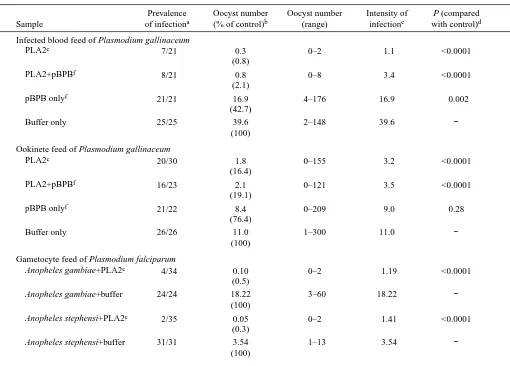
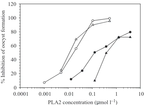
[image:4.612.51.561.93.459.2]When fed to Aedes aegypti mosquitoes as part of an infected blood meal, the PLA2 strongly inhibits oocyst formation (Table 1). This effect does not require the hydrolytic activity of the enzyme; when PLA2 was preincubated with the irreversible inhibitor p-bromophenacyl bromide (pBPB), oocyst formation was still strongly reduced. The inhibitor alone had some effect on oocyst numbers (Table 1). However, the enzyme in the presence of the inhibitor was completely inactive in our assays. The effect of the PLA2 is concentration-dependent (Fig. 1) both in the presence and in the absence of pBPB. We determined that the lowest concentration of phospholipase that resulted in a statistically significant reduction in oocyst frequency when feeding infected blood is Table 1. Effects of Crotalus adamanteus PLA2 on oocyst formation when feeding mosquitoes with infected blood or mature
ookinetes
Prevalence Oocyst number Oocyst number Intensity of P (compared
Sample of infectiona (% of control)b (range) infectionc with control)d
Infected blood feed of Plasmodium gallinaceum
PLA2e 7/21 0.3 0–2 1.1 <0.0001
(0.8)
PLA2+pBPBf 8/21 0.8 0–8 3.4 <0.0001
(2.1)
pBPB onlyf 21/21 16.9 4–176 16.9 0.002
(42.7)
Buffer only 25/25 39.6 2–148 39.6 −
(100)
Ookinete feed of Plasmodium gallinaceum
PLA2e 20/30 1.8 0–155 3.2 <0.0001
(16.4)
PLA2+pBPBf 16/23 2.1 0–121 3.5 <0.0001
(19.1)
pBPB onlyf 21/22 8.4 0–209 9.0 0.28
(76.4)
Buffer only 26/26 11.0 1–300 11.0 −
(100)
Gametocyte feed of Plasmodium falciparum
Anopheles gambiae+PLA2e 4/34 0.10 0–2 1.19 <0.0001 (0.5)
Anopheles gambiae+buffer 24/24 18.22 3–60 18.22 −
(100)
Anopheles stephensi+PLA2e 2/35 0.05 0–2 1.41 <0.0001 (0.3)
Anopheles stephensi+buffer 31/31 3.54 1–13 3.54 −
(100)
aNumber of infected mosquitoes/total number of mosquitoes dissected.
bGeometric mean of the oocyst numbers in gravid female mosquitoes; in parentheses, geometric mean as a percentage of the buffer control. cGeometric mean of the oocyst numbers in infected mosquitoes only.
dP-values were computed using Mann–Whitney rank-sum analysis.
ePLA2 was mixed into the infected blood at a final concentration of 0.5µmol l−1in infected blood feeds, 0.6µmol l−1in gametocyte feeds and 2.5µmol l−1in ookinete feeds.
21 nmol l–1 (Fig. 1). The PLA2 was also fed to Anopheles
gambiae and Anopheles stephensi together with cultured
gametocytes of the human malaria parasite Plasmodium
falciparum. Similar levels of inhibition were seen in both
anopheline species (Table 1), and the concentration-dependence of PLA2 inhibition was similar to that observed in
P. gallinaceum.
To determine whether the inhibition of oocyst formation occurs before or after the maturation of ookinetes, we fed PLA2 to mosquitoes together with purified, mature ookinetes suspended in naïve chicken blood. The PLA2 significantly inhibited oocyst formation, although the effect was less dramatic than when feeding infected blood (Table 1). The inhibition was also concentration-dependent but, in comparison with feeds with infected blood, higher PLA2 concentrations were necessary to achieve a given level of inhibition (Fig. 1). The lowest PLA2 concentration needed to achieve significantly different oocyst numbers compared with the control was 0.38µmol l–1. Once again, hydrolytic activity
of the enzyme was not required for inhibition.
PLA2 has no effect on ookinete development or viability
To determine whether any pre-ookinete developmental stage is sensitive to the PLA2, we added the enzyme during the course of exflagellation and purification of macrogametes and zygotes during routine preparation of parasites from
infected chicken blood. There was no difference in the number of exflagellation events in the presence or absence of PLA2, nor did the PLA2 have a significant effect on the total yield of macrogametes/zygotes obtained at the end of the purification (Table 2). The ookinete yields from the treated parasites were also unaffected. When the PLA2 was added to purified zygotes, which were then incubated in ookinete medium (for 24 h) to allow transformation into ookinetes, there was no effect on ookinete development in terms of either frequency (Table 3) or timing (data not shown). Some of the samples listed in Table 3 had PLA2 present continuously from exflagellation through ookinete formation and showed no significant differences in ookinete numbers compared with the controls. Furthermore, the ookinetes in all the samples displayed normal motility on glass slides. We conclude from these data that the PLA2 does not affect parasite development in the blood meal.
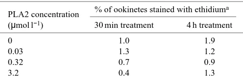
The effect of PLA2 on the viability of ookinetes was also determined. PLA2 was added to ookinete suspensions at various concentrations. At two times after PLA2 addition, a sample of ookinetes was removed and stained with ethidium homodimer, a dye that specifically stains necrotic or dead cells (Kaneshiro et al., 1993; Papadopoulos et al., 1994; Shahabuddin et al., 1998). There was no effect of the PLA2 treatment on the fraction of ookinetes staining with ethidium
PLA2 concentration (µmol l–1) 0.001
0.0001 0.01 0.1 1 10
%
I
n
h
ib
ition o
f ooc
ys
t
for
m
ation
[image:5.612.58.290.71.241.2]0 20 40 60 80 100 120
Fig. 1. Oocyst numbers from membrane feeds of purified Crotalus adamanteus PLA2. Open circles, the PLA2 was mixed with washed, infected chicken blood (16 % parasitemia) and fed to mosquitoes. Open triangles, the PLA2 was pretreated with p-bromophenacyl bromide (pBPB) at a final concentration of 100µmol l–1 and then mixed with washed, infected chicken blood (16 % parasitemia) and fed to mosquitoes. Filled circles, the PLA2 was added to an ookinete suspension in naive chicken blood and fed to mosquitoes. Filled triangles, the PLA2 was pretreated with pBPB, then added to an ookinete suspension in naive chicken blood and fed to mosquitoes. Oocyst numbers were scored 8 days after the membrane feed. Each data point represents the geometric mean of oocyst numbers from at least 20 mosquitoes expressed as percentage inhibition relative to control mosquitoes fed either with infected blood plus buffer or with infected blood plus inhibitor alone.
Table 2. Crotalus adamanteus PLA2 has no effect on parasite development when added during exflagellation and zygote
formation
Parasite numbersa
Experiment 1b Experiment 2b (0.28µmol l−1) (0.33µmol l−1)
Parasite stage +PLA2 −PLA2 +PLA2 −PLA2
Microgametesc 9.8×10⁷ 9.4×10⁷ 2.8×10⁸ 3.1×10⁸ Zygotes/macrogametesd 4.0×10⁷ 4.3×10⁷ 108 9.5×10⁷ Ookinetese 6.2×10⁶ 4.7×10⁶ 2.5×10⁷ 2.2×10⁷
C. adamanteus PLA2 was added to exflagellating, washed chicken
blood at the beginning of a parasite purification procedure. The numbers of microgametes were determined 15 min later by counting exflagellation centers; the total numbers of macrogametes or zygotes were counted at the end of the parasite purification, and the ookinete numbers were determined 1 day later, after ookinete development had been completed.
aNumbers of parasites obtained from parasite preparations treated with PLA2 or left untreated; PLA2 treatment occurred during exflagellation gradient purification; parasite densities in all samples were counted in quadruplicate and averaged.
bDifferent PLA2 concentrations were used in the two experiments (as indicated).
cTotal number of microgametes (exflagellation centers) during exflagellation.
dTotal number of macrogametes and zygotes obtained at the end of the parasite purification.
[image:5.612.316.570.109.207.2]homodimer (Table 4), showing that the enzyme does not affect ookinete viability.
As a final test of whether the PLA2 has an effect on the parasite, ookinetes were treated with 1.2µmol l–1PLA2 for 4 h.
The parasites were then washed to remove the enzyme, resuspended in naive chicken blood and fed to mosquitoes. Untreated ookinetes from the same batch were handled in the same manner and used as controls. Midguts were dissected and oocysts counted 8 days after feeding. There was no significant difference in oocyst numbers between treated and untreated ookinetes (Table 5), suggesting that PLA2 treatment does not affect the ability of the ookinetes to continue their development and form oocysts.
Inhibition of ookinete adhesion to mosquito midguts by PLA2
The data presented above argue that the PLA2 acts on the midgut surface, rather than on the ookinete, in blocking a critical step of the migration of the parasite. To test whether the PLA2 affects the ability of ookinetes to adhere to midguts,
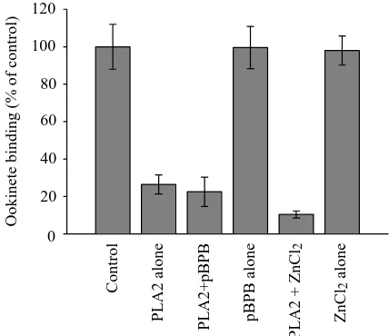
we pretreated midgut epithelia with PLA2 in vitro, and then used these epithelia in adhesion assays. Ookinetes adhering to midguts were counted, and the numbers were normalized and averaged. As shown in Fig. 2, pretreatment of midguts with PLA2 significantly lowered the number of midgut-bound ookinetes. The effect was the same when pBPB-inactivated enzyme is used, but pBPB by itself had no effect on adhesion. A second inhibitor of the active site of PLA2s, ZnCl2, was used
to duplicate the results with pBPB (Fig. 2). In the presence of 100µmol l–1 pBPB or 100µmol l–1 ZnCl
2, no enzymatic
activity (hydrolytic activity) could be detected. We conclude that the PLA2 inhibits the association of ookinetes with the midgut surface and that this effect is dependent on a structural feature of the phospholipase rather than on its catalytic activity. We also tested the inhibition of adhesion at various concentrations of PLA2 and found the lowest concentration at which significant inhibition was observed to be 0.5µmol l–1.
In the adhesion protocol described above, the PLA2 was diluted in the assay rather than being washed away after pretreatment of the midguts. To determine whether the inhibition would still occur after removing all excess PLA2, the midguts were thoroughly washed after pretreatment and then used in adhesion assays. Control midguts showed a level of ookinete binding of 100.0±2.3 %, ookinete binding of midguts pretreated with PLA2 alone was reduced to 70.3±5.0 % and ookinete binding of midguts pretreated with pBPB-inhibited PLA2 was reduced to 67.8±8.0 % (normalized means ± S.E.M., N=6). The differences between the PLA2-treated midguts and the control midguts are statistically significant in both cases. In the converse experiment, ookinetes were treated with 3.2µmol l–1of PLA2 for 1 h, after which they
[image:6.612.44.561.85.216.2]were washed and compared with mock-treated ookinetes (treated only with buffer) in adhesion assays. The treated ookinetes adhered to midguts at the same levels (99.9±4.7 %) Table 3. Crotalus adamanteus PLA2 has no effect on the transformation of zygotes to ookinetes
Ookinete densities (parasites ml−1)a
PLA2 concentration Experiment 1 Experiment 2
in zygote treatment +PLA2 in −PLA2 in +PLA2 in −PLA2 in
(µmol l−1) exflagellationb exflagellationb exflagellationb exflagellationb
0 − 1.3×10⁶ 2.8×10⁶ 2.4×10⁶
0.08 − 106 2.5×10⁶ 2.3×10⁶
0.16 − 106 2.6×10⁶ 2.3×10⁶
0.32 − 9.9×10⁵ 2.2×10⁶ 2.3×10⁶
0.63 − − 2.4×10⁶ 2.8×10⁶
1.6 − − 2.4×10⁶ 2.3×10⁶
Zygotes purified from chicken blood were incubated with various concentrations of ookinetes during ookinete development. The columns referring to + or –PLA2 in exflagellation refer to whether or not the zygotes used for ookinete development were derived from parasite purifications that had been treated with PLA2 during the exflagellation (i.e. from treated samples in Table 2); PLA2 treatment of the zygotes occurred continuously for 24 h during maturation of ookinetes from zygotes.
aOokinete densities in duplicate samples that were either treated with the PLA2 concentration indicated in the left column or treated with buffer only (0µmol l−1); parasite densities in all samples were counted in quadruplicate and averaged.
bZygotes were taken from parasite preparations that had either received PLA2 treatment (0.74µmol l−1) during exflagellation and gradient purification as described in Table 2 or not, as indicated.
Table 4. Crotalus adamanteus PLA2 has no effect on ookinete viability
PLA2 concentration % of ookinetes stained with ethidium a
(µmol l−1) 30 min treatment 4 h treatment
0 1.0 1.9
0.03 1.3 1.2
0.32 0.7 0.9
3.2 0.4 1.3
[image:6.612.44.292.603.687.2]as the mock-treated ookinetes (100.0±3.0 %) (normalized means ± S.E.M., N=4). On the basis of these results, we
conclude that the enzyme changes the properties of the midgut epithelium, but does not affect the adhesiveness of ookinetes. The adhesion assays performed in the presence and absence of PLA2 allowed us to observe shedding of lipids from the posterior ends of ookinetes, which can be detected as the capping of fluorescent stain on the posterior end of the parasite, and which correlates with ookinete motility. In certain ookinete preparations, all parasites bound to the midgut surface
show capping after the adhesion is complete and the fixed midguts are being examined for the number of bound ookinetes. In 10 pairs of midgut sheets where capping was observed, there were never any visible differences in the posterior capping of fluorescent stains in the presence or absence of PLA2. It is likely, therefore, that the PLA2 does not interfere with ookinete motility.
Adhesion assays using PLA2s from other organisms were used to determine whether the ability of the C. adamanteus enzyme to inhibit ookinete/midgut binding is a general feature of PLA2s. As shown in Fig. 3, PLA2s from the venoms of various snakes, as well as honeybee venom PLA2 and mammalian pancreatic PLA2s, were used to pretreat midguts in adhesion assays. The results (Fig. 3) indicate that the PLA2s derived from snake and insect venoms strongly inhibit ookinete binding, while the mammalian pancreatic enzymes do not. The extent of inhibition of oocyst formation by the different PLA2s in oocyst formation assays mirrors their inhibition of in vitro adhesion (data not shown).
Fluorescently labeled PLA2 binds to mosquito midguts but not to ookinetes
The results from the adhesion assays suggested that the PLA2 affects the mosquito midgut, but not ookinetes. To test whether the enzyme interacts with midguts, we prepared fluorescently labeled C. adamanteus PLA2 and measured its ability to bind to midguts and parasites. The labeled enzyme retained some of its hydrolytic activity and was identical to unlabeled enzyme in its ability to block oocyst formation when mixed with infected blood and fed to mosquitoes (data not shown).
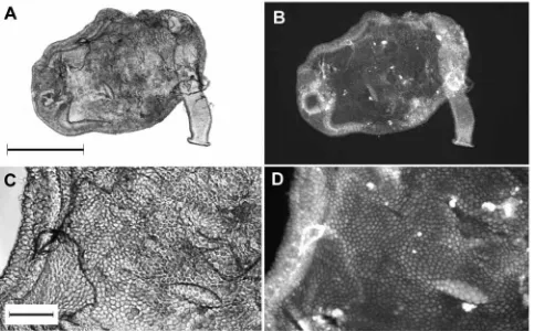
[image:7.612.67.568.86.162.2]The labeled PLA2 was used to stain isolated midguts and ookinetes. Staining was performed in the presence of 10 % chicken serum, as a non-specific competitor, and the stained tissues or cells were washed extensively after staining to remove non-specifically bound PLA2. The protein readily bound to midgut epithelia and showed a diffuse staining on both the luminal surface and the basement membrane on the other side of the epithelium (Fig. 4). The staining of the luminal surface was often irregular, with certain areas or individual cells staining more brightly for unknown reasons. Higher magnifications clearly showed surface staining of the
Fig. 2. Midguts were pretreated with 3.2µmol l–1 PLA2 in M199 medium for 10 min, and the treated guts were then used in adhesion assays. Control, midguts treated with buffer only; PLA2 alone, midguts treated with active PLA2; PLA2+pBPB, midguts treated with PLA2 in the presence of 100µmol l–1 p-bromophenacyl bromide (pBPB), an inhibitor of the enzyme; pBPB alone, midguts pretreated with the inhibitor pBPB only; PLA2+ZnCl2, midguts pretreated with PLA2 in the presence of 100µmol l–1 ZnCl
2, an inhibitor of the enzyme; ZnCl2 alone, midguts treated with 100µmol l–1ZnCl
2. Each column is the mean of 1–2 experiments, each performed in quadruplicate. The error bars represent ±S.E.M. (N=4–8). P values for each column are as follows: PLA2 alone, P=0.0002; PLA2+pBPB, P<0.0001; pBPB alone, P=0.98; PLA2+ZnCl2, P=0.004; ZnCl2alone, P=0.91.
Control
PLA2 a
lon
e
PLA2
+pB
P
B
pB
P
B
a
lon
e
PLA2
+
Z
nCl
2
Z
nCl
2
a
lon
e
O
o
ki
n
et
e
bi
n
di
n
g
(%
o
f
control
)
[image:7.612.67.281.389.574.2]0 20 40 60 80 100 120
Table 5. Ookinetes pretreated with PLA2 form oocysts at normal frequencies
Prevalence Oocyst number P (compared
Sample of infectiona (% of control) Oocyst numberb with control)c
Ookinetes pretreated with PLA2d 24/24 21.8 0–400 0.66
(80.7)
Ookinetes pretreated with buffer 24/24 27.0 0–300 −
(100)
aNumber of infected mosquitoes/total number of mosquitoes dissected.
bGeometric mean of the oocyst numbers in gravid female mosquitoes; in parentheses, geometric mean as a percentage of the buffer control. cP-values were computed using Mann–Whitney rank-sum analysis.
midgut cells (Fig. 4D), although the lack of resolution made it difficult to determine whether the PLA2 was associating with the microvilli-associated network observed on the midgut surface in earlier studies (Zieler et al., 1998, 2000). Although the unstained midguts are not shown for comparison, the degree of autofluorescence was low compared with the stained midgut and resulted in a very dim image at the lamp intensity and gain settings used for the digital camera. We also
demonstrated that the binding of labeled PLA2 to the midguts could be inhibited by an excess of unlabeled enzyme. In contrast to the midgut staining by the labeled PLA2, however, labeled PLA2 completely failed to bind to ookinetes. In addition, the PLA2 failed to label isolated peritrophic membranes, suggesting that its ligand is found only on the midgut surface (data not shown).
C. adamanteus and honeybee venom PLA2s are suitable
candidates for refractory genes
The C. adamanteus and honeybee venom PLA2s were fed to mosquitoes to test the premise that the genes encoding these proteins would be suitable as refractory genes. The percentage of mosquitoes that fed on chicken blood containing the PLA2s or on control blood was determined and was not significantly different on any of the feeds (Table 6). No difference in viability or in egg production was observed between mosquitoes fed the PLA2 compared with the control group (Table 6). We conclude that the PLA2s have no strong effect on the abilities of mosquitoes to engorge or reproduce. These results suggest that it may be feasible to express PLA2 genes in transgenic mosquitoes in a midgut-specific fashion.
Discussion
We have shown that the Crotalus adamanteus PLA2 greatly reduces the efficiency of oocyst formation of malaria parasites. This enzyme blocks the development of Plasmodium
falciparum as efficiently as of the model organism P. gallinaceum, enhancing the significance of our finding and
[image:8.612.67.267.73.245.2]implying that a conserved part of the plasmodial life cycle is affected. Furthermore, the reduction in oocyst numbers correlates with an inhibition of ookinete adhesion to the midgut surface in vitro. The PLA2 has no effect on any of the mosquito stages of the malaria parasite in vitro, nor does it alter the adhesion ability of ookinetes or their ability to form oocysts after pretreatment with the enzyme. The gliding motility of treated ookinetes on glass slides was also normal, and ookinetes shed their surface lipids normally in the presence of the enzyme. These results, together with the observation that fluorescently labeled PLA2 binds to mosquito midguts but not to ookinetes, suggest that the phospholipase binds to or
Fig. 3. Midguts were pretreated with different PLA2s (final concentration 10µmol l–1) in M199 medium for 10 min and then used in adhesion assays. Control, midguts treated with buffer only. The PLA2s used in treatments were as follows: Bovine pancr., bovine pancreatic PLA2; Porcine pancr., porcine pancreatic PLA2; Bee venom, honeybee venom PLA2; Crotalus d. t., Crotalus durissus terrificus venom PLA2 (crotoxin); Crotalus ad., Crotalus adamanteus venom PLA2 (7µmol l–1); Naja moss. I, Naja mossambica mossambica venom PLA2 isozyme I (pI=6.5); Naja moss. II, Naja mossambica mossambica venom PLA2 isozyme II (pI=8.8); Naja moss. III, Naja mossambica mossambica venom PLA2 isozyme III (pI=9.6); Naja naja, Naja naja venom PLA2. The PLA2s were dialyzed extensively before use in the assays. Each column represents the results of a single experiment, performed in quadruplicate. Error bars represent ±S.E.M. (N=4). P values for each column are as follows: Bovine pancr., P=0.23; Porcine pancr., P=0.05; Bee venom, P=0.001; Crotalus d. t., P=0.001; Crotalus ad., P=0.002; Naja moss. I, P=0.003; Naja moss. II, P=0.004; Naja moss. III, P=0.02; Naja naja, P=0.003.
Control B o vi n e pa n cr . P or ci n e pa n cr . Bee v eno m Crot al us d . t. Crot al us a d . N aj a m o ss . I N aj a m o ss . II N aj a m o ss . III N aj a n aj a O o k in et e b in d in g (% o f c ontrol ) 0 20 40 60 80 100 120
Table 6. Crotalus adamanteus PLA2 has no effect on mosquito viability or fecundity
% Engorgement Viability 4 days Egg count per
PLA2 fed to mosquitoes (number fed/total)a post blood feed (%)b gravid femalec
+1.2µmol l−1Crotalus PLA2d 52.3 (45/86) 88.9 55.0±7.8
+0.2µmol l−1Crotalus PLA2 51.6 (32/62) 90.6 57.3±6.0
+1.5µmol l−1bee venom PLA2 69.1 (47/68) 89.4 55.8±11.3
Control (+buffer only) 59.4 (37/69) 90.2 56.7±12.6
aPercentage of mosquitoes with visible blood meals in their abdomens immediately after membrane feeding on blood with or without PLA2. bPercentage of the blood-fed mosquitoes still alive 4 days after membrane feeding.
[image:8.612.40.563.611.687.2]obscures an essential ligand on the midgut surface that is needed for the ookinete successfully to traverse the midgut and form oocysts. The development of P. gallinaceum occurs in mosquitoes of the genus Aedes and that of P. falciparum in mosquitoes of the genus Anopheles. Our results suggest that a similar midgut ligand is used by both parasite species to recognize the midgut of their mosquito hosts and that this molecule is essential for the parasite to identify its target.
PLA2s from the venoms of other snakes and from honeybees appear to have the same property as the PLA2 from Crotalus
adamanteus (see Fig. 3). In contrast, the mammalian pancreatic PLA2s do not efficiently block adhesion. The data in Fig. 3 suggest a correlation between the membrane-inserting abilities of a PLA2 and its ability to block ookinete adhesion and oocyst formation. The mammalian pancreatic PLA2s tend to have a very low membrane-inserting ability, while the honeybee venom PLA2 has the highest inserting ability measured in any phospholipase (Demel et al., 1975; Waite, 1987). This result is consistent with our finding that the hydrolytic activity of the PLA2 is not an essential part of its antiparasitic activity and suggests that the inhibition of ookinete adhesion by the PLA2 is primarily dependent on the enzyme’s property of binding to exposed membrane lipids.
We have previously reported a novel membranous structure on the midgut surface that we have termed the microvilli-associated network (MN) and which seems to be the primary
structure with which ookinetes associate on the midgut surface (Zieler et al., 1998, 2000). It is likely that the PLA2 binds to the MN. Unfortunately, this structure is difficult to see using light microscopy, and in our fluorescence labeling experiments we were unable to determine whether the labeled PLA2 does indeed bind to MN strands.
The inhibition by the PLA2 of ookinete association with the midgut surface could be a result of its direct binding to the same periodate-sensitive carbohydrate ligand (Zieler et al., 1999) recognized by the ookinete. In this case, the ookinete ligand would probably be a glycolipid or a highly periodate-sensitive phospholipid such as phosphatidylglycerol. We were unable to test whether periodate treatment of midguts affects PLA2 binding, since extensive cell damage occurs during periodate treatment and the PLA2 binds more efficiently to the perturbed membranes in damaged cells (Dawson et al., 1984; Thuren et al., 1987).
[image:9.612.71.554.72.372.2]In contrast, it is unlikely that inhibition of ookinete adhesion by PLA2s is related to their ability to bind to heparin and other anionic glycosaminoglycans. First, this property of PLA2s is best-documented in the pancreatic enzymes (Diccianni et al., 1990, 1991), which are not very active in our assays. Second, we have tried heparin, as well as a variety of other anionic glycosaminoglycans, as competitors in adhesion assays and have been unable to detect any inhibition at concentrations as high as 500µg ml–1. Third, the Crotalus adamanteus PLA2
inhibits ookinete adhesion even in the presence of heparin (data not shown).
In the adhesion assay, we also tested human annexin V, a protein known to bind to acidic phospholipids (such as phosphatidylserine) normally present in the inner leaflet of the plasma membrane bilayer (Swairjo et al., 1995; Reutlingsperger and van Heerde, 1996), and found no inhibition of adhesion. Annexins are Ca2+-dependent
phospholipid-binding proteins that compete with PLA2s for binding to membranes (Davidson et al., 1987; Buckland and Wilton, 1998a,b). The phospholipid preference of PLA2s, or the manner in which they disrupt lipid bilayers during binding, is probably an important characteristic that enables these enzymes specifically to disrupt ookinete/midgut interactions.
The PLA2s have a number of unique properties that make them especially suitable for expression in mosquito midguts as refractory genes. First, they are exceptionally stable and resistant to protease cleavage, an important property in the degradative environment of the midgut lumen. Second, they are generally secreted as proenzymes, which are activated by trypsin cleavage (Pieterson et al., 1974; Waite, 1987). Proenzyme activation will occur after secretion into the midgut lumen, as happens with midgut chitinases (Shen and Jacobs-Lorena, 1997). The feasibility of expressing PLA2 enzymes as proenzymes in a variety of heterologous expression systems has already been demonstrated (Verheij and de Haas, 1991). Third, the hydrolytic activity of PLA2s is not required for inhibition of parasite development, implying that the midgut-secreted enzyme can be catalytically inactivated by mutation if necessary. We have shown with membrane feeds that ingestion of the active PLA2 has no measurable effect on the female mosquitoes (Table 6), arguing that a midgut-expressed PLA2 would be well-tolerated by transgenic mosquitoes. PLA2-expressing transgenic Aedes aegypti are currently being generated using the recently developed transformation methods for this organism (Coates et al., 1998; Jasinskiene et al., 1998). The PLA2 gene will thus be one of the first genes to be tested in transgenic mosquitoes for its ability to confer refractoriness to malaria parasites.
Our long-term studies are aimed at a complete characterization of the interaction between malaria parasites and mosquito tissues. In the process of this work, we will further our understanding of the biology of this parasite, and we hope that this knowledge will be a starting point for developing new methods of malaria control.
We are grateful to Dr Louis H. Miller for his encouragement and support. Special thanks go to Olga Muratova for technical assistance with P. falciparum cultures and to André Laughinghouse and Kevin Lee for technical assistance with raising mosquitoes and maintaining infected chickens. H.Z. was supported by the Vector Biology Network of the John D. and Catherine T. MacArthur Foundation.
References
Apiz-Castro, R., Jain, M. K. and de Haas, G. H. (1982). Origin of the
latency phase during the action of phospholipase A2 on unmodified phosphatidylcholine vesicles. Biochim. Biophys. Acta 688, 349–356.
Brunie, S., Bolin, J., Gewirth, D. and Sigler, P. B. (1985). The refined crystal
structure of dimeric phospholipase A2 at 2 Å. J. Biol. Chem. 260, 9742–9749.
Buckland, A. G. and Wilton, D. C. (1998a). Inhibition of human
cytosolic phospholipase A2 by human annexin V. Biochem. J. 329, 369–372.
Buckland, A. G. and Wilton, D. C. (1998b). Inhibition of phospholipase A2
by annexin. V. Competition for anionic phospholipid interfaces allows an assessment of the relative interfacial affinities of secreted phospholipases A2. Biochim. Biophys. Acta 1391, 367–376.
Burack, W. R., Yuan, Q. and Biltonen, R. L. (1993). Role of lateral phase
separation in the modulation of phospholipase A2 activity. Biochemistry 32, 583–589.
Coates, C., Jasinskiene, N., Miyashiro, L. and James, A. A. (1998). Mariner
transposition and transformation of the yellow fever mosquito, Aedes
aegypti. Proc. Natl. Acad. Sci. USA 95, 3748–3751.
Condrea, E., Yang, C. C. and Rosenberg, P. (1981). Lack of correlation
between anticoagulant activity and phospholipid hydrolysis by snake venom phospholipases A2. Thromb. Haemost. 45, 82–85.
Crampton, J. M., Warren, A., Lycett, G. J., Hughes, M. A., Comley, I. P. and Eggleston, P. (1994). Genetic manipulation of insect vectors as a
strategy for the control of vector-borne disease. Ann. Trop. Med. Parasitol.
88, 3–12.
Davidson, F. F., Dennis, E. A., Powell, M. and Glenney, J. R. J. (1987).
Inhibition of phospholipase A2 by ‘lipocortins’ and calpactins. An effect of binding to substrate phospholipids. J. Biol. Chem. 262, 1698–1705.
Dawson, R. M. C., Irvine, R. F., Bray, J. and Quinn, P. J. (1984).
Long-chain unsaturated diacylglycerols cause a perturbation in the structure of phospholipid bilayers rendering them susceptible to phospholipase attack.
Biochem. Biophys. Res. Commun. 125, 836–842.
Demel, R. A., Geurts van Kessel, W. S. M., Zwaal, R. F. A., Roelofsen, B. and van Deenen, L. M. (1975). Relation between various phospholipase
actions on human red cell membranes and the interfacial phospholipid pressure in monolayers. Biochim. Biophys. Acta 406, 97–107.
Dennis, E. A. (1973). Phospholipase A2 activity towards phosphatidylcholine
in mixed micelles: surface dilution kinetics and the effect of thermotropic phase transitions. Arch. Biochem. Biophys. 158, 485–493.
Dennis, E. A. (1983). Phospholipases. In The Enzymes, 3rd edn, vol. 16 (ed.
P. D. Boyer), pp. 307–353. New York: Academic Press.
Diccianni, M. B., Lilly-Staudermann, M., McLean, L. R., Balasubramaniam, A. and Harmony, J. A. K. (1991). Heparin prevents
the binding of phospholipase A2 to phospholipid micelles: importance of the amino-terminus. Biochemistry 30, 9090–9097.
Diccianni, M. B., Mistry, M. J., Hug, K. and Harmony, J. A. K. (1990).
Inhibition of phospholipase A2 by heparin. Biochim. Biophys. Acta 1046, 242–248.
Fleer, E. A., Verheij, H. M. and de Haas, G. H. (1981). Modification of
carboxylate groups in bovine pancreatic phospholipase A2. Identification of
aspartate-49 as Ca2+-binding ligand. Eur. J. Biochem. 113, 283–288.
Gelb, M. H., Cho, W. and Wilton, D. C. (1999). Interfacial binding of
secreted phospholipase A2: more than electrostatics and a major role for tryptophan. Curr. Opin. Struct. Biol. 9, 428–432.
Gerberg, E. J., Barnard, D. R. and Ward, R. A. (1994). Manual for Mosquito Rearing and Experimental Techniques. Lake Charles, LA:
American Mosquito Control Association.
Goormaghtigh, E., van Campenhoud, M. and Ruysschaert, J.-M. (1981).
Lipid phase separation mediates binding of porcine pancreatic phospholipase A2 to its substrate. Biochem. Biophys. Res. Commun. 101, 1410–1418.
Grabarek, Z. and Gergely, J. (1990). Zero-length cross-linking procedure
with the use of active esters. Anal. Biochem. 185, 131–135.
Gwadz, R. W. (1994). Genetic approaches to malaria control: how long the
road? Am. J. Trop. Med. Hyg. 50, 116–125.
Han, Y. S., Thompson, J., Kafatos, F. C. and Barillas-Mury, C. (2000).
Molecular interactions between Anopheles stephensi midgut cells and
Plasmodium berghei: the time bomb theory of ookinete invasion of
mosquitoes. EMBO J. 19, 6030–6040.
Hazlett, T. L. and Dennis, E. A. (1985). Aggregation studies on
fluorescein-coupled cobra venom phospholipase A2. Biochemistry 24, 6152–6158.
Higgs, S. and Beaty, B. J. (1996). Rearing and containment of mosquito
Hønger, T., Jørgensen, K., Stokes, D., Biltonen, R. L. and Mouritsen, O. G. (1997). Phospholipase A2 activity and physical properties of lipid-bilayer
substrates. Meth. Enzymol. 286, 168–190.
Jain, M. K., Gelb, M. H., Rogers, J. and Berg, O. G. (1995). Kinetic basis
for interfacial catalysis by phospholipase A2. Meth. Enzymol. 249, 567–614.
Jasinskiene, N., Coates, C. J., Benedict, M. Q., Cornel, A. J., Rafferty, C. S., James, A. A. and Collins, F. H. (1998). Stable transformation of the
yellow fever mosquito, Aedes aegypti, with the Hermes element from the housefly. Proc. Natl. Acad. Sci. USA 95, 3743–3747.
Kaneshiro, E. S., Wyder, M. A., Wu, Y.-P. and Cushion, M. T. (1993).
Reliability of calcein acetoxy methyl ester and ethidium homodimer or propidium iodide for viability assessment of microbes. J. Microbiol. Meth.
17, 1–16.
Keith, C., Feldman, D. S., Deganello, S., Glick, J., Ward, K. B., Jones, E. O. and Sigler, P. B. (1981). The 2.5 Å crystal structure of a dimeric
phospholipase A2 from the venom of Crotalus atrox. J. Biol. Chem. 256, 8602–8607.
Kensil, C. R. and Dennis, E. A. (1979). Action of cobra venom phospholipase
A2 on the gel and liquid crystalline states of dimyristoyl phosphatidylcholine vesicles. J. Biol. Chem. 254, 5843–5848.
Kini, R. M. and Evans, H. J. (1989). A model to explain the pharmacological
effects of snake venom phopholipase A2. Toxicon 27, 613–635.
Lehtonen, J. Y. A. and Kinunnen, P. K. J. (1995). Phospholipase A2 as a
mechanosensor. Biophys. J. 68, 1888–1894.
Mehlhorn, H., Peters, W. and Haberkorn, A. (1980). The formation of
kinetes and oocyst in Plasmodium gallinaceum (Haemosporidia) and considerations on phylogenetic relationships between Haemosporidia, Piroplasmida and other coccidia. Protistologica 16, 135–154.
Meis, J. F. and Ponnudurai, T. (1987). Ultrastructural studies on the
interaction of Plasmodium falciparum ookinetes with the midgut epithelium of Anopheles stephensi mosquitoes. Parasitol. Res. 73, 500–506.
Meis, J. F., Pool, G., van Gemert, G. J., Lensen, A. H., Ponnudurai, T. and Meuwissen, J. H. (1989). Plasmodium falciparum ookinetes migrate
intercellularly through Anopheles stephensi midgut epithelium. Parasitol.
Res. 76, 13–19.
op den Kamp, J. A. F., de Gier, J. and van Deenen, L. L. M. (1974).
Hydrolysis of phosphatidylcholine liposomes by pancreatic phospholipase A2 at the transition temperature. Biochim. Biophys. Acta 345, 253–256.
Papadopoulos, N. G., Dedoussis, G. V. Z., Spanakos, G., Gritzapis, A. D., Baxevanis, C. N. and Papamichail, M. (1994). An improved fluorescence
assay for the determination of lymphocyte-mediated cytotoxicity using flow cytometry. J. Immunol. Meth. 177, 101–111.
Pieterson, W. A., Vidal, J. C., Volwerk, J. J. and de Haas, G. H. (1974).
Zymogen-catalyzed hydrolysis of monomeric substrates and the presence of a recognition site for lipid–water interfaces in phospholipase A2.
Biochemistry 13, 1455–1460.
Reutlingsperger, C. P. M. and van Heerde, W. L. (1996). Annexin V and
cell surface-expressed phosphatidylserine: a revealing pas de deux. In
Annexins: Molecular Structure to Cellular Function (ed. B. A. Seaton), pp.
201–211. Austin, TX: Chapman & Hall.
Rosenberg, P. (1986). The relationship between enzymatic activities and
pharmacological properties of phospholipases in natural poisons. In Natural
Toxins: Animal, Plant and Microbial (ed. J. B. Harris), pp. 129–174.
Oxford: Clarendon Press.
Shahabuddin, M., Fields, I., Bulet, P., Hoffmann, J. A. and Miller, L. H.
(1998). Plasmodium gallinaceum: differential killing of some mosquito stages of the parasite by insect defensin. Exp. Parasitol. 89, 103–112.
Shahabuddin, M. and Pimenta, P. F. (1998). Plasmodium gallinaceum
preferentially invades vesicular ATPase-expressing cells in Aedes aegypti midgut. Proc. Natl. Acad. Sci. USA 95, 3385–3389.
Shahabuddin, S., Gayle, M., Zieler, H. and Laughinghouse, A. (1997).
Plasmodium gallinaceum: fluorescent staining of zygotes and ookinetes to
study malaria parasites in the mosquito. Exp. Parasitol. 88, 79–84.
Shen, Z. and Jacobs-Lorena, M. (1997). Characterization of a novel
gut-specific chitinase gene from the human malaria vector Anopheles gambiae.
J. Biol. Chem. 272, 28895–28900.
Sieber, K. P., Huber, M., Kaslow, D., Banks, S. M., Torii, M., Aikawa, M. and Miller, L. H. (1991). The peritrophic membrane as a barrier: its
penetration by Plasmodium gallinaceum and the effect of a monoclonal antibody to ookinetes. Exp. Parasitol. 72, 145–156.
Slotboom, A. J., Verheij, H. M. and de Haas, G. H. (1982). On the
mechanism of phospholipase A2. In Phospholipids (ed. J. N. Hawthorne and G. B. Ansell), pp. 359–434. Amsterdam: Elsevier Biomedical Press.
Staros, J. V., Wright, R. W. and Swingle, D. M. (1986). Enhancement by
N-hydroxysulfosuccinimide of water-soluble carbodiimide-mediated coupling reactions. Anal. Biochem. 156, 220–222.
Swairjo, M. A., Concha, N. O., Kaetzel, M. A., Dedman, J. R. and Seaton, B. A. (1995). Ca2+-bridging mechanism and phospholipid head group recognition in the membrane-binding protein annexin V. Nature Struct. Biol.
2, 968–974.
Syafruddin, Arakawa, R., Kamimura, K. and Kawamoto, F. (1991).
Penetration of the mosquito midgut wall by the ookinetes of Plasmodium
yoelii nigeriensis. Parasitol. Res. 77, 230–236.
Thuren, T., Tulkki, J. A., Virtanen, J. A. and Kinnunen, P. K. J. (1987).
Triggering of the activity of phospholipase A2 by an electric field.
Biochemistry 26, 4907–4910.
Tinker, D. O., Low, R. and Lucassen, M. (1980). Heterogeneous catalysis
by phospholipase A2: mechanism of hydrolysis of gel phase
phosphatidylcholine. Can. J. Biochem. 58, 898–912.
Torii, M., Nakamura, K., Sieber, K. P., Miller, L. H. and Aikawa, M.
(1992). Penetration of the mosquito (Aedes aegypti) midgut wall by the ookinetes of Plasmodium gallinaceum. J. Protozool. 39, 449–454.
Verger, R., Mieras, M. C. E. and de Haas, G. H. (1973). Action of
phospholipase A at interfaces. J. Biol. Chem. 248, 4023–4034.
Verheij, H. M. and de Haas, G. H. (1991). Cloning, expression and
purification of porcine pancreatic phospholipase A2 and mutants. Meth.
Enzymol. 197, 214–223.
Volwerk, J. J., Pieterson, W. A. and de Haas, G. H. (1974). Histidine at the
active site of phospholipase A2. Biochemistry 13, 1446–1454.
Waite, M. (1987). The Phospholipases. New York: Plenum Press.
Wells, M. A. (1973). Effects of chemical modification on the activity of Crotalus adamanteus phospholipase A2. Evidence for an essential amino
group. Biochemistry 12, 1086–1093.
White, S. P., Scott, D. L., Otwinowski, Z., Gelb, M. H. and Sigler, P. B.
(1990). Crystal structure of cobra-venom phospholipase A2 in a complex with a transition-state analogue. Science 250, 1560–1563.
Zieler, H. and Dvorak, J. A. (2000). Invasion in vitro of mosquito midgut
cells by the malaria parasite proceeds by a conserved mechanism and results in death of the invaded midgut cells. Proc. Natl. Acad. Sci. USA 97, 11516–11521.
Zieler, H., Garon, C. F., Fischer, E. R. and Shahabuddin, M. (1998).
Adhesion of Plasmodium gallinaceum ookinetes to the Aedes aegypti midgut: sites of parasite attachment and morphological changes in the ookinete. J. Euk. Microbiol. 45, 512–520.
Zieler, H., Garon, C. F., Fischer, E. R. and Shahabuddin, M. (2000). A
tubular network associated with the brush-border surface of the Aedes
aegypti midgut: implications for pathogen transmission by mosquitoes. J. Exp. Biol. 203, 1599–1611.
Zieler, H., Nawrocki, J. P. and Shahabuddin, M. (1999). Plasmodium gallinaceum ookinetes adhere specifically to the midgut epithelium of Aedes aegypti by interaction with a carbohydrate ligand. J. Exp. Biol. 202,