The limb postures of terrestrial tetrapods span a continuum from sprawling to fully upright, in which the limbs are held lateral to or beneath the body, respectively (Jenkins, 1971a; Gatesy, 1991; Reilly and Elias, 1998). Such differences in limb posture frequently are correlated with differences in limb kinematics. For instance, kinematic studies of lizards (Brinkman, 1980a; Brinkman, 1981; Jayne and Irschick, 1999; Irschick and Jayne, 1999) and crocodilians (Brinkman, 1980b; Gatesy, 1991) have demonstrated that axial rotation of the femur can be substantial during non-parasagittal locomotion in these lineages. These patterns contrast with those of tetrapods that employ parasagittal or near-parasagittal kinematics, in
which axial rotation of the limb bones is minimal (Jenkins, 1971a). Because the loads that a limb bone experiences correlate strongly with its orientation to the ground reaction force during stance (Biewener, 1983a; Biewener et al., 1983; Biewener et al., 1988), vertebrate limbs used in non-parasagittal and non-parasagittal locomotion could be exposed to very different loading regimes. Few studies have explicitly tested this possibility. However, such studies are necessary to evaluate the potential diversity of tetrapod limb bone loading patterns and to understand major aspects of evolutionary transitions in tetrapod locomotor mechanics.
Measurements of in vivo strain from the femur and tibia in JEB3037
In vivo measurements of strain in the femur and tibia of Iguana iguana (Linnaeus) and Alligator mississippiensis (Daudin) have indicated three ways in which limb bone loading in these species differs from patterns observed in most birds and mammals: (i) the limb bones of I. iguana and A. mississippiensis experience substantial torsion, (ii) the limb bones of I. iguana and A. mississippiensis have higher safety factors than those of birds or mammals, and (iii) load magnitudes in the limb bones of A. mississippiensis do not decrease uniformly with the use of a more upright posture. To verify these patterns, and to evaluate the ground and muscle forces that produce them, we collected three-dimensional kinematic and ground reaction force data from subadult I. iguana and A. mississippiensis using a force platform and high-speed video. The results of these force/kinematic studies generally confirm the loading regimes inferred from in vivo strain measurements. The ground reaction force applies a torsional moment to the femur and tibia in both species; for the femur, this moment augments the moment applied by the caudofemoralis muscle, suggesting large torsional stresses. In most cases, safety factors in bending calculated from force/video data are lower than those determined from strain data, but are
as high or higher than the safety factors of bird and mammal limb bones in bending. Finally, correlations between limb posture and calculated stress magnitudes in the femur of I. iguana confirm patterns observed during direct bone strain recordings from A. mississippiensis: in more upright steps, tensile stresses on the anterior cortex decrease, but peak compressive stresses on the dorsal cortex increase. Equilibrium analyses indicate that bone stress increases as posture becomes more upright in saurians because the ankle and knee extensor muscles exert greater forces during upright locomotion. If this pattern of increased bone stress with the use of a more upright posture is typical of taxa using non-parasagittal kinematics, then similar increases in load magnitudes were probably experienced by lineages that underwent evolutionary shifts to a non-sprawling posture. High limb bone safety factors and small body size in these lineages could have helped to accommodate such increases in limb bone stress.
Key words: locomotion, biomechanics, kinematics, force platform, bone stress, safety factor, posture, evolution, Sauria, Crocodylia, Lepidosauria, lizard.
Summary
Introduction
MECHANICS OF LIMB BONE LOADING DURING TERRESTRIAL LOCOMOTION IN
THE GREEN IGUANA (IGUANA IGUANA) AND AMERICAN ALLIGATOR
(ALLIGATOR MISSISSIPPIENSIS)
RICHARD W. BLOB1,* ANDANDREW A. BIEWENER2
1Department of Zoology, Division of Fishes, Field Museum of Natural History, 1400 South Lake Shore Drive, Chicago, IL 60605, USA and 2Concord Field Station, MCZ, Harvard University, Old Causeway Road, Bedford,
MA 01730, USA
*e-mail: rblob@fmnh.org
Iguana iguana and Alligator mississippiensis (Blob and Biewener, 1999) suggest that loading patterns in the limb bones of these species differ from those of previously examined terrestrial mammals and birds in two fundamental ways: (i) shear is a much more important mode of loading in I. iguana and A. mississippiensis than in most species that habitually use upright locomotion, and (ii) limb bone safety factors are higher in I. iguana and A. mississippiensis than in birds or mammals. Our data suggest a further distinction between A. mississippiensis and mammals: in contrast to interspecific patterns observed in mammals (Biewener, 1989; Biewener, 1990), bone loads did not decrease throughout the cortex when A. mississippiensis used more upright postures but, instead, increased at some locations (Blob and Biewener, 1999). Because limb bone loading mechanics have been examined in only two species that employ non-parasagittal locomotion, broad conclusions based on studies of I. iguana and A. mississippiensis must be viewed with caution. However, bone strain data from these species suggest preliminary hypotheses that substantial limb bone torsion, high limb bone safety factors, and increases in bone loading with the use of a more upright stance are common features of locomotor mechanics among tetrapods that use non-parasagittal kinematics and a non-upright limb posture.
As a first step in testing these hypotheses, we collected simultaneous three-dimensional kinematic and force platform data from iguanas and alligators. Integrated force and kinematic data provide an independent means of verifying the interpretations of load magnitudes and loading patterns derived from bone strain recordings. In addition, synchronized locomotor force and kinematic data allow analyses of joint equilibrium that document external and muscular forces and moments acting on limb bones. Although these analyses produce indirect estimates of load magnitudes, they provide insight into the mechanics underlying bone loading patterns (i.e. how forces are transmitted to bones) that direct in vivo strain measurements cannot supply (Biewener and Full, 1992). Therefore, in the context of our complementary study of limb bone strains in these species (Blob and Biewener, 1999), this study helps to clarify the mechanical differences between non-parasagittal and parasagittal locomotion and, thereby, evaluates the functional changes entailed in evolutionary transitions from sprawling to non-sprawling posture.
Materials and methods Experimental animals
Data were collected from four subadult green iguanas Iguana iguana (Linnaeus) (body mass 320–516 g) and one juvenile American alligator Alligator mississippiensis (Daudin) (body mass 1.98 kg) that also were used in experiments to measure in vivo limb bone strain (Blob and Biewener, 1999). Animal care and housing are described in the strain study. All experimental procedures followed University of Chicago IACUC guidelines (protocols 61341 and 61371). After bone strain recordings had been completed, the animals
were killed (Nembutal sodium pentobarbital, 200 mg kg−1 intraperitoneal injection) and frozen for later dissection and measurement of anatomical variables.
Collection of kinematic and force data
Animals were filmed using high-speed video (Kodak EktaPro, model 1012 image-intensified system) while running over a custom-built force platform inserted into a 6 m long wooden trackway (Biewener and Full, 1992). Video framing rate was 500 frames s−1for I. iguana and 250 frames s−1for A. mississippiensis. Between 18 and 34 frames (evenly spaced in time) were digitized for each step, depending on step duration (steps are defined as the period of foot contact with the ground). This produced effective framing rates of 71–167 frames s−1 for I. iguana and 25–50 frames s−1 for A. mississippiensis. To facilitate digitization of joint positions, dots of white latex were painted on the claw of the fourth digit, the metatarso-phalangeal joint, the ankle, knee and hip joints and the posterior ilium. Room temperature was 25 °C, but before and after trials animals were allowed to rest and bask under heat lamps, where the temperature was the same as in the enclosure in which they were housed (29–32 °C).
The surface of the force platform was flush with that of the track, and both were covered with thin rubber to give the animals sufficient friction to prevent slipping. A 1.1 m Plexiglas panel lateral to the force platform allowed the right side of the animal to be videotaped as it ran past the camera and over the platform. Dorsal and lateral views were filmed simultaneously using a mirror positioned over the force platform at 45 ° to the track surface. Joint positions were digitized from video frames in both views (Measurement TV; Updegraff, 1990). These two sets of coordinate data were used to calculate limb kinematics in three dimensions in a custom-designed Pascal program. Prior to kinematic calculations, coordinate data were filtered digitally with a zero-lag, second-order, low-pass Butterworth filter (cut-off frequency 4–6 times stride frequency) and corrected for parallax. The cut-off frequencies employed removed noise without attenuating signal peaks. Unless stated otherwise, reported angles are true angles (in three dimensions) between segments.
sampled through an A/D converter at 500 Hz and stored on computer. The plate was designed to accommodate a 50 N vertical load; amplifier gains for each channel were adjusted appropriately for the weight of the animal to allow more sensitive resolution of forces. Calibrations verified that the response of the platform to loads was linear over the range of forces recorded. The natural unloaded frequencies of the platform were 210 Hz (vertical), 150 Hz (anteroposterior) and 75 Hz (mediolateral), all sufficiently greater than the stride frequencies of the animals studied (less than 4 Hz in both species) so as not to confound the signal produced by the ground reaction force. Cross-talk between channels (<3 %) was corrected in the data-analysis software. Force data were filtered digitally with a zero-lag, second-order, low-pass Butterworth filter (cut-off frequency ⭓10.5 times stride frequency) prior to analysis. The point of application of the ground reaction force was calculated initially as half the distance between the toe and ankle. As the heel lifted off the substratum, the point of application was recalculated for each frame as half the distance between the toe and the most posterior part of the foot contacting the ground. Thus, the ground reaction force shifted anteriorly through the step so that it was applied at the end of the toes by the end of the support phase (Carrier et al., 1994). Steps in which the right hindfoot contacted the force platform in isolation were selected for analysis. However, the behavior of the animals was difficult to control, and such steps were uncommon for the alligator. Therefore, additional steps were analyzed in which the right hindfoot was in isolated contact with the platform prior to peak force (i.e. overlapping contact by the right forefoot did not influence measurements of peak forces). All steps with left limb contact or substantial tail contact with the plate were excluded. The Pascal program used for kinematic analysis also synchronized force and kinematic data, with the beginning of the force traces indicating the beginning of foot contact with the platform (i.e. the first video frame digitized). Calculations of force components in particular directions and joint moments due to the ground reaction force then were performed. Inertial and gravitational moments about the hindlimb joints were assumed to be negligible during stance because they are typically small relative to the moments produced by the ground reaction force during stance (Alexander, 1974; Biewener and Full, 1992).
Anatomical definitions
To facilitate calculations of locomotor stresses in the limb bones of A. mississippiensis and I. iguana, forces acting on their limbs were resolved into a frame of reference defined by the anatomical planes of the limb segments (Fig. 1). To distinguish between this anatomical frame of reference and the absolute frame of reference, descriptions of anatomical surfaces and directions of forces and moments in the anatomical frame of reference are placed in quotation marks. The anatomical ‘anteroposterior’ plane was defined as the plane including the long axes of the tibia and femur. The anatomical ‘dorsoventral’ plane then was defined as the plane including the long axis of the femur that is perpendicular to the
‘anteroposterior’ plane, and the anatomical ‘mediolateral’ plane was defined as the plane including the long axis of the tibia that is perpendicular to the ‘anteroposterior’ plane (Fig. 1A). Thus, the fibula is ‘posterior’ to the tibia in the anatomical frame of reference and the knee and ankle flex and extend within the anatomical ‘anteroposterior’ plane. Following these definitions, the direction of a motion or force is not the same as the plane in which the motion or force occurs. For instance, a dorsally directed force (tending to abduct the femur) will lie within the anteroposterior plane; similarly, during knee flexion, the tibia moves in a ‘medial’ direction, but within the ‘anteroposterior’ plane (i.e. the plane with anterior and posterior surfaces).
The anatomical planes change orientation (relative to the absolute frame of reference) through the step. For instance, axial rotation and retraction of the femur cause the anatomical ‘dorsoventral’ plane to rotate anteriorly and laterally in an absolute frame of reference; with sufficient rotation through stance, the anatomical ‘dorsal’ surface comes to face anteriorly and the ‘anterior’ surface to face ventrally (Fig. 1B). Consequently, moments at the joints and forces on the limb change in the anatomical frame of reference, even if the orientation of the ground reaction force remains constant. If the ground reaction force were oriented vertically throughout the step, it would exert a ‘dorsally’ directed force and an ‘abductor’ moment at the hip when the femur is in the position illustrated in Fig. 1A, but a ‘posterior’ force and ‘retractor’ moment at the hip when the femur is in the position illustrated in Fig. 1B.
Bone stress analyses
Both the ground reaction force and the forces exerted by limb muscles induce stress in the limb bones during locomotion. Stresses in the femur and tibia were calculated at midshaft, where empirical studies have shown that bending moments are typically greatest (e.g. Biewener and Taylor, 1986). To evaluate forces and moments acting at midshaft, the limb bones were modeled as beams; forces and moments acting at midshaft can be determined by ‘cutting’ each beam at that point and constructing a free body diagram of either half (Beer and Johnston, 1997). We constructed free body diagrams for the distal half of each bone (Alexander, 1974; Biewener et al., 1983). Therefore, only forces acting on the distal half of each bone, including the ground reaction force and forces exerted by muscles inserting distal to midshaft, entered directly into our calculations of peak bending stress. For the purposes of this analysis, limb muscle arrangements are essentially similar in I. iguana and A. mississippiensis (Fig. 2; Table 1). Anatomical differences between lizards and crocodilians (e.g. the lack of a calcaneal tuber and the presence of a pubotibialis muscle in lizards) affect specific calculations, but do not affect the underlying analyses of muscle forces and bone stresses.
acting to counter the rotational moment caused by the ground reaction force (i.e. antagonist muscles that would augment moments due to the ground reaction force were assumed to be inactive). Under these assumptions, the muscle forces (Fm)
necessary to maintain equilibrium at a joint can be calculated as:
Fm= (MGRF/rm) , (1)
where MGRFis the moment of the ground reaction force at the
joint and rm is the moment arm of the muscles countering
the moment of the ground reaction force (Alexander, 1974; Biewener, 1983a; Biewener, 1989). Where multiple muscles contributed to resisting rotation at a joint, a weighted mean moment arm was calculated for the muscle group on the basis of the cross-sectional areas of each muscle, which are assumed to be proportional to the contributions of each muscle to the total force the group exerts (Alexander, 1974; Biewener and Full, 1992). Muscle cross-sectional areas were calculated as described previously (Biewener and Full, 1992).
Limb muscles placing stress on the femur and tibia span the ankle, knee and hip joints. Because the ground reaction force generates a flexor moment at the ankle for nearly all of stance (see Results), only extensor muscles must be considered at the ankle, and calculation of the force they exert is straightforward. On the basis of anatomical relationships, Schaeffer (Schaeffer, 1941a) and Snyder (Snyder, 1954) proposed that the gastrocnemius, flexor digitorum longus and peroneus longus extend the ankle in lizards and crocodilians. Electromyographic (EMG) data from Sceloporus clarkii (Reilly, 1994/95; Reilly, 1998) support this interpretation for the gastrocnemius and peroneus longus in iguanian lizards, and EMG data from Caiman crocodilus (Gatesy, 1997) support this interpretation for the gastrocnemius in crocodilians. All three muscles were considered to be ankle extensors in the present study.
Evaluation of muscle forces acting on the femur is complicated by the multiple muscle groups crossing the hip and knee joints (Fig. 2). The model of muscle forces applied
D
V
L
M A
P
‘Posterior’ (retractor) moment ‘Dorsal’
(abductor) moment
B
Late stanceDirection of travel
Tibia Femur
Hip
D
V
L
M P
A
Tibia
Femur ‘Posterior’ (retractor) moment ‘Dorsal’
(abductor) moment
A
Early stanceDirection of travel
[image:4.612.73.534.67.435.2]Hip
to the femur is described fully in the Appendix; the primary features are as follows. (i) Muscle groups are assumed to act in the same anatomical plane throughout stance. (ii) The
caudofemoralis is the only muscle active during stance in the ‘anteroposterior’ direction; however, its main insertion is proximal to midshaft. Thus, ‘anteroposterior’ midshaft bending calculations are derived exclusively from the ground reaction force, using a free body diagram of the distal half of the femur. (iii) Hip adductors (the adductor femoris, puboischiotibialis, flexor tibialis internus, pubotibialis in lizards only) counter the ‘abductor’ (‘dorsally’ directed) moment of the ground reaction force at the hip early in stance and bend the femur to place its ‘ventral’ cortex in compression. Adductors spanning the knee joint also augment the knee flexor moment of the ground reaction force, but adductor activity ceases late in stance when rotation of the femur causes the moment of the ground reaction force to change direction (i.e. to become an ‘adductor’ moment). (iv) Knee extensors (the femorotibialis and iliotibialis) on the ‘dorsal’ surface of the femur counter the combined knee flexor moments of the ground reaction force and the hip adductors and ankle extensors that span the knee. The bending moment induced by knee extensors on the femur opposes that induced by hip adductors, placing the ‘dorsal’ cortex of the femur in compression. In addition, the iliotibialis (which spans the hip) is assumed to counter the ground reaction force moment at the hip when it becomes an ‘adductor’ moment late in stance. Because the actions of muscles crossing both the hip and knee oppose each other, there is no unique solution to muscle force calculations; however, the model applied in this study accounts for known co-activation of antagonist muscle groups to the extent that is possible. Muscle force calculations were made for every digitized frame of each step using a custom-designed Matlab (MathWorks) program. Ankle extensors
Adductor femoris Femorotibialis
Iliotibialis
adductors Biarticular
[image:5.612.55.297.74.315.2]Caudofemoralis
Fig. 2. Outline sketch (left lateral view) of the hindlimb skeleton of
Alligator mississippiensis illustrating the lines of action of the major
muscle groups contributing to stress in the femur and tibia during stance. Muscle forces could not be calculated for the caudofemoralis (dashed arrow; see text).
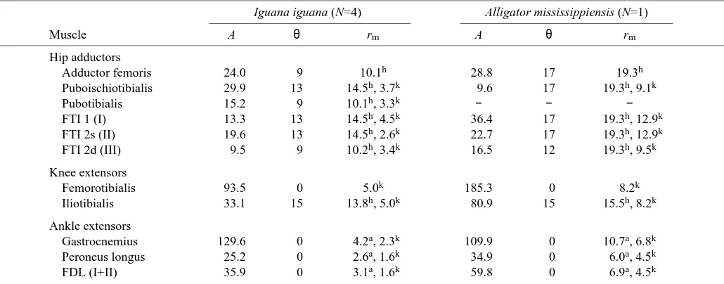
Table 1. Mean anatomical data from hindlimb muscles of experimental animals
Iguana iguana (N=4) Alligator mississippiensis (N=1)
Muscle A θ rm A θ rm
Hip adductors
Adductor femoris 24.0 9 10.1h 28.8 17 19.3h
Puboischiotibialis 29.9 13 14.5h, 3.7k 9.6 17 19.3h, 9.1k
Pubotibialis 15.2 9 10.1h, 3.3k − − −
FTI 1 (I) 13.3 13 14.5h, 4.5k 36.4 17 19.3h, 12.9k
FTI 2s (II) 19.6 13 14.5h, 2.6k 22.7 17 19.3h, 12.9k
FTI 2d (III) 9.5 9 10.2h, 3.4k 16.5 12 19.3h, 9.5k
Knee extensors
Femorotibialis 93.5 0 5.0k 185.3 0 8.2k
Iliotibialis 33.1 15 13.8h, 5.0k 80.9 15 15.5h, 8.2k
Ankle extensors
Gastrocnemius 129.6 0 4.2a, 2.3k 109.9 0 10.7a, 6.8k
Peroneus longus 25.2 0 2.6a, 1.6k 34.9 0 6.0a, 4.5k
FDL (I+II) 35.9 0 3.1a, 1.6k 59.8 0 6.9a, 4.5k
A, cross-sectional area of muscle (in mm2); θ, angle between the muscle and the long axis of the bone (in degrees); rm, moment arm of the
muscle (in mm) about the joint indicated by the superscript letter (h, hip; k, knee; a, ankle); FDL, flexor digitorum longus; FTI, flexor tibialis internus.
By convention, individual muscles in the FTI complex are identified by arabic numerals for Iguana iguana (s and d refer to superficial and deep muscles) and roman numerals for Alligator mississippiensis.
[image:5.612.51.566.464.666.2]Muscular contributions to limb bone torsion were not estimated. The tibial insertions of muscle groups active during stance are primarily in the plane of knee flexion and extension and are unlikely to promote substantial torsion; therefore, tibial torsion was assumed to be caused by the ground reaction force alone. However, the primary stance phase retractor of the femur in lizards and crocodilians, the caudofemoralis, inserts ‘ventrally’ on the femur and, thus, causes rotation of the femur about its long axis during retraction (Snyder, 1962; Gatesy, 1997). For most of stance, the caudofemoralis acts to produce moments at the hip that augment those due to the ground reaction force, both as a retractor and as a femoral rotator (see Results); therefore, calculations of caudofemoralis force based on joint equilibrium cannot be made without further assumptions about the activity of its antagonists. Rather than make such assumptions, torsional moments for the femur were calculated as though they had been induced by the ground reaction force alone. Torsional moments and stresses are almost certainly higher than indicated by these minimum estimates.
After calculation of muscle forces, the bending moments and axial and bending stresses acting in the limb bones were evaluated following published methods (Biewener, 1983a; Biewener and Full, 1992), with modifications for three-dimensional stress analysis. Anatomical data for skeletal elements from experimental animals are reported in Table 2. Compressive axial stresses (σax) in the midshafts of the femur
and tibia were calculated as:
σax= −[∑(Fmcosθax) + GRFax]/A , (2)
where θax is the weighted mean angle between each muscle
group and the long axis of the bone, ∑(Fmcosθax) is the sum
of muscle force components acting along the bone axis, GRFax
is the component of the ground reaction force acting along the bone axis and A is the cross-sectional area of the bone at midshaft. To calculate the cross-sectional area of each element, the limb bones excised from the killed animals were cut at midshaft, and a magnified tracing of each cross section was made. Endosteal and periosteal outlines from these tracings were entered into a computer using a digitizing tablet, and area calculations were made using a custom-designed QuickBasic routine.
To calculate bending stresses acting at the midshafts of the bones, bending moments induced by transverse components of the ground reaction force (GRFtr) and axial components of the
ground reaction force acting about the longitudinal curvature of the bone (GRFax), together with the net bending moment
induced by muscular forces (Fm), were calculated for each
bone in each of the two perpendicular planes illustrated in Fig. 1. Forces and moments considered in these calculations are illustrated in the ‘dorsoventral’ direction for the femur in Fig. 3. The moment due to the transverse component of the ground reaction force at midshaft (Mtr) was calculated as:
Mtr= GRFtr(L/2) , (3)
where L is the length of the bone. The bending moment
induced by the axial component of the ground reaction force due to bone curvature (Mc) was calculated as:
Mc= GRFaxrc, (4)
where rcis the moment arm of GRFaxdue to bone curvature
(Biewener, 1983a; Biewener, 1983b). Values of rc were
measured from outline sketches of the bones traced from photographs of each specimen in the defined anatomical views. Because the limb muscles insert on the bone cortices, muscular forces act at a distance from the central axis of the bone. Therefore, the midshaft bending moment induced by each muscle group (Mm) was calculated as the vector cross product
of the muscle force (Fm) and the moment arm of the muscles
about the midshaft centroid (Beer and Johnston, 1997): Mm= Fm(rbsinθb) , (5)
where rbis the distance between the centroid and the point of
application of muscle force on the bone surface, and θbis the
angle between rband the line of action of the muscle.
After calculating the net bending moment acting in each of the perpendicular anatomical directions for each bone (Mb/dir),
the bending stress in each direction (σb/dir) was calculated as:
σb/dir= Mb/dir(y/I) , (6)
[image:6.612.314.562.96.256.2]where y is the distance from the neutral axis of bending to the bone cortex in the direction under consideration, and I is the second moment of area for bending about that neutral axis.
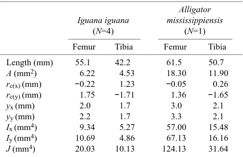
Table 2. Mean anatomical data from hindlimb bones of experimental animals
Alligator Iguana iguana mississippiensis
(N=4) (N=1)
Femur Tibia Femur Tibia
Length (mm) 55.1 42.2 61.5 50.7 A (mm2) 6.22 4.53 18.30 11.90
rc(x) (mm) −0.22 1.23 −0.05 0.26
rc(y) (mm) 1.75 −1.71 1.36 −1.65
yx (mm) 2.0 1.7 3.0 2.1
yy (mm) 2.2 1.7 3.3 2.1
Ix (mm4) 9.34 5.27 57.00 15.48
Iy (mm4) 10.69 4.86 67.13 16.16
J (mm4) 20.03 10.13 124.13 31.64
Subscript x denotes the anatomical ‘anteroposterior’ direction for the femur and tibia; subscript y denotes the ‘dorsoventral’ direction for the femur or the ‘mediolateral’ (flexion–extension) direction for the tibia (see Fig. 1 for definitions).
A, cross-sectional area of bone; rc, moment arm due to bone
curvature; y, distance from neutral axis to bone cortex; I, second moment of area; J, polar moment of area.
Data from the tibia are not corrected for the effects of the fibula. Curvature sign conventions: femurx, + = concave ‘posterior’, – =
concave ‘anterior’; femury, + = concave ‘ventral’, – = concave
‘dorsal’; tibiax, + = concave ‘posterior’, – = concave ‘anterior’;
Both y and I were calculated from the digitized limb bone sections used to calculate cross-sectional area (Table 2). The magnitude of net bending stress (σb/net) in the midshaft section
was calculated as the vector sum of the stresses in the two perpendicular anatomical planes for each bone. In the femur, for instance:
σb/net= (σb/DV2+ σb/AP2)0.5, (7)
where DV and AP refer to the ‘dorsoventral’ and ‘anteroposterior’ directions, respectively. The orientation of peak bending stress was also calculated. For the femur, the angular deviation of peak stress from the ‘anteroposterior’ axis (αb/net) is:
αb/net= tan−1(σb/DV/σb/AP) . (8)
The net neutral axis of bending is perpendicular to this axis. Net longitudinal stresses at the points of peak tensile and compressive bending stress then were calculated as the sum of axial and bending stresses. Torsional stress (τ) due to the ground reaction force was also calculated:
τ= T(yt/J) , (9)
where T is the torsional moment applied to the bone by the ground reaction force, yt is the distance from the centroid of
the bone to its cortex and J is the polar moment of area
(Wainwright et al., 1976). J was calculated from each digitized cross section, and yt for each section was calculated as the
average of the y values for the two perpendicular anatomical directions (Table 2).
In stress calculations for the tibia, additional steps were required to account for contributions of the fibula to load-bearing in the crus. In both I. iguana and A. mississippiensis, the fibula is robust, and the distance between the tibia and fibula is large; therefore, failure to consider the fibula could lead to substantial overestimation of tibial stresses. To evaluate the effects of the fibula on tibial stresses, calculations of y and I in the ‘anteroposterior’ and ‘mediolateral’ anatomical directions, and of ytand J, were made from a cross section of
an articulated tibia and fibula from an iguana similar in size to the experimental animals used in this study (Fig. 4). The ratio yt/J and the ratios y/I for each of the two anatomical planes
then were calculated to determine scaling factors to apply to the initial tibial stress calculations. The relative cross-sectional areas of the tibia and fibula also were calculated to assess the contribution of the fibula to resistance to axial stresses. These calculations indicated that, to account for stress resistance imparted by the fibula, tibial stresses should be multiplied by the following correction factors: ‘anteroposterior’ stress, 0.11; ‘mediolateral’ stress, 0.53; shear stress, 0.37; axial
Fkext
Fadd GRFDV
MGRF(DVhip)
radd rc
rb(add) rb(kext)
b(kext)
b(add)
GRFax
L/2
Dorsal
Ventral
Longitudinal axis
Tibia
θ
θ = – F
[image:7.612.66.558.63.311.2]addradd
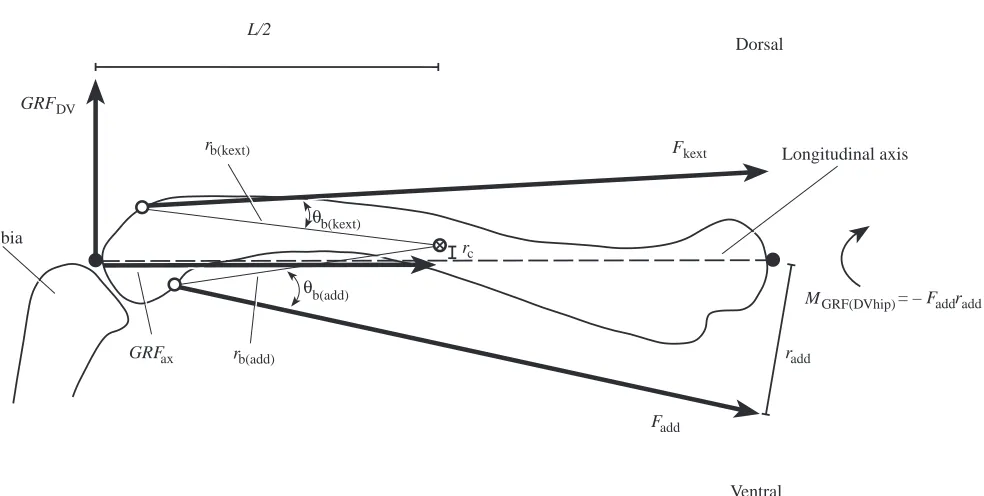
Fig. 3. Anterior view diagram of the femur and proximal tibia of Iguana iguana illustrating the forces and moments acting in the anatomical ‘dorsoventral’ direction. The centers of the proximal and distal articular surfaces are indicated by filled circles; points of application of adductor and knee extensor muscles (i.e. proximal extents of distal articular surface) are indicated by open circles; 丢 indicates the midshaft centroid of the femur. GRFax, the component of the ground reaction force acting along the long axis of the femur; rc, the bending moment arm of GRFax
due to bone curvature; GRFDV, the component of the ground reaction force acting transverse to the femur in the ‘dorsoventral’ direction; L/2,
the bending moment arm of GRFDVabout the midshaft of the femur (half femur length); MGRF(DVhip), the moment of the ground reaction force
about the hip; Fkext, the force exerted by the knee extensor muscles; rb(kext), the moment arm of the knee extensors about the midshaft centroid; θb(kext), the angle between rb(kext)and the line of action of the knee extensors; Fadd, the force exerted by the hip adductor muscles; radd, the
moment arm of the hip adductors about the hip; rb(add), the moment arm of the hip adductors about the midshaft centroid; θb(add), the angle
between rb(add)and the line of action of the hip adductors. Net lines of action of the knee extensors and hip adductors are illustrated. Force
compressive stress, 0.67. The effect of the fibula is to shift the net centroid of the combined section of the tibia and fibula ‘posteriorly’ and ‘medially’. Consequently, the ‘posterior’ and ‘medial’ cortices of the tibia contribute less to stress resistance than the ‘lateral’ and ‘anterior’ cortices. The effect of this shift in the neutral axis on values of y was taken into account and applied to calculations of stress for those surfaces (Fig. 4). These corrections were based on I. iguana, so they are a potential source of error for calculations of stress in the A. mississippiensis tibia. In addition, calculations of y, I, ytand J
from the combined tibial and fibular sections assume that no motion is possible between the tibia and fibula (i.e. that shear is transferred completely across both elements). Although some motion of the tibia relative to the fibula appears to occur in lizards and crocodilians (Rewcastle, 1980; Landsmeer, 1990; see also tracings from X-ray cine frames in Brinkman, 1980a; Gatesy, 1991), this motion is highly restricted. Therefore, it seems likely that conservative calculations accounting for fibular load bearing will be better estimates of actual stresses in the tibia than values that do not.
Calculation of limb bone safety factors
Safety factors were calculated for the femur and tibia in I. iguana and A. mississippiensis as the ratio of tensile yield stress to peak tensile locomotor stress. ‘Mean’ safety factors were calculated from the mean values of peak stress and mechanical properties. In addition, when error ranges for peak stress and
mechanical property values were available, ‘worst-case’ (i.e. lowest possible) safety factors were calculated on the basis of mean yield stress minus 2 S.D. and mean peak stress plus 2 S.D. Peterson and Zernicke (Peterson and Zernicke, 1987) report a value of 218 MPa (no error range) for the tensile yield stress of the tibia of Dipsosaurus dorsalis, an iguanian lizard closely related to I. iguana (Norell and de Queiroz, 1991; Petren and Case, 1997). We used this value to calculate safety factors for the femur and tibia of I. iguana. Currey (1988) reported a value of 12.0±2.4 GPa (mean ± S.D., N=6) for the stiffness of A. mississippiensis femur in bending. Estimates of tensile yield stress in bending were calculated by multiplying Currey’s (Currey, 1988) stiffness value by 6495µε(tensile yield strain for alligator femur; Blob and Biewener, 1999), producing a mean yield stress of 78.1 MPa and a ‘worst-case’ yield stress of 46.6 MPa. These yield stress estimates for A. mississippiensis appear low, but are reasonable when compared with Currey’s (Currey, 1990) published value for the ultimate stress of alligator femur (108 MPa) and are derived in part from independently collected data (Blob and Biewener, 1999).
Tests of correlations between limb bone stress and limb posture
Because of the small sample size of steps obtained from A. mississippiensis, correlations between limb bone stress and limb posture were tested only in I. iguana. Measurements of the angle of the femur below the horizontal at the time of peak stress were extracted from the kinematic calculations for each run. Peak tensile and compressive stresses in the femur, together with stresses calculated for the anterior and dorsal cortices, were then regressed (least-squares) on these posture angles to evaluate the relationship between limb posture and load magnitude and to assess the degree of correspondence with patterns observed during strain measurements. Reduced major axis (RMA) slopes were calculated for regressions with significant correlation coefficients. RMA is the most appropriate method of regression for the evaluation of structural relationships between variables when both are subject to error (McArdle, 1988; LaBarbera, 1989).
Analyses of bone strain in alligators (Blob and Biewener, 1999) suggested that tensile strains on the ‘anterior’ cortex of the femur decrease with more upright posture, but tensile strains on the ‘ventral’ cortex and compressive strains on the ‘dorsal’ cortex increase with more upright posture. ‘Anterior’ tensile strains (or stresses) could decrease if axial compression increased as posture became more upright. However, increases in ‘dorsal’ and ‘ventral’ femoral stresses (and strains) could result from one of two alternative mechanisms. (i) Hip adductor muscles (on the ‘ventral’ femur) bend the femur in the opposite direction from the ground reaction force; thus, if adductors exert less force during more upright steps, they would mitigate bending due to the ground reaction force less effectively, causing higher ‘dorsal’ and ‘ventral’ stresses. Reduced adductor force could result from decreases in the hip abductor moment of the ground reaction force during more ‘Lateral’
‘Anterior’
Tibia 1 mm
Fibula Tension
Compression
Neutral axis
yt
yc
[image:8.612.47.292.75.277.2]Net centroid
Fig. 4. Midshaft cross sections of the tibia and fibula of Iguana
iguana illustrating their relative positions in the crus and the
displacement of the neutral axis of bending away from the centroid of either bone (filled circles) to the net centroid of the section. Rotation of the neutral axis is based on the results of the experiments. Portions of the bones experiencing compression are shaded; portions experiencing tension are unshaded. Because of the displacement of the neutral axis to the net centroid, the distance from the neutral axis to the ‘medial’ cortex of the tibia (yc) is smaller than
the distance from the neutral axis to the ‘lateral’ cortex (yt), placing
upright posture, which would occur if the ground reaction force either acts closer to the hip or decreases in magnitude during more upright steps. (ii) Alternatively, an increase in the force exerted by the knee extensor muscles (on the ‘dorsal’ femur) with upright stance, rather than a decrease in hip adductor force, might cause higher dorsoventral stresses. Increased knee extensor force could result from increases in the flexor moment at the knee, which would occur if ground reaction force magnitude, its moment arm at the knee, or the forces exerted by the knee flexor muscles (e.g. biarticular ankle extensors) were greater in more upright steps. To differentiate between these mechanisms, we regressed the following variables (for each filmed step) on femoral posture: the moment arms of the ground reaction force at the hip, knee and ankle at peak stress; the magnitude of the ground reaction force; and the magnitudes of the forces exerted by each major muscle group.
Statistical notes
Differences between group means were tested using non-parametric Mann–Whitney U-tests unless noted otherwise. Error ranges reported for all measurements are one standard deviation (S.D.).
Results
Overview of loading mechanics for the femur and tibia of I. iguana and A. mississippiensis
The mechanics of skeletal loading in I. iguana and A. mississippiensis hindlimbs are sufficiently similar to be summarized together as follows. Detailed results are provided in the sections that follow.
At the beginning of stance, the tibia is oriented nearly vertically and the femur is directed with its distal end anterior and ventral to its proximal end (Fig. 5). The ground reaction force is directed posteriorly and medially, exerting flexor moments at the ankle and knee and ‘dorsal’ (‘abductor’) and ‘posterior’ moments at the hip (Figs 6, 7). The ground reaction force also exerts torsional moments tending to rotate the right femur counterclockwise about its long axis if viewed from its proximal end and the right tibia clockwise if viewed from its proximal end (Fig. 7). Ground reaction force moments are countered by ankle extensors at the ankle and by hip adductors at the hip. Both of these add to the flexor moment produced at the knee by the ground reaction force, which is countered by the knee extensors. These muscular forces add to the axial components of the ground reaction force, compressing both elements and, with transverse components of the ground reaction force, applying bending moments about the midshaft of each bone. The bending moment induced on the tibia by the ground reaction force is in the plane of flexion and extension, summing with the bending moment of the ankle extensors to place the ‘lateral’ (dorsiflexor) surface in tension and the ‘medial’ (plantarflexor) surface in compression (Figs 8, 9). Through the beginning of stance, the hip adductors exert a moment that bends the femur ‘ventrally’, but the summed moment of the knee extensors and ground reaction force is
larger and opposes the adductors, producing a net moment tending to bend the femur ‘dorsally’ (i.e. placing its ‘dorsal’ surface in compression; Figs 8, 9). In the ‘anteroposterior’ direction, the bending moment induced by the ground reaction force places the ‘posterior’ cortex of the femur in compression; thus, the femur bends about an ‘anterodorsal–posteroventral’ axis to place its ‘posterodorsal’ cortex in compression and its ‘anteroventral’ cortex in tension. In addition to bending, both bones experience torsion and axial compression.
Through stance, ground reaction force magnitude increases and its orientation shifts anteriorly, but its medial inclination is slight (Fig. 6). By the time of peak stress (approximately mid-stance), the proximal end of the tibia rotates anteriorly and laterally relative to its distal end and the heel lifts off the ground, raising the distal tibia with respect to the proximal tibia (Fig. 6). Bending moments acting on the tibia become large in the ‘mediolateral’ (flexor–extensor) direction as the tibia is oriented nearly perpendicular to the ground reaction force and the ankle extensors must exert larger forces to counter increases in the ankle flexor moment (Figs 6–8). The femur adducts slightly, retracts to nearly 90 ° to the body, and rotates almost 90 ° about its long axis, shifting its anatomical ‘dorsoventral’ plane towards the original location of its anatomical ‘anteroposterior’ plane. With the limb bones in these positions, the ground reaction force maintains a flexor moment at the knee and a torsional moment tending to rotate the tibia ‘laterally’ (Fig. 7). The ‘posterior’ bending moment on the femur is maintained as the anatomical ‘posterior’ direction rotates to face upwards (i.e. dorsal) in an absolute frame of reference, and a larger component of the nearly vertical ground reaction force comes to act in this direction (Fig. 8). However, as the femur rotates axially and retracts, the ground reaction force component in the ‘dorsoventral’ direction gradually decreases in magnitude, and then increases in the opposite sense from its original direction. Thus, the ground reaction force comes to exert an ‘adductor’ moment at the hip (although ‘adduction’ is now aligned close to posterior in an absolute frame of reference) and to bend the femur ‘ventrally’ (placing the ventral cortex in compression) (Figs 7–9). With these shifts, the bending moment induced by the ground reaction force now opposes that induced by the knee extensors, but the moment of the extensors is larger and maintains the initial orientation of bending (i.e. posterodorsal compression, anteroventral tension). Early in stance, as the hip moves forward over the foot, the line of action of the ground reaction force comes to pass posterior to the long axis of the femur and to exert a torsional moment that augments that of the caudofemoralis (Figs 6, 7).
are summarized to provide a context for ground reaction force data and analyses of limb bone loading (Fig. 5). All six trials from the alligator were fast walking trots (0.62±0.21 m s−1) requiring considerable exertion from the animals (gait designations follow the conventions of Hildebrand, 1976).
However, successful trials (isolated right hindfoot falls on the force platform) from iguanas included walking trots (1.29±0.45 m s−1, N=8), running trots (1.47±0.48 m s−1, N=8), single-foot diagonal sequence runs (1.92±0.32 m s−1, N=3) and bipedal running steps (2.00±0.31 m s−1, N=7). In some cases,
-80 -60 -40 -20 0 20 40 60 100 120 140 160 180
0 20 40 60 80 100 80 100 120 140
20 40 60 80
0 20 40 60 80 100 120
Alligator mississippiensis
0 0.2 0.4 0.6 0.8
B
Time (s)
Plantarflexion
Dorsiflexion
Dorsiflexion
Plantarflexion
Extension
Flexion
Protraction
Retraction
Adduction
Abduction
Horizontal
Vertical 180
100 120 140 160
0 0.12 0.16
80 100 120 140 60 80 100 120
20 40
Ankle
0 20 40 60 80 100 0 20 40 60 80 -80 -60 -40 -20 0 20 40 60
Iguana iguana
0.04 0.08
A
Knee
T
ibia AP
Femur AP
Femur D
V
MP
Angle (de
[image:10.612.225.565.69.671.2]grees)
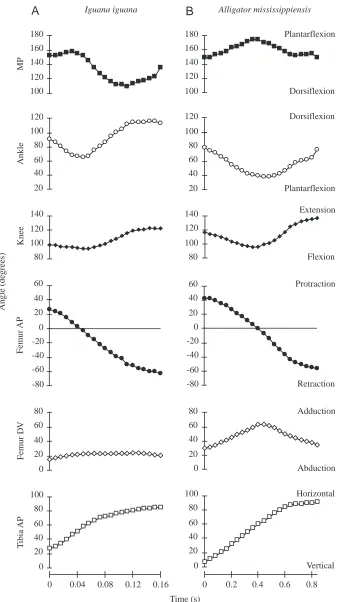
Fig. 5. Kinematic profiles (changes in joint angles over time) for representative steps from (A)
Iguana iguana and (B) Alligator mississippiensis.
joint angles at which particular movements begin or end (although not necessarily total angular excursions) differ significantly among gaits. However, the timing of particular joint motions (e.g. flexion or extension) generally do not. Furthermore, force measurements and bone stress calculations (see below) do not indicate significant correlations between bone loading and gait or speed over the ranges observed in I. iguana. In the context of ground reaction force data, therefore, a single, overall kinematic pattern is described for I. iguana. Thus, unless noted otherwise, samples sizes for mean values reported in the following sections are N=6 trials for A. mississippiensis and N=26 trials for I. iguana.
Kinematics of fast locomotion in I. iguana
The foot is placed flat on the ground at the beginning of stance in I. iguana, with the toes angled lateral to the direction of travel. The tibia is usually inclined slightly anteriorly (12±16 °) and medially (8±10 °) from vertical (i.e. the proximal end is anterior and medial to the distal end) (Fig. 5A). The femur is angled anteriorly 43±11 ° from the transverse plane, with the distal end depressed 19±10 ° relative to the proximal end. As Brinkman (Brinkman, 1981) described, initial flexions of the ankle and knee usually precede substantial movement at the hip or metatarso-phalangeal joint. The ankle and knee both form nearly right angles at the start of the step (89±18 ° and 105±19 °, respectively). Both joints are flexed for approximately the first third of the step, reaching angles of 59±15 ° (ankle) and 80±13 ° (knee), after which both joints are extended (to 123±13 ° and 121±14 °, respectively).
During flexion and extension of the knee and ankle, the tibia is inclined further laterally and anteriorly (Figs 5A, 6). Through the first 30–50 % of the step, these movements coincide with retraction and adduction of the femur, with maximum femoral adduction reaching 38±10 ° below horizontal. The femur is abducted as retraction continues after midstep, eventually reaching nearly the same dorsoventral position as at the start of the step (18±6 °). However, by midstep, dorsiflexion at the metatarso-phalangeal joint begins, raising the ankle off the substratum and raising the distal end of the tibia relative to the proximal end. As a result, the tibia continues to be inclined more anteriorly through the step (86±10 °), often placing the ankle higher than the knee late in stance. Lateral inclination of the tibia also increases through midstep, but as the knee is re-extended the tibia becomes more closely aligned with the direction of travel. These rotations of the tibia reflect substantial axial rotation of the femur, such that the anatomical ‘dorsal’ aspect of the femur comes to face anteriorly in an absolute frame of reference. By toe-off, the femur has typically been retracted through an arc of at least 80 °.
Kinematics of fast locomotion in A. mississippiensis
The alligator placed its foot with the toes lateral to the direction of travel (21±5 °), and with a slight upward arch at the metatarso-phalangeal joint (17±5 °) that was flattened through the early portion of the step. The tibia is initially
inclined slightly more anteriorly (18±10 °) and less medially (1±6 °) than in I. iguana (Fig. 5A,B), although only the difference in medial inclination was significant (P=0.030). The femur is initially 38±8 ° anterior to the transverse plane, similar to I. iguana; however, A. mississippiensis begins stance with a significantly more adducted femur (34±7 ° below horizontal) than I. iguana (P=0.003).
At the beginning of stance, the ankle is slightly more flexed (72±11 °) and the knee slightly more extended (109±11 °) in A. mississippiensis than in I. iguana (Fig. 5A,B). The ankle is flexed until approximately midstep, closing to 39±4 ° (smaller than I. iguana: P<0.001), before extending to nearly 90 °. In contrast to slow walking (Gatesy, 1991), the knee is flexed through the first third of the step during fast locomotion in A. mississippiensis. However, knee flexion is approximately 10 ° less than in I. iguana, leaving the knee at a significantly greater (P=0.003) obtuse angle (nearly 100 °) when its extension begins. As in I. iguana, the tibia becomes more laterally and anteriorly inclined through the first half of stance in A. mississippiensis, coinciding with femoral retraction and adduction (Fig. 5A,B). Femoral adduction reaches a maximum of 68±6 ° below horizontal at midstep, before abducting to return to its starting angle (36±4 °) by toe-off. As in I. iguana, flexion at the metatarso-phalangeal joint raises the heel off the ground and raises the distal tibia relative to its proximal end, leading to continued anterior inclination of the tibia throughout the step (maximum 93±3 °) that reflects axial rotation of the femur. Later in stance, the tibia becomes aligned more closely with the sagittal plane as the knee is extended. The femur retracts through an arc of at least 90 ° through stance. Ground reaction forces and limb orientation
The ground reaction force is oriented posteriorly when the foot is placed on the ground and shifts anteriorly through stance (Fig. 6), reflecting initial deceleration of the animal at foot contact, followed by reacceleration as the foot pushes off the substratum (e.g. Alexander, 1977; Cavagna et al., 1977). The ground reaction force is also oriented medially at the beginning of the step in I. iguana (Fig. 6). However, the vertical component of the ground reaction force increases much more rapidly than the medial component through stance, with medial component magnitude averaging less than 15 % of vertical component magnitude by midstep. As a result, the medial inclination of the ground reaction force decreases considerably through the first 10–15 % of the step and averages only 8±7 ° when peak limb bone stress is developed near midstep. In A. mississippiensis, the ground reaction force often is oriented slightly laterally at the start of the step and can remain so at peak stress; however, this inclination is very small (3±7 °), indicating an essentially vertical force. Both vertical and medial components of the ground reaction force reach maxima near midstep.
essentially parallel to the ground and the ground reaction force shifts more vertically, the angle between the two (from the distal end of the bone) increases, reaching 62±6 ° in A. mississippiensis and 74±17 ° in I. iguana at peak stress. Thus, in spite of the near-vertical orientation of the ground reaction force at midstep, the kinematics of these species result in a large component of the reaction force acting normal to the tibia (‘posteromedially’ in the anatomical frame of reference), indicating substantial bending loads. Peak forces normal to the tibia in these species are usually approximately equal to or slightly greater in magnitude than forces acting along the tibial axis. In addition, the lateral deflection of the foot, combined with an essentially vertical ground reaction force, suggests that the ground reaction force will usually act lateral to the axis of the lower leg.
In both species, the femur begins the step directed anteriorly and subhorizontally. While the femur is retracted through the step (i.e. the knee moves posteriorly with respect to the hip), the hip is also moving forwards. The femur therefore passes over the foot and, thus, over the point from which the ground reaction force originates. As the inclination of the ground reaction force shifts anteriorly through the step and becomes more vertically directed, the angle between it and the femur
generally decreases until midstep, but then increases as the leg is further extended until toe-off (Fig. 6). Thus, in contrast to the tibia, the femur is often most closely aligned with the ground reaction force at peak stress. Nonetheless, this angle is large in both species (A. mississippiensis, 38±7 °; I. iguana, 40±16 °). As for the tibia, the magnitudes of ground reaction force components acting transverse to the femur roughly equal those acting along the axis of the femur for much of the step in both species, suggesting that the femur also experiences substantial bending. This bending is initially directed ‘dorsoventrally’; however, as the femur rotates about its long axis, the ground reaction force (directed almost vertically for most of the step) comes to act closer to the anatomical ‘anteroposterior’ direction. In addition, despite the anterior shift in the orientation of the ground reaction force through stance, forward movement of the hip causes the line of action of the ground reaction force to act posterior (in an absolute frame of reference) to the femur for much of the step, including the time of peak stress.
Moments exerted by the ground reaction force In both species, the ground reaction force exerts moments Hip
Knee
Ankle MP IP1 Toe 0.68 N
Frame 16 (75%) 2.38 N
A
Frame 5 (20%) Frame 11 (50%) 5.77 N Iguana: lateral view
Direction of travel
5.77 N
Hip
Knee
Ankle
MP Toe
B
Frame 11 (50%) Iguana: anterior view
16 0
1 2
11 21
-1 5
Medial -1
0 1 2 3 4 5 6
11 21
5 16
Vertical
-1 0 1 2
11 16 21 5
Horizontal
Frame 0.8
0 0.16
Time (s)
C
Femur
Tibia
F
[image:12.612.50.558.78.394.2]orce (N)
tending to flex (collapse) both the knee and ankle for most of stance (Fig. 7). For equilibrium to be maintained at these joints, the knee and ankle extensor muscles must be active to counteract these moments. Both knee and ankle moments are small at the beginning of stance, when ground reaction force
magnitudes are low. The moments at both joints increase as the ground reaction force magnitudes increase towards midstep, but then decline as ground reaction force magnitudes decrease towards the end of the step (Fig. 7).
Moments about the hip are most simply described with -0.2
0 0.2
-0.1 -0.05 0 0.05
-0.4 -0.2 0 0.2
Plantarflexion
Dorsiflexion
-0.2 0 0.2
-0.1 -0.05 0 0.05
0.16 0.12 0.08 0.04
0 0 0.2 0.4 0.6 0.8
Time (s)
Rt. prox. clock. Rt. prox. counter.
T
orsion (tibia)
-0.2 0 0.2
Rt. prox. clock. -0.05
0 0.05 0.1
Rt. prox. counter.
T
orsion (femur)
Ankle
-0.05 0 0.05 0.1
Flexion
Extension
Knee
Moment (N
m)
-0.15 -0.1 0 0.05 0.1
-0.05
-0.6 -0.4 -0.2 0 0.2 0.4
‘Ventral’
‘Dorsal’
Hip (‘D
V’)
-0.6 -0.4 -0.2 0 0.2
-0.2 -0.15 -0.1 -0.05 0 0.05
Alligator mississippiensis
B
Iguana iguana
A
‘Anterior’
‘Posterior’
Hip (‘
[image:13.612.220.572.72.661.2]AP’)
reference to the two anatomical planes of the femur defined in Fig. 1. With few exceptions, the ground reaction force exerts a moment at the hip that tends to rotate the femur ‘posteriorly’ about the axis of the anatomical ‘anteroposterior’ plane throughout stance (Fig. 7). However, through the step, axial rotation of the femur and elevation of the heel off the substratum shift the ‘anteroposterior’ plane so that its ‘anterior’ surface comes to face ventrally in an absolute frame of reference. Thus, after a 90 ° rotation, a ‘posterior’ moment would be a tendency to rotate the femur upwards (dorsally) in an absolute frame of reference (Fig. 1). The magnitude of this moment generally increases until midstep, after which it steadily decreases (Fig. 7). With respect to the anatomical ‘dorsoventral’ plane, the ground reaction force initially exerts an ‘abductor’ moment (i.e. it rotates the distal end of the femur dorsally relative to its proximal end: Fig. 7). However, as the femur rotates about its long axis and the hip moves forwards through the step, this ‘abductor’ moment reaches a maximum early in the step, then decreases to zero and increases in the opposite direction, becoming an ‘adductor’ moment tending to rotate the femur ‘ventrally’ (Fig. 7). After 90 ° axial rotation of the femur, such a tendency to rotate the femur ‘ventrally’ would equal a tendency to rotate the femur posteriorly (backwards) in an absolute frame of reference (Fig. 1). Further, it indicates that hip abductor muscles, rather than adductors, must be activated late in stance to counter this moment.
The ground reaction force also exerts torsional moments about the long axes of the femur and tibia (Fig. 7). As stance begins, the ground reaction force often briefly acts anterior to the femur, producing torsional moments that tend to rotate the femur posteriorly (i.e. rotate the right femur clockwise if viewed from its proximal end). However, as the femur retracts and the hip moves forward, much larger, anteromedial torsional moments develop (tending to rotate the right femur counterclockwise if viewed from its proximal end) that peak after midstep (Fig. 7). Torsional moments for the tibia were more variable; however, for 19 of the 26 trials in I. iguana (73 %), the greatest torsional moments indicated lateral rotation of the tibia (clockwise looking down on the proximal end of a vertically oriented right tibia: Fig. 7). Two of the six trials for A. mississippiensis exhibited similar tibial torsion, although the other four showed medially directed torsional moments.
Hindlimb muscle forces
Because of the large moments exerted by the ground reaction force about the hindlimb joints, hindlimb muscle forces are also large and make substantial contributions to axial and bending stress in the femur and tibia of I. iguana and A. mississippiensis. The bending moments induced by the limb muscles usually exceed those induced by the components of the ground reaction force acting transverse to the limb (Fig. 8). For example, the knee extensors originate on the ilium (iliotibialis) and on the ‘dorsal’ femur (femorotibialis); forces exerted by these muscles bend the distal end of the femur
dorsally towards the proximal end, placing its ‘dorsal’ aspect in compression. Hip adductors act in an opposite fashion, bending the distal femur ventrally and placing the ‘dorsal’ surface in tension. Because the line of action of the adductors is at a greater net angle to the long axis of the femur than the line of action of the knee extensors (Fig. 3; Table 1), a relatively greater component of adductor force than of knee extensor force acts transverse to the femur. However, the knee extensors must exert force to counter not only the flexor moment of the ground reaction force but also flexor moments produced by the ankle extensors and hip adductors that span the knee. Further, adductor force tends to decrease by midstep as the femur rotates about its long axis and the ground reaction force comes to generate an ‘adductor’ moment itself. As a result of these factors, and the long moment arm of the adductors at the hip compared with that of the knee extensors at the knee, peak knee extensor forces can be 4–6 times greater than peak adductor forces. Consequently, muscles spanning the length of the femur generate a large net bending moment that places the ‘dorsal’ cortex in compression (Fig. 8). At the beginning of the step, bending moments due to the ground reaction force also bend the femur ‘dorsally’ and the muscular and external moments are additive. Through the step, however, axial rotation of the femur causes the ground reaction force bending moment to change direction and bend the femur ‘ventrally’. Nonetheless, because the knee extensors must still counter a large flexor moment at the knee, they continue to exert substantial force and produce a bending moment that exceeds that due to the ground reaction force. Muscular forces thus act to maintain a ‘dorsally’ directed bending moment throughout stance.
‘mediolateral’ bending moment produced by the ankle extensors deflects the axis of tibial bending closer to the ‘mediolateral’ plane.
Femoral and tibial stresses
Stress calculations for the femur of I. iguana and A. mississippiensis confirm that it is loaded in a combination of axial compression, bending and torsion in both species (Table 3). In both species, the ‘posterodorsal’ cortex of the femur is placed in compression whereas the ‘anteroventral’
cortex is placed in tension (Fig. 9). The direction of femoral bending changes little for the first two-thirds (or more) of stance, with the neutral axis typically oriented 30–40 ° from the anatomical ‘anteroposterior’ axis. Later in stance, neutral axis orientation gradually shifts, keeping the net direction of bending closer to dorsoventral (in an absolute frame of reference) by tracking axial rotation of the femur and rotating towards the anatomical ‘dorsoventral’ axis (Fig. 9). Axial compression is superimposed on bending in the femur of both species (−2.4±0.4 MPa in A. mississippiensis, −4.9±2.0 MPa in I.
GRFtr (‘AP’)
GRFtr (‘DV’)
-0.15 -0.1 -0.05 0 0.05 0.1 0.15
Adductors (‘DV’)
Knee extensors (‘DV’)
Bending moment (N
m)
A
Femur Iguana iguana
0.04 0.08 0.12 0.16 0
Time (s)
GRFtr (‘ML’) GRFax (‘ML’) GRFtr (‘AP’)
-0.15 -0.1 -0.05 0 0.05 0.1 0.15
GRFax (‘AP’)
Ankle extensors (‘ML’)
Bending moment (N
m)
Tibia Iguana iguana
[image:15.612.214.565.74.609.2]B
Fig. 8. Comparison of bending moments exerted in the femur (A) and tibia (B) ofIguana iguana by muscle forces and
components of the ground reaction force. Anatomical directions in which moments act are indicated as: ‘AP’, anteroposterior; ‘DV’, dorsoventral (femur); ‘ML’, mediolateral (tibia).
GRFax, the bending moment exerted by
the axial component of the ground reaction force due to bone curvature in the direction indicated; GRFtr, the
iguana); thus, peak compressive stresses are greater than peak tensile stresses (Fig. 9). Peak femoral stress was not significantly correlated with speed in either species or with gait in I. iguana (non-parametric Kruskal–Wallis test). However, mean peak stresses in the femur of I. iguana (+27.1±10.8 MPa ‘anteroventral’, −37.0±14.2 MPa ‘posterodorsal’) were significantly higher than those in A. mississippiensis (+11.7±1.5 MPa ‘anteroventral’, −16.4±2.2 MPa ‘posterodorsal’) (P<0.001 for both comparisons; Table 3).
In both I. iguana and A. mississippiensis, the tibia is loaded in bending, with the ‘lateral’ (dorsiflexor) surface in tension and the ‘medial’ (plantarflexor) surface in compression (Table 3; Fig. 9). The neutral axis is aligned close to the anatomical ‘anteroposterior’ plane for almost all of stance, although a slight ‘anterolateral–posteromedial’ inclination (5–10 °) increases at the end of the step. Axial compression is superimposed on tibial bending in both species (−2.3±0.5 MPa in A. mississippiensis,
−5.2±2.7 MPa in I. iguana). However, the fibula shifts the neutral axis of crural bending to place the tibia in net tension (i.e. peak tension exceeds peak compression) (Figs 4, 9). Peak stresses are lower in A. mississippiensis (+28.8±4.3 MPa ‘anterolateral’, −10.9±1.7 MPa ‘posteromedial’) than in I. iguana (+43.1±18.8 MPa ‘anteroventral’, −18.6±8.5 MPa ‘posterodorsal’), but only peak compression differs significantly (P=0.016; Table 3). For both species, mean peak tensile stresses in the tibia are significantly greater than mean peak tensile stresses in the femur (non-parametric paired sign tests, P<0.001 for I. iguana, P=0.031 for A. mississippiensis).
Shear stresses in the femur due to the ground reaction force average 1.9±0.5 MPa in A. mississippiensis and 5.8±2.8 MPa in I. iguana (Table 3). In the tibia, shear stresses (for steps exhibiting clockwise torsion of the tibia, viewed from its proximal end) averaged 0.7±0.3 MPa in A. mississippiensis and 2.6±1.3 MPa in I. iguana. As noted in the Materials and methods section, these values are minimum
estimates that do not account for torsion induced by the limb muscles.
Safety factor calculations
‘Mean’ safety factors in bending for the femur and tibia in I. iguana are 8.0 and 5.1, respectively (Table 3). These decrease to 4.5 (femur) and 2.7 (tibia) in the ‘worst-case’ estimates. For A. mississippiensis, ‘mean’ safety factors in bending for the femur and tibia are 6.7 and 2.7, respectively, and ‘worst-case’ estimates decrease to 3.2 (femur) and 1.3 (tibia) (Table 3). Safety factors in shear were not calculated because of the uncertainties in shear stress estimates from the force platform data.
Correlations of stress magnitudes with limb posture in I. iguana
Peak tensile and peak compressive stresses in the femur both tend to be higher during upright steps in I. iguana (Table 4). Stresses on the ‘anterior’ and ‘dorsal’ femoral cortices (at the time of overall peak stress) are also correlated with femoral posture: ‘anterior’ stress decreases significantly with more upright posture, whereas ‘dorsal’ stress increases significantly in more upright steps (Table 4). These trends are consistent with observations from A. mississippiensis in limb bone strain experiments (Blob and Biewener, 1999), verifying that tests of the alternative mechanisms proposed to explain these loading patterns (see Materials and methods) are warranted.
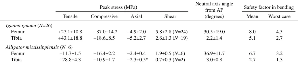
[image:16.612.44.562.99.213.2]Axial stress (at the time of overall peak stress) shows a significant trend to be higher among more upright steps (Table 4). However, neither the moment arm of the ground reaction force about the hip, nor the force exerted by the adductor muscles, is significantly correlated with femoral posture (Table 4). Instead, knee extensor force increases significantly among more upright steps (Table 4; Fig. 10). Besides adductor force (which did not change significantly with limb posture), both the ground reaction force and the Table 3. Mean peak stresses and safety factors calculated from force platform data for the femur and tibia of Iguana iguana and
Alligator mississippiensis
Neutral axis angle Peak stress (MPa)
from AP Safety factor in bending Tensile Compressive Axial Shear (degrees) Mean Worst case
Iguana iguana (N=26)
Femur +27.1±10.8 −37.0±14.2 −4.9±2.0 5.8±2.8 (N=24) 30.5±19.0 8.0 4.5 Tibia +43.1±18.8 −18.6±8.5 −5.2±2.7 2.6±1.3 (N=19) 2.2±1.4 5.1 2.7
Alligator mississippiensis (N=6)
Femur +11.7±1.5 −16.4±2.2 −2.4±0.4 1.9±0.5 (N=6) 36.9±11.7 6.7 3.2 Tibia +28.8±4.3 −10.9±1.7 −2.3±0.5* 0.7±0.3 (N=2) 3.0±0.8 2.7 1.3
Shear stresses are reported for counterclockwise rotation of the right femur and clockwise rotation of the right tibia, both bones viewed from their proximal end (sample sizes in parentheses); for all other other variables, N=26 for Iguana iguana and N=6 for Alligator mississippiensis.
Deviations of the neutral axis from the ‘anteroposterior’ (AP) axis of each bone are clockwise. See text for details of mean and worst-case safety factor calculations.
ankle extensors can potentially contribute to the flexor moment at the knee and, thus, require the knee extensors to exert higher countering forces. Neither the net ground reaction force nor its moment arm at the knee increased significantly among more upright steps (Table 4). However, ankle extensor force is significantly greater in more upright steps (Table 4; Fig. 10) because of increases in ankle joint moment (Table 4). The increase in ankle moment results from an increase in the moment arm of the ground reaction force about the ankle (i.e.
the moment arm of the force that the ankle extensors must counter to maintain equilibrium) (Table 4; Fig. 10) that is probably related to earlier lifting of the ankle from the substratum in more upright steps. Maximum metatarso-phalangeal flexion occurs earlier in upright steps (Table 4), shifting the origin of the ground reaction force further anteriorly along the foot at peak stress and increasing its moment arm and the joint moment at the ankle during upright locomotion (Fig. 10).
‘Anterior’
0 0.2 0.4 0.6 0.8 0 0.04 0.08 0.12 0.16
-60 -40 -20 0 20 40
Tibia
t
c
-60 -40 -20 0 20 40
Tibia
t
c
Stress (MP
a)
Neutral axis Neutral axis
‘Posterior’
‘Lateral’
‘Medial’
Time (s) 0
-60 -40 -20 20 40
t
c
Femur
Neutral axis
‘Posterior’
Neutral axis ‘Anterior’
‘Dorsal’
‘Ventral’ -60
-40 -20 0 20 40
c t
Femur
Stress (MP
a)
Iguana
[image:17.612.184.569.67.616.2]