• No results found

MR Imaging, Single photon Emission CT, and Neurocognitive Performance after Mild Traumatic Brain Injury

N/A
N/A
Protected

Academic year: 2020

Share "MR Imaging, Single photon Emission CT, and Neurocognitive Performance after Mild Traumatic Brain Injury"

Copied!
9
0
0

Loading.... (view fulltext now)

Full text

Figure

TABLE 1: Demographic data, trauma characteristics, imaging, and neurocognitive results of the mTBI patients
FIG 2.Images obtained from a 23-year-old man who was involved in a car accident.A, SPECT image shows a left frontal perfusion deficit.B, FLAIR image shows no lesions at this location.C, Owing to diffuse axonal injury, T2*-weighted image shows two deep hemorrhagic lesions.
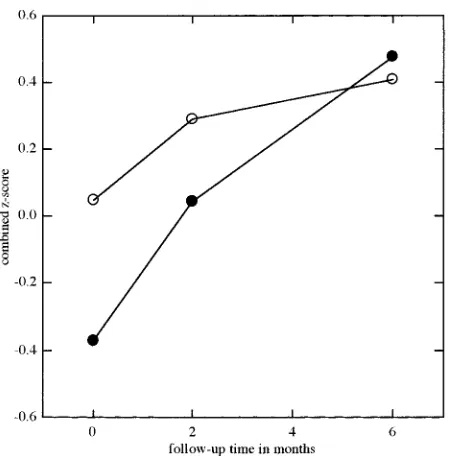
FIG 4.Combined z scores of the patients with normal (�) andabnormal (●) MR findings over the 6-month follow-up period.

References

Related documents