VORTEX FORMATION DURING TETHERED FLIGHT OF
FUNCTIONALLY AND MORPHOLOGICALLY TWO-WINGED
INSECTS, INCLUDING EVOLUTIONARY CONSIDERATIONS ON
INSECT FLIGHT
DMITRY L. GRODNITSKY ANDPAHVEL P. MOROZOV
V. N. Sukachev Institute of Forest, Siberian Branch of the Russian Academy of Sciences, Krasnoyarsk 660036, Russia
Accepted 31 March 1993
Summary
Tethered flight of six insect species (two pentatomid bugs, a moth, a butterfly, a muscid fly and a crane fly) was studied using several modifications of a dust flow visualization procedure. The spatial structure of the near vortex wake of flying specimens was reconstructed on the basis of two-dimensional flow pictures. The dynamics of the wake was followed during a stroke cycle, revealing interspecific differences in vortex formation.
It is suggested that insects create a single vortex ring during each stroke. Therefore, the hypothesis of double vortex chains advanced by Brodsky is not verified. The same is true of the jet hypothesis of Bocharova-Messner. While pronating at the top of their trajectory, the flapping wings throw air masses off their lower surfaces, but there is not a jet from between their upper sides. Flow separation from leading edges was found to be a rare phenomenon, taking place irregularly during the stroke cycle. That is why, contrary to widespread theoretical expectations, the Weis-Fogh fling mechanism is not likely to contain a leading edge separation bubble, which must follow stalling at the front part of the wings.
It is suggested that flying animals possess special mechanisms for extracting energy back from the near vortex wake. Some hypothetical adaptations for such an extraction in insects are put forward. Possible pathways for the evolution of insect flight are described.
Introduction
Progress in animal flight science during the last decade has been closely connected with the development of a new research field dealing with investigation of the structure and dynamics of the vortex wake behind flying specimens (Hummel and Goslow, 1991; Rayner, 1991). Interesting results have been published on birds (Kokshaysky, 1979; Kokshaysky and Petrovsky, 1979; Spedding, 1986, 1987; Spedding et al. 1984), bats (Rayner and Aldridge, 1985; Rayner et al. 1986) and insects (Ellington, 1980; Brodsky, 1985, 1986, 1988, 1991; Brodsky and Ivanov, 1983, 1984; Ivanov, 1990). Comparative analysis of the
data has created a background for fruitful thinking about some original aspects of fla p p i n g wing functioning (Brodsky and Ivanov, 1983, 1984; Brodsky, 1986) and about possible pathways of the evolution of insect flight (Brodsky, 1988, 1991; Rayner, 1988).
The expansion of our knowledge of the vortex wake is inevitably connected with the appearance of many views, which sometimes differ substantially. That is why further experimental research is necessary. Development of new experimental techniques and investigation of more insect species are both highly desirable as well.
In an earlier paper, we reported on vortex formation during a stroke cycle in a green lacewing – an insect with four uncoupled wings, which flap independently during fli g h t (Grodnitsky and Morozov, 1992). This species belongs to the so-called functionally four-winged insects. The work we present now was carried out with functionally and morphologically two-winged species using some new modifications of the former techniques. It is a common opinion that a functionally four-winged condition has been a primary characteristic of flying insects. During the evolution of insects, many unrelated groups have independently developed special adaptations for connecting the fore- and hindwing pairs, so that the functionally two-winged condition was achieved. Next, in many groups the hindwings lost their independence and began to diminish in size. In some taxa the hindwings became too small to play an aerodynamic role, and thus only the forewings produced useful forces. The latter are called morphologically two-winged insects. This state is characteristic of some tropical syntomid moths, some hymenopterans (Mymaridae: Mymar; Mymarommatidae), caddis flies (some Limnephilidae) and species from the superfamily Coccoidea (Hemiptera).
Some other groups have evolved a morphologically two-winged condition while reducing the forewings. These are the Strepsiptera and some of the Coleoptera, Phasmoptera, Gryllotalpidae, Tetrigidae and Tridactylidae. These insects have never had coupled fore- and hindwings. Their condition is derived directly from functionally four-winged forms.
It is easy to presume that such reconstructions of the flight apparatus led to some evolutionary changes in flight aerodynamics. Moreover, it has been just the functional features of insect wings which have caused their morphological evolution. Hence, the historical development of the flight apparatus is closely connected with changes in the structure and dynamics of the vortex wake. The data presented here provide a basis for constructing a hypothesis concerning this problem. Brodsky (1985, 1986, 1988) recently proposed a very interesting new hypothesis on vortex wake structure in insects that flap their wings at high (more than 70Hz) frequency. The model described by Brodsky contains some contradictions to the information on vortex formation during the flapping flight of insects (Grodnitsky et al. 1988). To resolve these contradictions, we undertook experiments with one of the insects studied by Brodsky.
Materials and methods
Species selected
Noctuidae), the European skipper Thymelicus lineola Ochsenheimer (Lepidoptera: Hesperiidae), the muscid fly Musca domestica L. (Diptera: Muscidae) and the crane fly
Tipula oleracea L. (Diptera: Tipulidae). Bugs, crane flies and skippers were caught in the
wild. Moths and muscid flies were raised from larvae, taken from laboratory populations in the Research Institute of Molecular Biology (Koltsovo town, Novosibirsk region) and in the Institute of Chemical Kinetics and Flame (Siberian Branch of the Russian Academy of Sciences, Novosibirsk).
Tethering
Specimens were tethered to fine entomological pins with the help of quickly drying ‘Moment’ glue. The moths, butterflies and dipterous flies were glued by the base of their abdomen, the bugs by the upper side of their prothorax. Before the tethering procedure, the crane flies, butterflies and moths were anaesthetized with ether vapour; the muscid flies were anaesthetized by cooling. The bugs were glued without anaesthesia.
Study of wing kinematics
Wing movements during tethered flight were analysed from high-speed ciné films, taken with an SCS-1M-16 camera. 16-mm negative KN-1 films (15 ASA) were used. The fil m s were taken at 3000–4000 frames per second. Flying specimens were situated between the camera and a KV-1000 lamp (power 1kW). Ciné films were taken in front, side and top view. Frame-by-frame analysis of the films let us determine several kinematic variables: wing positional angle f, stroke period T, downstroke-to-upstroke duration ratio Td/Tu,
longevity of different stroke phases (downstroke and upstroke, pronation and supination). Wingbeat frequency was measured with a stroboscope after each 15min of tethered fli g h t .
Visualization
A detailed description of the visualization procedure was given in our earlier paper (Grodnitsky and Morozov, 1992). Lycopodium spores served as visualizing dust. They were poured out of a container situated above the tethered, flying insect, under the influence of oscillations from a vibrator.
Photographs of the visualized airflow were taken using four different techniques. (1) Illumination of the vortex wake by a flat laser beam. This method gave images of only those dust particles that were struck by the light while moving in the beam plane. The three-dimensional vortex wake structure was then reconstructed from a series of photographs with different beam positions, and the changes in the wake during the stroke were described. This technique only worked on insects with a rather low wingbeat frequency. At higher flapping frequencies, the dust particles move faster and their photographic images become too thin for analysis.
This technique increased the amount of information contained in the photographs, and it also provided an opportunity to work on a fast-flapping insect such as Musca domestica. Nevertheless, the technique was not successful with the cotton moth, which demonstrated a rapid manoeuvre as soon as it heard the sound of an opening camera shutter. The moth seems to confuse this sound with the echolocation impulses of hunting bats. As a result, it was impossible to interpret the flow pictures.
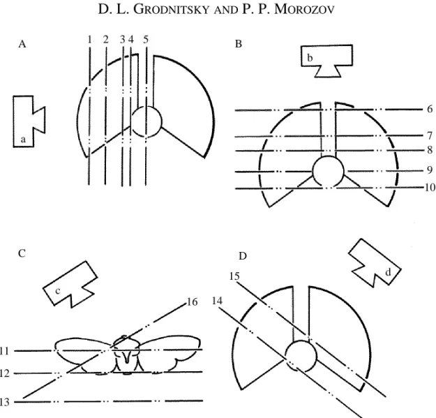
A three-dimensional reconstruction of the wake based upon photographs taken with techniques 1 and 2 demands pictures of different parts of the flow in different views. We therefore changed the position of the light plane during the experiments. The total number of positions was 16: five views from the side (Fig. 1A), five from above (Fig. 1B), three back views (Fig. 1C), two from the side and above (Fig. 1D) and one from the side and the front (Fig. 1C). The views and light plane positions used for each species are shown in Table 1.
(3) Illumination of the entire flow field using a stroboscopic flash lamp. A device had been made specially for this purpose, which gave 1–4 flashes at arbitrarily set intervals. Pictures obtained with this method contain comma-like particle images because of the rapid growth and rather slow decay of each flash. Therefore, it was possible to determine the direction of particle movement directly, judging by the tail of the comma. Results from techniques 1 and 2 are easier to interpret, but in some cases they lack information on the direction of flow.
While using this method, the camera was focused on one of the light plane positions from procedures 1 and 2. Eight photographic films taken with the help of a similar technique (using not stroboscopic, but single, flashes) have been obtained by Drs V. D. Ivanov and M. V. Kozlov in their tethered flight studies of crane fly aerodynamics, and were kindly presented to us.
(4) Illumination of the wake using the KV-1000 lamp. This method showed the succession of vortex structures found in experiments with techniques 1 and 2.
The methods outlined above were used to a varied extent in the studies of different model species. Detailed information on this is given in Table 1. In all the experimental situations specimens flew in still air.
Definitions of terms
In describing vortex structures we use the same terms as in the earlier paper (Grodnitsky and Morozov, 1992). As an aerofoil begins to move, the fluid will accelerate around the leading and trailing edges. The steep velocity gradients will generate a high vorticity. The separation point, initially on the top of an aerofoil, will gradually move backwards as vorticity is shed from the trailing edge into the fluid. This process stops when the circulation on the aerofoil is such that the Kutta-Joukovsky condition is satisfied and the flow leaves the trailing edge smoothly. The shed vorticity rolls up to form the
starting vortex at a point near the original position of the aerofoil. The circulation of the
tube represents a starting vortex. One end of the starting tube is attached proximally to the wing, the other to the tip of the wing. A flat section of the tube close to the tip reveals a tip
vortex. We can say that the starting tube is linked to the wing tip through the tip vortex.
Stopping tubes are created below the rear edges after the latter decelerate at the bottom of their trajectory and begin upward movement (Fig. 2B). A cross section of this tube by light plane shows the stopping vortex, which is a mirror image of the starting vortex. The stopping vortex of the downstroke is at the same time the starting vortex of the upstroke. Starting and stopping vortex tubes join each other through the tip vortex and thus form a vortex ring (Fig. 2C). Hence, a vortex tube and a vortex ring are volume structures, while vortices are flat images of their cross sections made with the flattened beam.
Results
Kinematics
The wingbeat frequency of each specimen was measured stroboscopically several times during a single flight. The results show that this variable was maximal at the beginning of flight, but then decreased during the experiment. This phenomenon has been noted in earlier works (Sotavalta, 1947; Ward and Baker, 1982; Grodnitsky and Kozlov, 1987). In some cases the decrease in frequency was quite significant, for example from 107 to 37 H z in P. juniperina ( T a b l e 1). Because of this dramatic decrease in wingbeat frequency, we tried to register flow patterns only during first 5–10min of each single fli g h t .
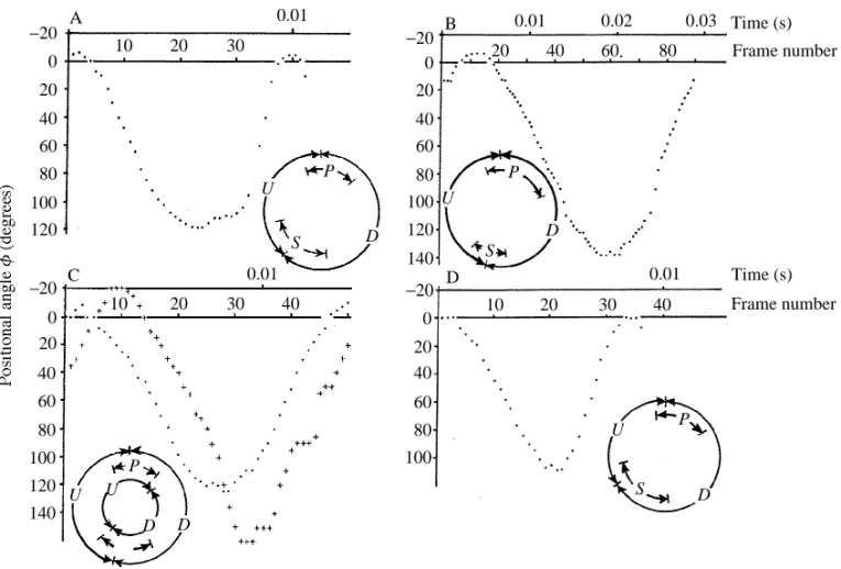
Considering also the earlier data on T. lineola wing kinematics (Brodsky and Grodnitsky, 1985), we can conclude that the curve of wing positional angle fagainst time is very similar in all investigated species (Fig. 3). Most different are the Td/Turatios
(Table 1). These may vary because the ratio is involved in the regulation of flight power for many species from different groups (Betts, 1986; Betts and Wootton, 1988; Grodnitsky, 1992). Hence, if a specimen tries to free itself from the tether it would possibly change the Td/Turatio to a considerable extent. Accepting this, the magnitude
and sign of Td/Tuchanges might depend on the component of total aerodynamic force (lift
or thrust) that the insect tries to alter.
Among the species studied, only the moth and the butterfly demonstrated a typical Weis-Fogh clap. The upper surfaces of their wings touch each other at the top of their trajectory. Following this contact, the wings open rather slowly, beginning with the leading edges; this is the fling phase. At the beginning and end of each stroke cycle, the wings of the moth and the butterfly undergo a kinematic pause, with the wings held still above the body for some time (Fig. 3B). The wings of the other three species do not clap at the end of the stroke cycle, but perform pronation without mutual contact. Therefore, there is no pause at the top of their movement (Fig. 3A,B,D). This is a so-called ‘near clap-and-fling’. Thus, there are two alternative patterns of kinematics in the upper part of the wing path: a smooth pronation without a clap and an interrupted pronation with a clap pause.
plane). Among the species in our study, the moth and the butterfly belong to the first group. The second one is represented by the bugs and dipterous flies (Table 1).
Kinematic analysis of the tethered crane flies allows us to determine the synchronization between wings and halteres. It was found that the halteres perform strokes with an amplitude higher than that of the wings (Fig. 3C). Phase relationships between the wings and the halteres strongly resemble those in functionally four-winged species, e.g. green lacewings (Ellington, 1984; Grodnitsky and Morozov, 1992). In these insects, the forewings lead the hind ones during the stroke cycle.
Aerodynamics
The beginning of the stroke
The flow visualization results for tethered flapping flight have shown an absence of stalling at the leading edges. The airflow moves smoothly around the leading edges without any formation of a separation vortex. All the species explored are alike in this respect, and all the techniques gave the same result, including straight visual observation of stroboscopic flow patterns.
B
C D
A
a
c
d b
1 2 3 4 5
6
7 8
9 10
11
12
13
16 15
14
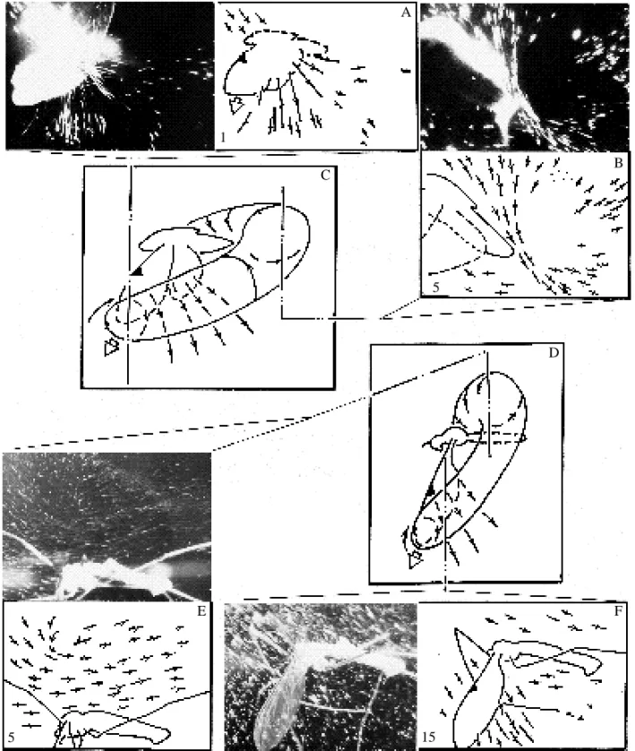
Some interspecific differences are observed later, when the movement of the trailing edges causes the generation of starting vortex tubes above the rear parts of the wings ( F i g . 4). In bugs and dipterous flies, this process is followed by the tubes’ formation along the whole length of the trailing edges (Fig. 4A–C). The distal ends of the two tubes are borne close to the forewing tips (Fig. 4D). Cross sections of the tubes show clearly visible tip vortices near the apical parts of the forewings (Fig. 4E). The proximal ends of the starting tubes stick to the base of the trailing edges at the beginning of a stroke. Transverse diameter and speed of rotation are not uniform along a starting tube. The most intensive vortex formation has been found near the most rapidly moving wing parts, namely the distal third. Vortices near wing hinges are less well developed.
Table 1. Morphological and kinematic variables of the species studied
Pitedia Heliothis Thymelicus Tipula Musca
Wing/wing couple aspect ratio 2.2 1.8 1.4 5.4 3.2 Stroke frequency (Hz) 37–107 30–40 25–35 38–70 90–120 Stroke plane Inclined Vertical Vertical Inclined Inclined
Td/Tu 1.60 1.2 − 1.18 2.00
Registration methods involved 2, 3 1 4 2, 3 2 Light plane positions involved 1–3, 5, 2, 5–8, 5 1–9, 1, 2, 5,
7–15 11–16 11, 13–15 7, 9, 11 13–15 Total number of frames 1000 700 2 films 800 600
Td, duration of downstroke; Tu, duration of upstroke.
B C
A
U
w
w
w
v.r. st.v.t. st.v.t.
sp.v.t.
t.v. st.v. sp.v.
Downstroke
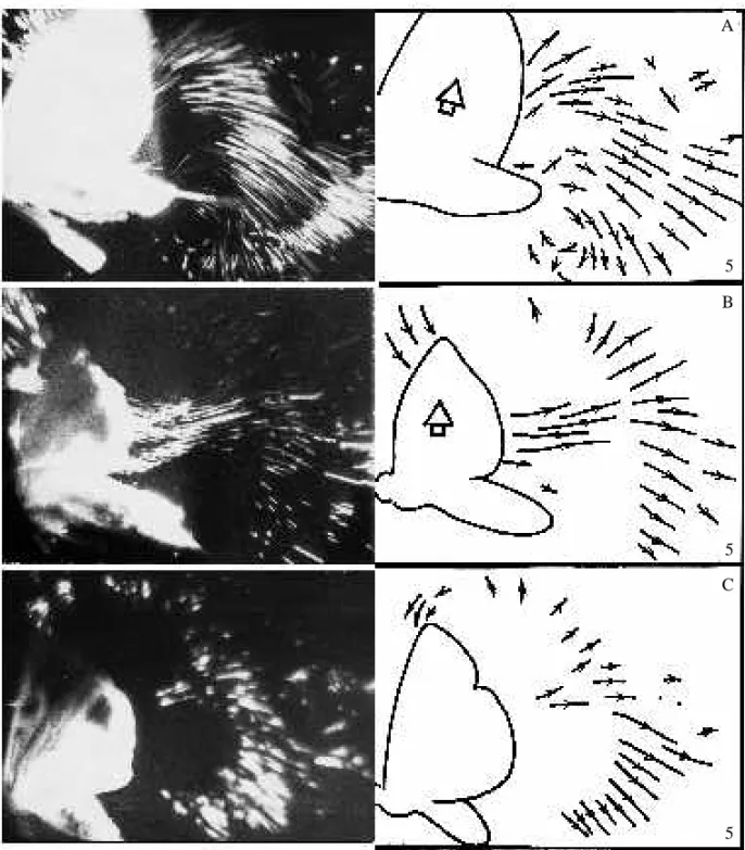
As the wings accelerate, the starting tubes increase their volume and rotational speed. At the same time, the tubes elongate and their proximal ends move along the trailing edges towards the insect body. They connect, and a common single vortex bubble is formed. It looks like a U-shaped vortex tube situated above the body and stuck to the wing tips by means of tip vortices (Fig. 5). Merging of the starting tubes, giving birth to the U-shaped bubble, is likely to occur after the wings have reached a positional angle of 45–60˚. After the U-shaped bubble has formed, distribution of vorticity inside the vortex tubes becomes more uniform. Flow sections made in the longitudinal sagittal plane show the presence of a large vortex situated just above the insect body (Fig. 5A,B). This vortex is interpreted to be a result of the coalescence of the starting tubes above the insect. It is a so-called dorsal vortex (Brodsky and Ivanov, 1983, 1984). It drives the airstream above the body towards the tip of the abdomen (Fig. 5C). At this moment, tip vortices are also clearly visible where the U-shaped bubble attaches to the wing tips (Fig. 5E–G).
As outlined above, the moth and the butterfly wings, unlike those of the other species studied, touch each other at the top of the stroke. We failed to obtain flow pictures
C −20 0 20 40 60 80 100 120 140 −20 20 0 40 60 S P P D D U U U D 80 100 120 A 0.01 0.01 10
10 20 30
20 30 40
B D −20 −20 0 20 40 60 80 100 0 20 40 60 80 100 120 140 U D U P P S S D 0.01
20 40 60 80 0.02 0.03
0.01 10 20 30 40
Time (s) Frame number
Time (s) Frame number
Fig. 3. Wingbeat kinematics in (A) Pitedia juniperina, (B) Heliothis armigera, (C) Tipula
oleracea, (+) movement of halteres, and (D) Musca domestica. Angular diagrams show the
resembling Fig. 4A–C for the butterfly and the moth, which is probably due to their specific kinematic patterns at the end and beginning of the stroke cycle. The start of the downstroke in these insects is followed by a gradual movement apart of the trailing edges, which open like a zip; the most distal point of contact between the wings slides along the trailing edges towards the metathorax. Hence, it is possible for an early connection to be made between the right and left starting tubes, compared to the other, non-clapping species. The upper U-shaped vortex bubble is thus likely to be formed during the fling.
In moths, butterflies and bugs, the flow induced by the wing movement blows the U-shaped bubble back towards the tip of the abdomen. This process is easy to observe in ciné films of the European skipper flying in a cloud of dust. As a result, the dorsal vortex is situated not above, but behind, the body at the end of downstroke (Fig. 6B). At the same time, in dipterous flies the dorsal vortex keeps its position approximately over the wing hinges (Fig. 6E). This observation leads to the conclusion that, for some reason, the dorsal vortex in crane and muscid flies is not blown backwards.
Supination
In all the species studied, supination causes a distinct mass of air to be thrown off the dorsal surfaces of the wings. The airstreams coming off the wings tend to follow the directions of veins, and those of flexion and foldlines, which diverge from the hinges in a fan-like manner. Therefore, this phenomenon could be termed ‘fan-like throw-off’. The aerodynamic process during this throw-off is likely to resemble that near an oscillating fan at the points of stroke reversal. The fan-like throw-off was detected by analysis of data provided by all the photographic techniques (Fig. 6A,F) and seen most clearly on ciné films.
The wing supination starts before and ends after the beginning of the upstroke (see angular diagrams in Fig. 3). This prolongs the downward movement of the trailing edges even after the wing tip has begun moving upwards. As we have already seen, the aerodynamic processes determining the flow around the wing occur near the trailing edge. Thus, flow pictures characteristic of the upstroke occur not after its beginning, but some time later after the finish of supination. In effect, the downstroke of the trailing edge is longer than that of the wing tip and the leading edge. This is distinctly seen on photographs taken in stroboscopically glimmering light (see Fig. 8C in Grodnitsky and Morozov, 1992).
Upstroke: the bugs, the muscid fly and the lepidopterans
As a result of such a prolonged downstroke, the generation of stopping vortex tubes is delayed until the end of supination. These tubes are formed after the beginning of the upstroke. They either coincide temporally with, or occur slightly after, the fan-like throw-off.
B
C
D
E
14 7 7 7
A
During the subsequent upstroke, the stopping tubes gain vorticity shed from the wings and thus elongate, shifting their proximal ends along the trailing edges towards the body. Finally, they meet under the body and connect to each other, giving birth to a complete vortex ring (Fig. 8D). This is characteristic for almost all the species studied, although the connection of stopping tubes under the body takes place at different moments in different species.
In the bugs, the moth, the butterfly and the muscid fly, a complete ring is created shortly after the end of supination, presumably before the wings reach a positional angle of 90˚. During the rest of the upstroke a vortex is seen below their abdomen, rotating in the opposite sense to that of the dorsal vortex (Fig. 8A–C). This is a so-called ‘ventral vortex’. Its diameter is substantially smaller than that of the dorsal vortex.
During the main part of the upstroke the wings move close to the vortex ring situated just behind them. The upper two-thirds of the ring are represented by the upper (starting) U-shaped vortex bubble, the lower third by the stopping one. The ring is clearly visible from both the side (Fig. 8A–C,G) and the top (Fig. 8E,F) views. Formation of a new ring corresponding to the second half-stroke has never been observed. If this were to occur, not one but two sequential vortices would be seen from above at each side of the body.
End of the stroke
Before the end of the stroke, insect wings perform pronation – rotation around their hinge-tip axes – when their leading edges move downwards. Pronation results in a fan-like throw-off of air from the lower surfaces, in a manner similar to that during supination (Fig. 9). This time the vortex ring is not joined to the wings. Therefore, the throw-off blows the ring backwards and thus vacates a space for the formation of new starting tubes at the beginning of a new stroke.
Upstroke: the crane fly
Vortex formation during the upstroke of crane flies is somewhat different (Fig. 10). We failed to detect the presence of a vortex below the crane fly body, and believe it to be absent (Fig. 10A), so it is necessary to conclude that stopping vortex tubes do not connect with each other under the body. This is probably because crane fly wings have a very narrow base; they are attached to the pterothorax by thin stalks. The small stalk diameter does not allow them to accelerate air effectively and actively to participate in vortex generation. Thus, the proximal ends of stopping tubes stick to the rear basal part of the broad wing plate and do not spread further down the stalks to meet each other (Fig. 10D). As a result, the vortex ring stays unclosed over most of the upstroke. The stopping tubes are strongly curved at this moment, so that horizontal light planes occupying light plane positions 8–10 strike every tube twice. That is why two vortices rotating in opposite
B C
D
Fig. 5
G F
E
9 14
8
A
directions are observed behind each wing. One vortex is located near the distal part of the wing, while the other is near the proximal part (Fig. 10B,C).
Connection of the stopping tubes, and hence closure of the vortex ring, takes place rather late for crane flies. It probably happens when the wings come close to each other at the end of the stroke (Fig. 10E). This results in the appearance of two opposite vortices above the body (Fig. 10G). These vortices are to be interpreted as the dorsal and ventral ones. Curiously, the ventral vortex in crane flies is situated above the body, and consequently the entire ring is located over the body.
It is difficult to say whether this result corresponds to the vortex wake of freely flying crane flies. Self-closing of the ring in natural flight possibly happens above the body and is also due to the characteristic wing planform. Nevertheless, the wings of some brachycerous (Orthorrhapha+Cyclorrhapha) flies have a similar shape, while the ventral vortex is clearly observed under the body of the muscid fly. Moreover, we detected the ring closing above the body at times in a broad-hinged species, the cotton moth (Fig. 11). Faced with this evidence, we think that the transition of the ventral vortex to the upper side of a crane fly is probably the result of a stroke with low amplitude, when the wings open to a maximal positional angle of approximately 90–100˚ or even less. We suppose that, in natural flight, the stopping tubes connect below the body, as in the other insects.
The problem of leading edge separation
To finish the description of vortex generation during a stroke, we should like to point out the lack of regularly observed flow separation from the frontal wing parts. Vortices that should follow such a separation have not been detected either (Fig. 7). Hence, it seems likely that the flow moves smoothly about the leading edges during the entire stroke cycle, with neither separation nor stalling. It is possible that leading edge bubbles exist but are too small to detect using our techniques, so some more experimental data are necessary before drawing final conclusions.
The wake dynamics during the stroke
In summary, these results for tethered flight show that each functional wing (or single wing for dipterous flies) generates its own starting vortex tube at the beginning of each stroke. The tube is tied to the wing tip at its distal end and to the trailing edge at the proximal end. During the downstroke, the starting tubes grow to meet each other and finally connect, giving rise to a single upper U-shaped vortex bubble which is tied to the wing tips. In the Lepidoptera studied, the connection of the tubes occurs especially early, long before the right and left wings lose mutual contact.
After supination, the stopping vortex tubes are created following the deceleration and cessation of the trailing edges’ downward movement. The distal ends of the stopping tubes stick to the wing tips and thus connect to the upper U-shaped bubble. The proximal ends of the tubes are joined to the basal parts of the rear wing edges. During the upstroke,
the stopping tubes meet under the insect body, so that a self-closed vortex ring is formed. The ring keeps its connection with the wing tips during the rest of the stroke cycle. After the end of the cycle, the ring loses its contact with the wings and travels backwards under the influence of a fan-like throw-off that takes place during pronation.
Thus, each wing stroke results in the generation of a single vortex ring. During the
B A
C
D
F E
5 15
1
5
Fig. 6. Position of the upper U-shaped bubble at the end of the downstroke. (A–C) The bug
process of ring formation, its shape is rather different from toroidal. Moreover, different parts of the ring are characterized by different transverse diameters and rotational velocities. Hence, parts of the ring differ according to impulse values. This is due to a pronounced unsteadiness in the wing movement. Vortices born in the downstroke are stronger because of large aerodynamic angles of attack. These angles are much smaller during the upstroke, so the intensity of vortex generation decreases.
Next, the wings pass the upper point of their movement close to, or even touching, each other. This condition is not even approached at the bottom of the trajectory. This explains why the starting tubes reach mutual connection rather early, whilst the same process in the stopping tubes is delayed. As a result, the starting tubes intensify each other, while the stopping ones are deprived of this effect. All of this results in a s i g n i ficant non-uniformity in vorticity distribution inside the ring during the fir s t moments of its existence.
The results of flow visualization show that the ring becomes more uniform and assumes a more regular shape after it has lost connection with the wings. During this, the diameter of the ring reduces markedly; squeezing of vortex rings was also observed by Brodsky (1991).
Discussion
Formation of the vortex ring
Demoll (1918, cited in Rohdendorf, 1949) was the first to carry out a successful visualization experiment with a tethered flying insect. He described a total flow picture averaged over a whole stroke cycle. With modern techniques it is possible to study the wings’ movement in much finer detail (Ellington, 1980; Brodsky and Ivanov, 1983, 1984; Brodsky, 1986, 1988, 1991). These data provided an opportunity to follow how the flow picture develops during a single stroke and to study the intimate features of the interaction between the flow and wing movement. However, fewer than a dozen insect species have so far been studied using these techniques, and the results of different experiments are often contradictory because authors usually use different methods.
We should like to emphasize that the leading edge separation bubble, whose existence has long been accepted by many authors, was not detected in our experiments. The absence of this vortex structure is also revealed by earlier studies (Ivanov, 1990; Grodnitsky and Morozov, 1992). It is likely to be due to a smooth flow around the leading edges, originating from the wing surface microstructure effect (Vogel, 1967) or from flexible deformation, which adjusts the wings to a contrary wind (Ellington, 1984). We think that the problem of the leading edge separation is not yet finally resolved. We will accept, for the time being, that this separation is absent.
B
C
D A
1
1
2
2
Our results show that the distribution of vorticity over vortex tubes and bubbles is very non-uniform. The tubes’ transverse diameter and rotation velocity are higher in those places and at those moments where the impulse coming from the moving wings to the flow is greater. The most intensive air rotation is observed near the wing’s distal third during the downstroke.
Starting and tip vortices are the most strongly developed structures of the first quarter of the stroke. After the wings have reached a positional angle of 45–60˚, the left and right starting tubes meet and connect, forming the upper U-shaped bubble. This seems to cause some redistribution of vorticity and an enhancement of the proximal part of the tubes. Before this connection, the basal ends of the tubes have been tied to the wing hinges and, thus, decelerated. After the connection, they start to accelerate and this leads to a decrease of vortex structures and of energy losses that originate from viscid e f f e c t s .
A vortex tube represents a zone of low pressure. While situated above an insect it must create a rarefaction and a corresponding additional lift. It is therefore to the insect’s advantage to maintain the tube impulse and to minimize negative viscid effects that decelerate the tube. Mechanisms that lead to an early closing of the starting tubes will therefore be selected for. As we have already seen in the cotton moth, the clap and subsequent opening of the wings at the top of their trajectory result in the early formation of the upper U-shaped bubble. Hence, the Weis-Fogh clap-and-fling could be an adaptation enabling the insect to control the vortex wake structure.
The clap-and-fling mechanism has been found in tiny wasps (Weis-Fogh, 1973), various Diptera (Ellington, 1984; Ennos, 1989), lacewings (Antonova et al. 1981) and
Trialeurodes vaporariorum (Wootton and Newman, 1979). Larger insects, such as
locusts (Cooter and Baker, 1977), bush crickets, mantids (Brackenbury, 1990, 1991a) and butterflies (Brodsky, 1991; Brackenbury, 1991b), tend to use clap-and-peel, a flexible modification of this mechanism. It is probably necessary to allow the more quick-starting tubes to connect. Nevertheless, we should not overestimate the importance of this mechanism, since a great variety of excellent fliers do not let their wings touch each other in the upper part of a stroke. The useful effect of clap-and-fling is likely to be limited by the need to stop the wings and then accelerate them again, which will lead to overall losses in aerodynamic force.
As a result, an insect resorting to clap-and-fling profits at the beginning of each cycle but loses at the end, where the wings are fully stopped. According to a proposal of A. P. Rasnitsyn (personal communication), some additional losses from the clap could possibly be due to a waste of energy stored in elastic pterothorax structures. When the wings stop moving, the elastic energy is dissipated. An alternative to the clap-and-fling is provided by wing movement not involving a pause. This pattern is associated with a comparatively late formation of the upper U-shaped bubble, but at the same time it economizes on energy and time by maintaining stroke continuity.
Both kinematic patterns can occur in the same insect species. For example, butterflies
B C
D
E
F G H
A
5 5 5
9
8 3 10
can demonstrate a typical clap-and-fling. Nevertheless, they often avoid it when flying freely (Brackenbury, 1991b; Grodnitsky, 1992).
During the downstroke, a powerful U-shaped bubble creates a rarefaction above the body. We propose that the insect could gain additional force benefit if it were to prolong the action of the bubble. A probable adaptation for such a prolongation could be the wing kinematics observed at the bottom of the trajectory. Supination here starts before and ends after the beginning of the second half-stroke. As a result, the formation of the stopping tubes is delayed until the supination finishes. So the ‘aerodynamic’ downstroke appears to be longer than the ‘kinematic’ one. As all the vorticity generation that determines the wake structure takes place near the trailing edges, we should set the border between half-strokes at the moment of reversal of the trailing edge, rather than the wing tip.
Supination activates a fan-like throw-off of air from the dorsal surfaces of the wings, whose aerodynamic significance is not entirely clear. It may be that the throw-off provides additional impulse directed forwards and upwards. If so, we could consider it to be a special propulsive mechanism. It could be enhanced by fine wing movements resembling a ‘swinging edge’, as described by Nachtigall (1979, 1985). Nevertheless, we would need to do much more detailed work to provide a serious basis for such a hypothesis. The throw-off may also be an adaptation for blowing away the vortices generated during the previous half-stroke of the wing.
At the end of the downstroke, the dorsal vortex travels towards the tip of the abdomen in some species. This phenomenon has been described in butterflies flying tethered in still air (Brodsky and Grodnitsky, 1985) and in the presence of a slight contrary wind (Brodsky, 1991), in caddis flies (Ivanov, 1990), in the cotton moth and in bugs. All these species possess broad-hinged wings. Green lacewings lack the dorsal vortex transition (Grodnitsky and Morozov, 1992). The same is true of the dipterous flies studied in the present paper. Both lacewings and flies have wings with comparatively narrow bases. Hence, migration of the dorsal vortex is likely to be caused by a flow induced by the proximal parts of the wings. Narrow hinges are rather inactive aerodynamically. That is why the vortex stays in the same place above the body until the very end of the stroke.
Stopping tubes situated below the body and the wings are likely to create a negative aerodynamic force during the upstroke. We hypothesize that insects may somehow be able to diminish the parasitic influence of the stopping tubes. It is possible to do this having shortened the duration of the upstroke, reduced the aerodynamic angle of attack and delayed the connection of the tubes, following formation of the lower U-shaped bubble under the body. The shortened upstroke and its reduced angle of attack have already become common knowledge. Delayed lower U-shaped bubble formation is referred to for the first time.
From our point of view, the late connection of the stopping tubes originates from an incomplete stroke amplitude. The stroke angle usually does not exceed 120–150˚. As a result, the proximal ends of the stopping tubes are very far apart. To become connected
B
C
8
Fig. 9
A
they need additional time. So the incomplete stroke is probably a functional adaptation to preserve the stopping tubes from an early connection.
Diptera have evolved an additional adaptation to delay the formation of the lower U-shaped bubble. Their wings are attached very narrowly to the pterothorax. Because of this, the distance between the proximal ends of the tubes has been increased. As a result, the lower U-shaped bubble can remain unconnected during a considerable part of the upstroke and can finally connect to itself above the body.
While performing a stroke, insect wings create vortices that accompany the establishment of circulatory flow about the flapping planes. This flow establishes the aerodynamic force that supports and propels the insect through the air. It is important to point out that the circulation mechanism might not be enough to supply an insect with the power necessary for flight. We do not know yet what percentage of the total power balance of a flying insect is taken up by the circulation force. According to Uldrick (1968), animals have probably evolved mechanisms which enable them to control the gain and loss of energy to the vortex wake. Such adaptations might considerably increase the total efficiency of the flight apparatus.
On the basis of the results described above, we suggest that insects have evolved some special mechanisms for utilizing their own vortex wake during flight. This is essential in animals that fly by flapping, because the vortex structures have to be renewed every time a new stroke starts. In insects with high wingbeat frequencies, the vortices have no time to dissipate under the influence of viscid effects or to travel far from the insect. All this enables the insect to interact with its own wake during the whole stroke cycle, and thus to gain additional energy.
Adaptations of insects to control their wake can be both kinematic and morphological. The first group includes (1) making the upper part of the stroke path complete and the lower part incomplete and (2) prolonging the downstroke by supination. The second group of adaptations includes the development of narrow-hinged wings.
Vortex wake structure
According to the data presented here and published earlier (Grodnitsky and Morozov, 1992), a flying insect creates a single vortex ring per stroke, irrespective of the number of coupled or separated wings it has. The aerodynamic wake of an insect thus consists of a series of sequential independent vortex rings. The rings are approximately parallel to the wings’ stroke plane and to each other. This agrees well with the results of Ellington (1980) and with the results of visualization experiments on slowly flying birds (Kokshaysky, 1979; Kokshaysky and Petrovsky, 1979; Spedding et al. 1984; Spedding, 1986).
Such a wake structure contradicts the conclusions of some authors (Brodsky and Ivanov, 1983, 1984; Brodsky, 1988, 1991; Ivanov, 1990), who consider the wake to consist of vortex rings connected by their upper and lower parts in a chain. We used to think so too (Brodsky and Grodnitsky, 1985) but further development of experimental
F i g . 9. Vortex formation at the end of the upstroke. (A) The muscid fly, (C) the bug
P. juniperina. (B) Frontal three-dimensional representation of the vortex ring. Symbols as in
B C
D
E
F
G A
5 8 9
9
Fig. 10
techniques has made us abandon our former position. Comparison of a vast number of photographs taken with different light plane positions in various views has driven us to the conclusion that there is no place near the wings for a new ring to be formed during an upstroke. Backward transition of the dorsal vortex at the end of the downstroke, which is observed in the broad-hinged species, cannot lead to the formation of a separate ring associated with the upstroke. After the stopping tubes have connected with each other, a ring thus formed does not lose contact with the wings, so there is simply no free space for the generation of the next ring. The final argument that the rings are in fact connected could be that our experiments were conducted in still air. It is possible to hypothesize that a contrary air flow would drive the upper U-shaped bubble far behind the insect and thus leave the space free for an upstroke ring to be created. Flow visualization experiments with an insect flying in moving air are necessary to settle this finally.
The ‘double chain’ wake model
Brodsky (1985, 1986, 1988, 1991) considers that an increase in flapping frequency (over about 70Hz) results in the generation of a separate vortex chain behind each wing. Brodsky described this phenomenon in a sesiid moth and a crane fly, and named it the ‘double chain model’. In an earlier publication (Grodnitsky et al. 1988), we outlined some contradictions contained in Brodsky’s wake model. Indeed, the presence of independent vortex chains at each body size would inevitably induce a flow above the insect moving towards its head. However, such a flow reversal has been detected neither by Brodsky nor in our previous attempt to repeat his experiments.
Analysis of the extensive data obtained by us clearly indicates that flow around a tethered flying crane fly is not characterized by such strikingly different features as was proposed by Brodsky (1985, 1986, 1988, 1991). Moreover, even a very high flapping frequency (up to 100–120Hz in Musca domestica and Pitedia juniperina) does not generate double vortex chains creating a single common ring per stroke.
The available data allow us to suppose that double chain generation, as described by Brodsky, was inferred from those flow pictures that showed two vortices at each side of the body seen from above (as in Fig. 10B,C here and Fig. 4 in Brodsky, 1986). The evidence for a separate ring at each side of the fly seems clear. Nevertheless, a more detailed wake study demonstrates a clearly visible dorsal vortex above the crane fly’s body (Fig. 6E). This contradicts the model proposed by Brodsky. Having compared the pictures containing double vortices at each side with the other photographs, we interpret them as the result of cross-sectioning two strongly curved tubes, so that a horizontal plane cuts each tube twice, giving images of four flat vortices (Fig. 10D,E). If these tubes formed two distinct rings, a picture similar to Fig. 10D,E would be obtained with every horizontal plane position. Nevertheless, it has been observed with light planes 8–10 only. In light planes 6–7 there is a single vortex behind each wing (Fig. 10E). In addition, light plane position 10 indicates that double vortices also occur in some other species (Fig. 8H). Thus, we see no strong evidence to suggest that the vortex wake in crane flies
is, in principle, different from that in insects with higher wingbeat frequencies. We emphasize once more that the aerodynamic vortex wake of all the species studied so far consists of consequent parallel non-connected rings. The wake is probably not very long. We have never registered more than three rings at a time. This is an underestimate, because of the low resolving capacity of the films we used: in reality, the wake is somewhat longer. Visual observation showed 5–6 vortex rings behind the insects. Thus, the rings seem to dissipate within at least 0.2s under the influence of viscid effects. The
B
5
C
5 A
5
time taken for wake dissipation given in our previous paper (0.1s) (Grodnitsky and Morozov, 1992) therefore needs some correction.
The evolution of insect flight
Rasnitsyn (1976, 1981) considers that the ancestors of flying insects lived in tree crowns and fed on the liquid contents of sporangia. These animals may have regulated their thoracic temperature by using paranotal lobes on the three thoracic segments (Kingsolver and Koehl, 1985). While searching for food and escaping from predators, these animals could jump from one branch to another, and there might then have been a selective advantage for longer lobes, enabling the first insects to jump further. The lobes might thus have begun to play a role in enabling the insects to glide. Folding of the proto-wings would have enabled the ancestors of modern Pterygota to stow away their proto-wings whilst walking along branches and in vegetation. Having evolved the ability to fold their wings, insects also became able to change their angle of incidence while flying and thus to prolong the flight or change its direction. Stroke movements possibly originated after this. At first, insects performed strokes with low amplitude in order to enhance starting vortex tubes. Then, the strokes became more energetic, and thus flapping flight originated.
We suggest that in the earlier stages of the evolution of flight, during the gradual transition from gliding to flapping, the fore- and hindwings performed non-synchronized oscillations. Such a kinematic pattern was not very efficient, because each wing pair created its own vortex wake. Vortex rings that had come off the fore- and hindwings interfered with each other and thus decelerated. This probably prevented an effective circulation and corresponding increases in lift. Such a primitive condition (an aerodynamic four-wingedness according to Brodsky, 1988) served as a basis for the further evolution of flight in winged insects: this would have involved optimization of the vortex wake structure and the development of mechanisms for vortex control and energy extraction from the wake.
In modern flying insects, only the Odonata show aerodynamic four-wingedness. The fore- and hindwings of dragonflies flap in opposite directions to each other, in contrast to the situation in all other functionally four-winged species. Such kinematics is sometimes supposed to be an adaptation for the enhancement of aerodynamic output (Brodsky, 1988). Nevertheless, in situations when additional force is needed (for example, during take off, when accelerating, when transporting prey, when additional weight is placed on the wings in experimental conditions, when flying in-copula, and during tethered flight), wings of dragonflies always operate with a phase shift typical for all the four-winged insects (Alexander, 1984; Azuma and Watanabe, 1988; Azuma et al. 1985; Somps and Luttges, 1985; Rüppell, 1989). Therefore, the synchronized wingbeats are more effective aerodynamically than fore- and hindwings beating out of phase.
Opposing wing strokes are likely to diminish or even stop these oscillations, thus increasing the effectiveness of hunting in Odonata.
The wings of dragonflies can operate independently of each other. The left and right wings can even work with different frequencies (Rüppell, 1989). This is a unique phenomenon among insects. Increasing the wings’ degrees of freedom gives dragonflies a surprising manoeuvrability, of which most higher insects are not capable. For example, insects usually change flight direction in a series of sequential strokes accomplished by a roll to the turn side (Kammer, 1985). Dragonflies can turn 3–4 times faster. During the turn, the stroke plane of the forewings is inclined in the turning direction, and that of the hindwings to the opposite side. Thus, the forewings turn the front of the body in the necessary direction, while the hindwings simultaneously turn the tail in the opposite direction. As a result, a dragonfly slews around its vertical axis during a single stroke (Alexander, 1986).
By having independently moving wings, dragonflies waste energy but gain manoeuvrability. They have evolved rather elongated wings. It is known that increasing flapping plane area is associated with decreasing cost of flapping flight (Weis-Fogh, 1964; Heinrich, 1974; Casey et al. 1985).
The unique kinematics of dragonfly flight could have originated from an ancestral non-synchronized condition. Alternatively, the contrary wingbeats could have developed later, after the strokes had become partially synchronized. The latter is more probable, since the ancestors of Odonata are thought to have originated from early Paraneoptera+Oligoneoptera after their divergence from the Polyneoptera (Rohdendorf and Rasnitsyn, 1980). Odonata have become specialists in manoeuvrability rather than in energetic and aerodynamic efficiency of the flight apparatus. This is presumably associated with their mode of life as aerial predators. As a result, the aerodynamic four-wingedness of dragonflies is characterized by a series of very advanced features. It is to be interpreted as an extreme plesiomorphic specialization. This is a rather widespread phenomenon in organisms that have separated early on from the common pathway of evolution of their group.
The alternative way of developing flying capacities is demonstrated by the other insects from the Paraneoptera+Oligoneoptera and Polyneoptera infraclasses. For these, the functionally four-winged condition is primitive. At the beginning of a stroke each of the four wings generates its own vortex tube. All the four tubes then coalesce, giving birth to a single upper U-shaped bubble (Grodnitsky and Morozov, 1992). The energy losses associated with this process provided the selective basis for the origin of the coupling of the fore- and hindwings. These losses are presumably rather high, since wing-coupling mechanisms evolved independently in many unrelated orders. This scenario is supported by the fact that caddis flies (Ivanov, 1985) and moths (Grodnitsky and Kozlov, 1991) were initially functionally four-winged, as well as by the strong variation in coupling structures seen in many functionally two-winged insects.
initial evolutionary steps. It was after this that the coupling of the fore- and hindwings originated.
The following scenario might also have accompanied wake optimization. A shortening of the lower part of the wingbeat is a common feature of the kinematics in all the functionally two-winged insects. As a result, their wings usually do not clap below the body. We assume that this decreases the negative aerodynamic effect of the lower U-shaped bubble. Because there is no clap under the body, the left and right stopping tubes do not connect just after the start of the upstroke. Thus, the formation of the lower U-shaped bubble is delayed, and its rotation velocity and total volume decrease. This is expressed in the rather late appearance and smaller size of the ventral vortex in comparison wirh the dorsal one. In some cases, the ventral vortex is absent. All these effects diminish the negative influence of the stopping vortices upon the magnitude of the total aerodynamic force created during the upstroke.
The climax of the morphological and functional evolution of the insect flight apparatus is the morphologically two-winged state characteristic of the Diptera. Reduction of the hindwings and development of halteres is connected with the appearance of a range of new adaptations for flapping flight. First, the halteres become navigation organs (Pringle, 1948). Second, the forewings became more free, not being limited in their movement by the hind ones. This enabled the Diptera to enhance in-flight manoeuvrability and to reach the level of dragonflies, although by an entirely different mechanism. The flight manoeuvres of flies are strikingly varied. This is achieved not only by high wing mobility, but also by using a range of special adaptations in pterothorax and wing-hinge construction, which allow different kinematic wing operational patterns (Nachtigall, 1985; Nalbach, 1989). Third, narrowing the proximal part of the wing resulted in the enhancement of its turning ability. So the wings of flies can work with near-zero angles of attack during the upstroke.
Finally, developing narrow-hinged wings turned out to be a mechanism for the delayed self-closing of the stopping part of the vortex ring. This has weakened the lower U-shaped bubble and thus decreased its negative contribution to the total average aerodynamic force created by the wings.
We have described the main hypothetical phases in the evolution of insect flapping flight, all of which are connected with the development of mechanisms for vortex wake control. We have not really touched upon the problems of flight evolution in beetles, which fly mainly with their hindwings. The main reason for this is an absolute absence of information about the vortex wake of flying beetles.
manuscript. The installations used in experiments were designed and made by Dr Lev T. Soukhov and Pyotr I. Geck (The ‘Nauka’ Specialized Constructor Bureau, Siberian Branch of the Russian Academy of Sciences, Krasnoyarsk). Dr Grigory B. Kofman (Institute of Forest, Krasnoyarsk) supplied us with a personal computer, which greatly diminished the time spent preparing the manuscript. The research was financially supported by a grant from the Siberian Branch of the Russian Academy of Sciences.
References
ALEXANDER, D. E. (1984). Unusual phase relationships between the forewings and hindwings in flying dragonflies. J. exp. Biol. 109, 379–383.
ALEXANDER, D. E. (1986). Wind tunnel studies of turns by flying dragonflies. J. exp. Biol. 122, 81–98. ANTONOVA, O. A., BRODSKY, A. K. ANDIVANOV, V. D. (1981). Wing beat kinematics of five insect
species. Zoologicheskii Zhurnal 60, 506–518 (in Russian).
AZUMA, A., AZUMA, S., WATANABE, T. ANDFURUTA, T. (1985). Flight mechanics of a dragonfly. J. exp.
Biol. 116, 79–108.
AZUMA, A. ANDWATANABE, T.(1988). Flight performance of a dragonfly. J. exp. Biol. 137, 221–252. BETTS, C. R.(1986). The kinematics of Heteroptera in free flight. J. Zool., Lond. 1, 303–315.
BETTS, C. R. ANDWOOTTON, R. J. (1988). Wing shape and flight behaviour in butterflies (Lepidoptera:
Papilionoidea and Hesperioidea): a preliminary analysis. J. exp. Biol. 138, 271–288.
BRACKENBURY, J. (1990). Wing movements in the bush-cricket Tettigonia viridissima and the mantis
Ameles spallanziana during natural leaping. J. Zool., Lond. 220, 593–602.
BRACKENBURY, J. (1991a). Wing kinematics during natural leaping in the mantids Mantis religiosa and
Iris oratoria. J. Zool., Lond. 223, 341–356.
BRACKENBURY, J. (1991b). Kinematics of take-off and climbing flight in butterflies. J. Zool., Lond. 224,
251–270.
BRODSKY, A. K. (1985). Some new principles of insect flight. Dokl. Acad. Nauk SSSR 283, 1491–1495 (in Russian).
BRODSKY, A. K.(1986). Flight of insects with high wing beat frequency. Entomologicheskoye Obozrenie
65, 269–279 (in Russian).
BRODSKY, A. K. (1988). Flight Mechanics in Insects and the Evolution of their Wing Apparatus. Leningrad University Press. 208pp. (in Russian).
BRODSKY, A. K. (1991). Vortex formation in the tethered flight of the peacock butterfly Inachis io L. (Lepidoptera, Nymphalidae) and some aspects of insect flight evolution. J. exp. Biol. 161, 77–92. BRODSKY, A. K. ANDGRODNITSKY, D. L. (1985). Tethered flight aerodynamics of the European skipper
Thymelicus lineola Ochs. (Lepidoptera, Hesperiidae). Entomologicheskoye Obozrenie 64, 484–492
(in Russian).
BRODSKY, A. K. ANDIVANOV, V. D. (1983). Visualization of the air flow around a flying insect. Dokl.
Acad. Nauk SSSR 271, 742–745 (in Russian).
BRODSKY, A. K. ANDIVANOV, V. D.(1984). Role of vortices in insect flight. Zoologicheskii Zhurnal 63, 197–208 (in Russian).
CASEY, T. M., MAY, M. L. ANDMORGAN, K. R.(1985). Flight energetics of euglossine bees in relation to
morphology and wing stroke frequency. J. exp. Biol. 116, 271–290.
COOTER, R. J. ANDBAKER, P. S. (1977). Weis-Fogh clap and fling mechanism in Locusta. Nature 269, 53–54.
DEMOLL, R.(1918). Der Flug der Insekten und der Vogel, 170pp. Jena.
ELLINGTON, C. P. (1980). Vortices and hovering flight. In Instationare Effekte an schwingenden
Tierflugeln, pp. 64–101. Wiesbaden: F. Steiner.
ELLINGTON, C. P. (1984). The aerodynamics of hovering insect flight. Phil. Trans. R. Soc. Ser. B 305, 1–181.
ENNOS, A. R. (1989). The kinematics and aerodynamics of the free flight of some Diptera. J. exp. Biol.
142, 49–85.
GRODNITSKY, D. L. (1992). Free and tethered flight of butterflies (Papilionida: Papilionoidea).
GRODNITSKY, D. L. AND KOZLOV, M. V. (1987). Dependence of tethered flight kinematics on
experimental conditions in the codling moth Laspeyresia pomonella (Lepidoptera, Tortricidae).
Zoologicheskii Zhurnal 66, 1314–1320 (in Russian).
GRODNITSKY, D. L. ANDKOZLOV, M. V. (1991). Evolution and functions of wings and their scale
covering in butterflies and moths (Insecta: Papilionida=Lepidoptera). Biol. Zbl. 110, 199–206. GRODNITSKY, D. L., KOZLOV, M. V. ANDNEHSINA, M. V.(1988). Problem of creating a well-fitted model
of insect flight apparatus. I. Overview. Uspekhi sovremennoi Biologii 105, 284–299 (in Russian). GRODNITSKY, D. L. ANDMOROZOV, P. P. (1992). Flow visualization experiments on tethered, flying
green lacewing Chrysopa dasyptera McL. (Neuroptera: Chrysopidae). J. exp. Biol. 169, 143–163. HEINRICH, B.(1974). Thermoregulation in endothermic insects. Science 185, 747–756.
HUMMEL, D. ANDGOSLOW, G. E. (1991). Concluding remarks: bird flight. Acta 20 Congr. Int. Ornithol.,
Christchurch, 2–9 December 1990. 2, 748. Wellington.
IVANOV, V. D. (1985). A comparative analysis of wingbeat kinematics of caddis flies (Trichoptera).
Entomologicheskoe Obozrenie 54, 273–284 (in Russian).
IVANOV, V. D. (1990). A comparative analysis of flight aerodynamics of caddis flies (Insecta: Trichoptera). Zoologicheskii Zhurnal 69, 46–60 (in Russian).
KAMMER, A. E. (1985). Flying. In Comprehensive Insect Physiology, Biochemistry and Pharmacology, vol. 5 (ed. G. A. Kerkut and L. J. Gilbert), pp. 491–552. Oxford: Pergamon Press.
KINGSOLVER, J. ANDKOEHL, M. A. R. (1985). Aerodynamics, thermoregulation and the evolution of insect wings: differential scaling and evolutionary change. Evolution 39, 488–504.
KOKSHAYSKY, N. V. (1979). Tracing the wake of a flying bird. Nature 279, 146–148.
KOKSHAYSKY, N. V. ANDPETROVSKY, V. I.(1979). Preliminary data on the vortex wake of a flying bird.
Dokl. Acad. Nauk SSSR 244, 1248–1251 (in Russian).
NACHTIGALL, W. (1979). Rasche Richtungsänderungen und Torsionen schwingender Fliegenflügel und Hypothesen über Zugeordnete instationäre Strömungseffekte. J. comp. Physiol. A 133, 351–355. NACHTIGALL, W. (1985). Calliphora as a model system for analysing insect flight. In Comprehensive
Insect Physiology, Biochemistry and Pharmacology, vol. 5 (ed. G. A. Kerkut and L. I. Gilbert), pp.
571–605. Oxford: Pergamon Press.
NALBACH, G. A. (1989). The gear change mechanism of the blowfly (Calliphora erythrocephala) in tethered flight. J. comp. Physiol. A 65, 321–331.
PRINGLE, J. W. S. (1948). The gyroscopic mechanism of the halteres of Diptera. Phil. Trans. R. Soc.
Lond. B 233, 347–384.
RASNITSYN, A. P. (1976). On the early evolution of insects and on the origin of Pterygota. Zhurnal
obstchei Biologii 37, 543–555 (in Russian).
RASNITSYN, A. P. (1981). A modified paranotal theory of insect wing origin. J. Morph. 168, 331–338. RAYNER, J. M. V.(1988). The evolution of vertebrate flight. Biol. J. Linn. Soc. 34, 269–287.
RAYNER, J. M. V.(1991). Wake structure and force generation in avian flapping flight. In Acta 20 Congr.
Int. Ornithol., Christchurch, 2–9 Dec., 1990 2, 702–715. Wellington.
RAYNER, J. M. V. ANDALDRIDGE, H. D. J. N. (1985). Three-dimensional reconstruction of animal flight paths and the turning flight of microchiropteran bats. J. exp. Biol. 118, 247–266.
RAYNER, J. M. V., JONES, J. AND THOMAS, A. (1986). Vortex flow visualizations reveal change in upstroke function with flight speed in bats. Nature 321, 162–164.
ROHDENDORF, B. B. (1949). Evolution and classification of insect flight apparatuses, 176pp. Moscow, Leningrad: Acad. Sci. Pr. (in Russian).
ROHDENDORF, B. B. AND RASNITSYN, A. P. (eds) (1980). Historical Development of Insects, 272pp. Moscow: The Nauka Publishing House (in Russian).
RÜPPELL, G. (1989). Kinematic analysis of symmetrical flight manoevres of Odonata. J. exp. Biol. 144, 13–42.
SOMPS, C. ANDLUTTGES, M. (1985). Dragonfly flight: novel uses of unsteady separated flows. Science
228, 1326–1329.
SOTAVALTA, O.(1947). The flight-tone (wing-stroke frequency) of insects. Acta ent. Fenn. 4, 1–117. SPEDDING, G. R. (1986). The wake of a jackdaw (Corvus monedula) in slow flight. J. exp. Biol. 125,
287–307.
SPEDDING, G. R. (1987). The wake of a kestrel (Falco tinnunculus) in flapping flight. J. exp. Biol. 127, 59–78.
ULDRICK, J. P. (1968). On the propulsion efficiency of swimming flexible hydrofoils of finite thickness.
J. Fluid Mech. 32, 29–53.
VOGEL, S.(1967). Flight in Drosophila. III. Aerodynamic characteristics of fly wings and wing models.
J. exp. Biol. 46, 431–443.
WARD, J. P. ANDBAKER, P. S.(1982). The tethered flight performance of a laboratory colony of Triatoma
infestans (Klug.) (Hemiptera: Reduviidae). Bull. ent. Res. 75, 17–28.
WEIS-FOGH, T. (1964). Biology and physics of locust flight. VIII. Lift and metabolic rate of flying
locusts. J. exp. Biol. 41, 257–272.
WEIS-FOGH, T. (1973). Quick estimates of flight fitness in hovering animals, including novel
mechanisms for lift production. J. exp. Biol. 59, 169–230.
WOOTTON, R. J. ANDNEWMAN, D. J. S. (1979). Whitefly have the highest contraction frequencies yet