Radiologic-Pathologic Correlation:
Hemangioblastoma
Vincent B. Ho, James G. Smimiotopou/os, 1 Frances M. Murphy, and Elisabeth J. Rushing
From the Department of Radiology, Madigan Army Medical Center, Tacoma, WA (VBH);
Department of Radiology and Nuclear Medicine, The Uniformed Services University
of the Health Sciences, Bethesda, MD (VBH, JGS); Department of Radiologic Pathology, Armed Forces Institute of Pathology, Washington, DC (JGS); Department of Neurology,
Georgetown University Hospital; Washington, DC (FMM); Department of Pathology,
Armed Forces Institute of Pathology, Washington, DC (EJR)
History
A 56-year-old man, with a history of multiple prior abdominal operations, presented for this follow-up screening head MR.
Case Discussion
Findings (see Fig. 1)
Figure 1
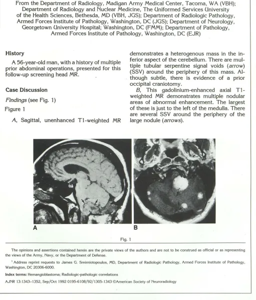
A, Sagittal, unenhanced T1-weighted MR
A
demonstrates a heterogenous mass in the in-ferior aspect of the cerebellum. There are mul-tiple tubular serpentine signal voids (arrow)
(SSV) around the periphery of this mass. Al-though subtle, there is evidence of a prior occipital craniotomy.
B, This gadolinium-enhanced axial T1-weighted MR demonstrates multiple nodular areas of abnormal enhancement. The largest of these is just to the left of the medulla. There are several SSV around the periphery of the large nodule (arrows).
B
Fig. 1
The opinions and assertions contained herein are the private views of the authors and are not to be construed as official or as representing the views of the Army, Navy, or the Department of Defense.
1 Address reprint requests to James G. Smirniotopoulos, MD, Department of Radiologic Pathology, Armed Forces Institute of Pathology,
Washington, DC 20306-6000.
Index terms: Hemangioblastoma; Radiologic-pathologic correlations
AJNR 13:1343-1352, Sep/Oct 1992 0195-6108/92/1305-1343 ©American Society of Neuroradiology
[image:1.612.53.564.138.737.2]1344 HO
Discussion
The differential considerations for a cerebel-lar lesion in an adult patient include metastasis,
hemangioblastoma, cavernous hemangioma, abscess, lobar hemorrhage, and infarction. The hypointensity of the lesion seen on the T1-weighted image (Fig. 1A) and intense pattern of enhancement following gadolinium admin-istration (Fig. 18) favor metastasis, hemangiob-lastoma, cavernous hemangioma, and abscess. Multiplicity of lesions, as noted in this case, may be seen with metastasis,
hemagioblas-toma, cavernous hemangioma, or abscess. The apparent lack of supratentorial lesions would, however, make multiple hemangioblastomas more likely, because metastases or abscesses are both hematogenously disseminated and would most likely also involve the cerebrum if
they were multiple, paralleling blood flow.
Cav-ernous hemangiomas, although commonly oc-curring in multiple locations, are less common than hemangioblastomas in the adult cerebel-lum. Hemangioblastomas may be the most common primary cerebellar neoplasm in the adult with an incidence at least as common as that of metastasis (1).
The presence of SSV as seen on Figure 1 A in the periphery of a lesion is very suggestive of a vascular lesion, especially cerebellar he-mangioblastoma. The cavernous hemangioma, although also a vascular lesion, is not usually associated with SSV. A rim of peripheral hy-pointensity may be seen with a cavernous hemangioma; however, this results from prior hemorrhage and hemosiderin deposition (2).
The detection of the patient's prior craniot-omy (Fig. 1 B) further supports the diagnosis of hemangioblastomas. Craniotomy is seldom performed for metastasis and rarely for multi-ple central nervous system (CNS) lesions. He-mangioblastomas, however, are resectable and potentially curable. In this case, the presence of multiple lesions confined to the cerebellum and radiographic evidence of prior tumor re-moval is very suggestive for multiple heman-gioblastomas. The patient's history of prior abdominal operations suggests that the he-mangioblastomas are in association with von
Hippel-Lindau disease (VHL), a disease with multisystem involvement. In this particular
AJNR: 13, September /October 1992
case, the patient had had bilateral renal cell carcinomas and multiple pancreatic cysts, both manifestations of VHL. He had previously undergone bilateral nephrectomies, cadaveric renal transplant, and numerous drainage pro-cedures for his pancreatic cysts.
Diagnosis
Multiple Hemangioblastomas (in VHL)
Hemangioblastoma
The hemangioblastoma (Lindau tumor, cap-illary hemangioblastoma, hemangioendothe-lioma, angioreticuloma, angioblastoma) is a be-nign tumor of the CNS. These tumors may be predominantly solid or have both solid and cystic areas. Hemangioblastomas account for 1%-2.5% of all intracranial neoplasms (3, 4). Although also found in the spinal cord (3%
-13%), medulla (2%-3%) and cerebrum (1.5%), hemangioblastomas. most commonly occur in the cerebellum (83%-86%), where they com-prise 7%-12% of primary posterior fossa tu-mors (3-6).
Hemangioblastomas typically present in the third through fifth decades of life; however,
they have been reported at all ages ranging from 1-75 years old (3-1 0). There is a slight male predilection for hemangioblastomas, with male:female ratios ranging from 1.3:1 to 2.6:1 (3, 4, 8, 9, 11).
Patients with hemangioblastomas usually have a long history (6-10 months) of minor neurologic symptoms that are often followed by a sudden exacerbation, forcing them to seek medical attention (3, 6-8). The most common presenting symptoms of intracranial heman-gioblastomas are headache (70% ), disequili-brium (50%), nausea/vomiting (37% ), and diz-ziness/vertigo (26%) (5). On neurologic ex-amination, patients most frequently demon-strate cerebellar signs (38% ), papilledema (33%) and nystagmus (20%) (5). At the time of presentation, as many as 50% of patients manifest signs and symptoms of increased
AJNR: 13, September /October 1992
2
3
HEMANGIOBLASTOMA 1345
4
Fig. 2. Cerebellar hemangioblastoma, operative photograph. The cerebellum has been decorticated, and we are peering into the cyst surrounding the tumor. There is a deep red nodule (arrow) at the upper portion of the lesion that represents the neoplasm itself. The cyst is external to the neoplasm and lined by normal cerebellar tissue.
Fig. 3. Cerebellar hemangioblastoma, subgrossed specimen. This hematoxylineosin-stained specimen illustrates normal cerebellar folia at the top of the photograph. The nodule of the hemangioblastoma is in a subpial location. Within this solid nodule, are numerous large tubular structures representing vascular sinusoids (large arrows). There are pale pink areas representing collections of stromal cells. The nodule is located on the right side of the cyst. Note that the neoplasm does not form the wall of the cyst but rather, the cyst "wall" is composed of compressed normal cerebellar tissue (small arrows).
Fig. 4. Cyst fluid, specimen. This test tube contains the aspirated fluid from a cyst surrounding a hemangioblastoma, demonstrating the
5
typical xanthochromic appearance. The fluid is benign and does notcontain neoplastic cells.
Fig. 5. Cerebellar hemangioblastoma, photomicrograph (hematoxylineosin stain, 350X). There is a relatively sharp demarcation between the hemangioblastoma and the adjacent brain (arrows). Most of the field demonstrates pale pink, polygonal, foamy-appearing stromal cells. In between the stromal cells is a lace-like pattern of capillaries lined by plump endothelial cells. There are several "microcysts" or lakes of proteinaceous fluid (asterisks) within the tumor.
Hemangioblastomas may produce polycy-themia in some cases. Up to 40% of hemangio-blastomas have been reported to secrete eryth-ropoietin (7). The polycythemia usually re-solves following resection of the tumor but may return with tumor recurrence. The poly-cythemia is more commonly associated with solid hemangioblastomas (3, 4, 7, 9, 12, 13).
Pathology
Gross
The hemangioblastoma is typically a well-circumscribed tumor that forms a solid "mural
nodule" within a larger cyst cavity (60%) (Figs. 2 and 3) (3). The tumor nodule commonly
abuts the pial surface (Fig. 3). The cyst fluid is often xanthochromic (Fig. 4), but rarely may be rusty brown if hemorrhage into the cyst has occurred (7, 14). The "cyst" around the mural nodule is a collection of fluid and is not truly part of the neoplasm. Therefore, the cyst and its lining need not be resected unless there is evidence of tumor involvement. Less
fre-quently, the hemangioblastoma has no sur-rounding cyst and is purely solid (40%) (3, 6,
7).
[image:3.615.54.558.76.450.2]Addi-1346 HO
tionally, the cyst fluid has been found to con-tain erythropoietin (13). Note that cysts may also occur within a mural nodule or solid tumor; the cysts in this instance are part of the tumor mass (3, 7, 16).
A
c
DB
AJNR: 13, September/October 1992
Microscopic
The hemangioblastoma is composed of a cluster of thin-walled, tightly packed blood vessels lined by plump endothelial cells on a
E
Fig. 6. Cerebellar hemangioblastoma, typical solid nodule with surrounding cyst. A lateral film (A) from a vertebral angiogram demonstrates a faint area of enhancement (arrow) corresponding to the nodule seen on the gadolinium-enhanced sagittal T1-weighted MR (B, arrow). Note the nodule's subpial location. The remainder of the lining of the cyst is not composed of neoplastic tissue and, therefore, does not enhance. Unenhanced T 1-weighted axial MR (C) demonstrates a homogenous "cystic" mass involving most of the cerebellar hemisphere. The cyst fluid is slightly hyperintense compared to CSF. The proton-density image (D) demonstrates an increase in signal intensity of the cyst fluid, which becomes even brighter on the T2-weighted pulse sequence
[image:4.613.56.561.85.644.2]AJNR: 13, September /October 1992
background of abundant connective tissue (Fig. 5). The blood vessels vary in size from capillary to cavernous and are interspersed with polygonal, lipid-laden "stromal" cells. The cytologic features of the endothelial and stromal cells is usually benign without mitotic figures. Necrosis and hemorrhage may occur,
but are uncommon (3, 7, 12, 17).
The cyst wall, if present, is composed of compressed adjacent brain parenchyma or re-active neuroglial cells. This is in distinction to a cyst found within a tumor nodule that is part of the hemangioblastoma which may represent dilated vascular spaces or regions of necrosis within the neoplastic tissue of the hemangiob-lastoma (3, 7, 12, 17).
Radiology
Angiography
Angiographically, the hemangioblastoma may be identified as a dense tumor nodule (Figs. 6A and 7 A) or as a heterogeneous net-work of tangled vessels fed by a dilated artery (4, 14, 18-20). Dilated draining veins (Fig. 7 A) have also been reported (21). If the tumor sits within a cyst, it may appear as a vascular nodule within an avascular region, secondary to displacement of the surrounding vessels by the avascular cyst (Figs. 6A and 7 A).
Angiog-A 8
HEMANGIOBLASTOMA 1347
raphy continues to be a crucial tool in the evaluation of hemangioblastoma, inasmuch as,
in several cases, it has detected tumors not otherwise identified on computed tomography (CT) or magnetic resonance (MR) (11, 22, 23).
CT
On unenhanced CT, the hemangioblastoma most commonly appears as a small isodense nodule within a well-circumscribed, thin-walled hypodense cyst (Figs. 8A and 9A) (17, 19, 21,
22, 24). Following the administration of intr a-venous contrast, the mural nodule will enhance
homogeneously (Figs. 88 and 98). The cyst wall generally does not enhance (Fig. 88).
Should the wall enhance, neoplastic extension along the cyst wall should be suspected (Fig.
98) (16). Solid hemangioblastomas are usually isodense but are occasionally hyperdense on precontrast CT, and show prominent homo-geneous enhancement (11, 20). Ring enhance-ment of solid hemangioblastomas has also been reported (20). On CT, the secondary fea-tures of hemangioblastoma such as hydro-cephalus (Figs. 8A and 88) and edema are well visualized.
MR
The most common MR patterns (Fig. 1 0) of hemangioblastoma are the traditionally
de-Fig. 7. Cerebellar hemangiob
-lastoma, typical solid nodule with
surrounding cyst. The lateral view
of a vertebral angiogram (A) dem
-onstrates the vascular tumor nod
-ule (solid arrow) in the posterior
cerebellum in a relatively
avascu-lar region that corresponds to the surrounding cyst. A dilated drai
n-ing vein is also appreciated on this angiogram (open arrows). There
-lationship of the hemangioblas
-toma abutting the posterior aspect
of the cyst is better appreciated on the gadolinium-enhanced sagittal Tl-weighted MR image (B) that
illustrates the densely enhancing
1348 HO
scribed solid "mural nodule" with an adjacent nonenhancing surrounding cyst (Figs. 68, 78, 8C, and 8D) (1/3) or purely solid (Figs. 1A, 18, 11A, and 118) (1/3) (5, 11). In some cases the tumor may also appear completely "cystic" (without a mural nodule detected on MR, but seen on angiography, CT, or at resection). Sometimes, there will be a mural nodule asso-ciated with an enhancing cyst wall (Figs. 9C and 90), or a solid mass with internal cysts. Overall, roughly 55% of hemangioblastomas have a surrounding cyst and the other 45% are predominantly solid (5, 11). This radio-graphic distribution correlates well with the pathologic spectra of 60% cystic and 40% solid described by Rubinstein (3).
The typical hemangioblastoma is hypo- to isointense on short T1-weighted MR (Figs. 1A
AJNR: 13, September/October 1992
and 6C) and hyperintense on proton- (Fig. 6D) and T2-weighted MR (Figs. 6E and 118). Oc-casionally, hemangioblastomas may be heter-ogeneous on T1-weighted MR (Fig. 11A), with foci of increased signal intensity seen within the solid portion of the tumor. These regions of T1 signal shortening may represent lipid within stromal cells or methemoglobin from hemorrhage within the tumor ( 11, 17, 23).
The cyst fluid surrounding the neoplasm will be slightly hyperintense compared to cerebro-spinal fluid (CSF) on T1-weighted (Fig. 6C) and proton-weighted images (Fig. 6D) and much more hyperintense on T2-weighted images (Fig. 6E). These characteristics of the fluid are attributable to its high protein content (25, 26). Following intravenous administration of gad-olinium, the neoplastic tissue markedly
Fig. 8 Cerebellar hemangioblastoma, typical solid nodule with surround-ing cyst. A "cystic" mass is noted in the area of the cerebellar vermis on the noncontrast CT scan (A). Following contrast infusion, a faint nodule of enhancement is seen along the right lateral border of this cystic mass (8,
arrow). Tl-weighted MR after gadolinium infusion in the coronal (C) and sagittal (D) planes illustrate only a small nodule (arrows) of enhancement. Again, this nodule is in a subpial location and the majority of the tissue
lining the cyst does not enhance and is not composed to neoplastic tissue.
[image:6.612.60.552.330.724.2]AJNR: 13, September /October 1992 HEMANGIOBLASTOMA 1349
A
B
c
Fig. 9 Supratentorial hemangioblastoma, mural nodule with extension into the
cyst wall. The axial precontrast (A) and postcontrast (B) CT scans demonstrate a
rounded abnormality in the high parietal cortex. The mass is of relative homoge-neous low attenuation before contrast but demonstrates dense enhancement of
the central nodule (arrowhead) with thick ring-enhancement of the cyst wall
(arrows) following iodine infusion. The corresponding gadolinium-enhanced axial
T1-weighted MR (C) also shows complete enhancement of the cyst wall (arrow).
On the gadolinium-enhanced coronal T1-weighted scan (D) the nonenhancing central cystic region is clearly seen to be surrounded by a rim of thick enhancement
(arrows). This appearance of ring-enhancement mimics necrosis in a malignant
tumor (for example, a metastasis).
D
enhances (Figs. 18, 68, 78, 8C, 8D, 9C, and 9D). Because of the high signal intensity of the surrounding cyst fluid on proton- and T2-weighted MR images, the tumor nidus is usually better visualized on gadolinium-enhanced T 1-weighted sequences (compare Fig. 68 to Fig. 6E). As seen on contrast-enhanced CT, the cyst wall usually does not enhance (Figs. 68,
78, 8C, and 8D). However, if the cyst is lined by neoplasm, the wall will enhance (Figs. 9C and 9D) (11, 19, 23, 25, 27).
On MR, hemangioblastomas commonly (60%-69%) have associated internal and/or peripheral SSV (Figs. 1A, 11A, and 118), con-sistent with the dilated afferent and efferent
vessels ( 11, 23, 27, 28) identified pathologi-cally. In fact, according to Lee et al, "the association of a peripheral cyst in the posterior fossa with a mural nodule supplied by enlarged vessels is virtually pathognomonic for heman-gioblastoma" (23).
Overall, MR is more sensitive than CT in the detection of hemangioblastomas (11, 19, 23,
[image:7.617.51.551.84.541.2]1350 HO
the effects of beam hardening and other
arti-facts commonly noted with CT.
Associations
Hemangioblastomas may be inherited as
sol-itary or multiple lesions or may occur in
con-junction with additional visceral tumors as part
of VHL (3). VHL is a hereditary disorder with
an autosomal dominant mode of transmission
and a greater than 90% penetrance (29). VHL
has been linked to a defect on chromosome 3
(30). The diagnosis of VHL may be established
by the presence of 1) more than one CNS
(including retinal) hemangioblastoma, 2) one
Pure Cyst-8% Mural Nodule-35% Cyst With Wall
Enhancement-6%
e)
QO
Cystic Nodule-6% Solid With Cyot-12 ,-, Solld-33%
Fig. 10. Radiologic patterns of hemangioblastoma. There are six morphologic types of hemangioblastoma as visualized
on MR and CT. (Modified from Murphy et al (5).) Fig. 11. Cerebellar heman
-gioblastoma, purely solid. This solid hemangioblastoma is seen on the unenhanced Tl-weighted (A) and T2-weighted (B) pulse se-quences. On the T1-weighted im-age, there is a heterogeneous solid mass involving the left cerebellar
hemisphere. Portions of this mass
have areas of focal hyperintensity
(arrows). There are also multiple curvilinear serpentine signal
avoids (SSV) around the periphery. On the T2-weighted image (B),
most of the nodule has become hyperintense, except for the cu
r-vilinear SSV (arrows).
A
AJNR: 13, September /October 1992
CNS hemangioblastoma with a visceral
mani-festation of VHL (Table 1), or 3) one
manifes-tation of VHL with a known family history (10,
25, 31-34). In Huson et al's series of 35 patients
with VHL, cerebellar hemangioblastomas
(83%), pancreatic cysts (69%), renal cell
car-cinoma (51%), renal cysts ( 49% ), and retinal
hemangioblastoma ( 46%) were the most
com-mon features noted at autopsy (10).
Solitary hemangioblastomas are associated
with VHL in 4%-40% of cases (average of
10%-20%) (3-6, 8, 1 0). The age of
presenta-tion of hemangioblastoma in VHL is usually one to two decades earlier than that of the
sporadic variety (6, 10, 32). The male:female
ratio of hemangioblastomas in VHL is closer to
1:1 (32).
Prognosis
The prognosis for cerebellar
hemangioblas-toma is quite good, with 85% of patients
sur-viving 5-20 years following surgical removal
of the tumor (3). The perioperative mortality is
between 7%-15% and related to postoperative
hemorrhage or increased intracranial pressure
(4, 6). Medullary hemangioblastomas have a
higher rate of morbidity (40%) (9). Spinal
he-mangioblastomas, which are frequently
asso-ciated with subarachnoid bleeding at
presen-tation, also have an increased morbidity (6).
Solid hemangioblastomas have been reported to have a higher incidence of complications (9,
20). Young and Richardson, in their series of
AJNR: 13, September /October 1992
TABLE 1: Manifestations of von Hippei-Lindau disease•
CNS
Kidney
Pancreas
Adrenal
Liver
Spleen
Lung
Bladder
Epididymis
Sympathetic chain
Hemangioblastoma (cerebellum,
eye, medulla, spinal cord,
cere-brum) Meningioma
Renal cell carcinoma (often cystic} Cyst
Hemangioblastoma
Hemangioma
Adenoma
Cyst Cystadenoma
Islet cell tumor
Carcinoma
Hemangioblastoma
Hemangioma
Pheochromocytoma Adenoma
Cyst
Cortical hyperplasia
Cyst
Adenoma
Hemangioma
Hemangioblastoma
Hemangioma
Cyst
Hemangioblastoma
Hemangioblastoma
Cyst
Hypernephroid tumor Cystadenoma• Paraganglioma
' Modified from References 25 and 31-36.
• Cystadenoma may also occur in the broad ligament in female patients.
14 solid hemangioblastomas, reported a 50%
rate of death or "poor result" postoperatively
(20). Patients with VHL may have a graver
prognosis, but perhaps secondary to the other
associated conditions, especially that of renal
cell carcinoma (32, 33).
The overall recurrence rate for
hemangiob-lastomas ranges from 8%- 16% (4, 9, 32). The
recurrent tumor may not necessarily have the
same morphology as the original tumor. Solid
hemangioblastomas may recur as cystic tu
-mors and vice versa (4).
HEMANGIOBLASTOMA 1351
References
1. Martin F, Lemmen LJ. Calcification in intracranial neoplasms. Am
J Pathol1952;28:1107-1131
2. Rapacki TFX, Brantley MJ, Furlow TW, Geyer CA. Toro VE, George ED. Heterogeneity of cerebral cavernous hemangiomas
diagnosed by MR imaging. J Comput Assist Tomogr 1990;
14:18-25
3. Rubinstein LJ. Tumors of the central nervous system. 2nd series,
Fascicle 6. Washington, DC: Armed Forces Institute of Pathology,
1972:235-24 I
4. Mondkar VP, McKissack W, Russell RWR. Cerebellar he
mangio-blastomas. Br J Surg 1967;54:45-49
5. Murphy FM, Smirniotopoulos JG, Parisi J. Hemangioblastoma:
radiologic-pathologic correlation. Paper presented at the 87th
Annual Meeting of the American Roentgen Ray Society, 1987
6. Neumann HPH, Eggert HR. Weigel K, Friedburg H. Wiestler OD,
Schollmeyer. Hemangioblastomas of the central nervous system:
a 1 0-year study with special reference to von Hippei-Lindau
syndrome. J Neurosurg 1989;70:24-30
7. Silver ML, Hennigar G. Cerebellar hemangioma (hemangioblas-toma): a clinicopathological review of 40 cases. J Neurosurg
1952;9:484-489
8. Jeffreys R. Clinical and surgical aspects of posterior fossa heman-gioblastoma. J Neurol Neurosurg Psychiatry 1976;38: 105-111 9. Constans JP, Meder F, Maiuri F. Donzelli R, Spaziante R, de
Divitiis E. Posterior fossa hemangioblastomas. Surg Neural
1986;25:269-275
10. Huson SM, Harper PS, Hourihan MD, Cole G, Weeks RD.
Comps-ton DAS. Cerebellar hemangioblastoma and von Hippei-Lindau
disease. Brain 1986;109:1297-1310
11. Smirniotopoulos JG, Murphy FM, Brown DC. MR imaging of
hemangioblastoma. Radiology 1989; 173(P}:85
12. Jeffreys R. Pathological and hematological aspects of posterior
fossa hemangioblastoma. J Neural Neurosurgy Psychiatry
1975;38:112-119
13. Waldmann TA, Levin EH, Baldwin M. The association of polycy-themia with a cerebellar hemangioblastoma: the production of an erythropoiesis stimulating factor by the tumor. Am J Med 1961;31:318-324
14. Coulam CM, Brown LR, Reese DF. Hippei-Lindau syndrome. Semin Roentgenol1976; 11 :61-66
15. Cumings JN. The chemistry of cerebral cysts. Brain 1950
;73:244-250
16. Maiuri F. Cysts with mural tumor nodules in the cerebral hemi
-spheres. Neurosurgery 1988;22: 703-706
17. Tan WS, Wilbur A, Spigos DG, Choi KH. Cystic mural nodule in cerebellar hemangioblastoma: CT demonstration. J Comput As-sist Tomogr 1984;8:1175-1178
18. Wolpert SM. The neuroradiology of hemangioblastomas of the
cerebellum. AJR 1970; 110:56-66
19. Filling-Katz MR. Choyke PL, Patronas NJ, Gorin MB, Barba D,
Chang R, Doppman JL, Seizinger B, Oldfield EH. Radiologic screening for von Hippei-Lindau disease: the role of Gd-DTPA
enhanced MR imaging of the CNS. J Comput Assist Tomogr
1989;13:743-755
20. Young S, Richardson AE. Solid hemangioblastomas of the po
s-terior fossa: radiological features and results of surgery. J Neural
Neurosurg Psychiatry 1987;50: 155-158
[image:9.613.73.548.98.735.2]1352 HO
22. O'Reilly GV, Rumbaugh CL, Bowens M, Kido DK, Naheedy MH. Supratentorial haemangioblastoma: the diagnostic roles of com-puted tomography and angiography. C/in Radio/
1980;32:389-392
23. Lee SR, Sanches J, Mark AS, Dillon WP, NormanD, Newton TH. Posterior fossa hemangioblastomas: MR imaging. Radiology
1989;171 :463-468
24. Ganti SR, Silver AJ, Hilal SK, Mawad ME, Sane P. Computed tomography of cerebellar hemangioblastoma. J Comput Assist Tomogr 1982;6:912-919
25. Braffman BH, Bilaniuk L T, Zimmerman RA. MR of central nervous system neoplasia of the phakomatoses. Semin Roentgenol
1990;25: 198-217
26. Kjos BO, Brant-Zawadzki M, Kucharczyk W, Kelly WM, Norman D, Newton TH. Cystic intracranial lesions: magnetic resonance imaging. Radiology 1985; 155:363-369
27. Sato Y, Waziri M, Smith W, et al. Hippel-Lindau disease: MR imaging. Radiology 1988; 166:241-246
28. Pont MS, Elster AD. Lesions of skin and brain: modern imaging of the neurocutaneous syndromes. AJR 1992; 158:1193-1203 29. Go RCP, Lamiell JM, Hsia YE, Yuen JWM, Paik Y. Segregation
and linkage analyses of von Hippei-Lindau disease among
220 descendants from one kindred. Am J Human Genet
AJNR: 13, September /October 1992
1984;36:131-142
30. Hosoe S, Brauch H, Latiff F, et al. Localization of the von Hippel-Lindau disease to a small region of chromosome 3. Genomics
1990;8:634-640
31. Melman KL, Rosen SW. Lindau's disease: review of the literature and study of a large kindred. Am J Med 1964;36:595-617 32. Michels VV. Von Hippei-Linadu disease. In: Gomez MR, ed.
Neu-rocutaneous diseases: a practical approach. Boston: Butterworth,
1987:53-66
33. Horton WA, Wong V, Eldridge R. Von Hippel-Lindau disease: clinical and pathological manifestations in nine families with 50
affected members. Arch Intern Med 1976;136:769-777 34. Lee KR, Wulfsberg E, Kepes JJ. Some important radiological
aspects of the kidney in Hippei-Lindau syndrome: the value of prospective study in an affected family. Radiology 1977; 122:649-653
35. Funk KC, Heiken JP. Papillary cystadenoma of the broad ligament in a patient with von Hippei-Lindau disease. AJR 1989;153:52 7-528
36. McGrath FP, Gibney RG, Morris DC, Owen DA, Erb SR. Case report: multiple hepatic and pulmonary hemangioblastomas-a new manifestation of von Hippei-Lindau disease. C/in Radio/