The majority of teleost fishes excrete nitrogen primarily in the form of ammonia (ammoniotely). However, the gulf toadfish Opsanus beta is one of only a few teleosts which can facultatively utilize metabolically expensive pathways and excrete the majority of its waste nitrogen as urea (ureotely) (Mommsen and Walsh, 1989, 1991). Ureogenesis in gulf toadfish is not linked to osmoregulation (Walsh et al. 1990), acid–base balance (Walsh et al. 1990; Barber and Walsh, 1993) or air-exposure during low tides (C. M. Wood, T. E. Hopkins and P. J. Walsh, unpublished data). In the laboratory, ureotely in gulf toadfish is inducible by exogenous ammonia, air-exposure or behavioural factors such as confinement and/or crowding (Walsh et al. 1990, 1994; Walsh and Milligan, 1995). Confinement/crowding also leads to a rapid (within 24 h) several-fold activation of hepatic glutamine synthetase (GNS) (Walsh et al. 1994), a central rate-limiting enzyme of
ureogenesis in toadfish (Mommsen and Walsh, 1991; Walsh and Milligan, 1995). These observations led Walsh et al. (1994) to hypothesize that a cortisol-related stress response may be involved in the transition from ammoniogenesis to ureogenesis.
Two points are now clear about the transition to ureotelism. First, the transition is signalled by an induction of GNS activity in the liver (Walsh et al. 1994), mostly in the cytosolic compartment (Walsh and Milligan, 1995). Second, the transition largely involves a reduction in ammonia-N excretion, with either no change or a modest increase in urea-N excretion (Walsh and Milligan, 1995). With this in mind, in the present study we examined the relationship between plasma cortisol levels, GNS activity and the form of nitrogen excreted (e.g. urea, ammonia) in undisturbed versus confined/crowded (stressed) gulf toadfish and specifically tested the hypothesis We examined the relationship between plasma cortisol
levels, hepatic glutamine synthetase (GNS) activity and the form of nitrogen excreted (e.g. urea, ammonia) in undisturbed versus confined/crowded (acutely stressed) gulf toadfish. Specifically, we tested the hypothesis that acute increases in plasma cortisol levels are required to trigger the increase in GNS activity induced by the confinement/crowding stress. Toadfish responded to the stress of confinement/crowding with an initial cortisol surge (approximately 37 ng ml21at 2 h), which was rapidly cleared and was indistinguishable from resting levels (approximately 10 ng ml21by 24 h). Treatment of fish with metyrapone (which inhibits cortisol synthesis) successfully blocked the acute 2 h confinement/crowding-induced surge in plasma cortisol levels. Additionally, GNS activity in confined fish 24 h after metyrapone injection also did not differ from that of control fish, indicating that acute GNS activation probably requires the earlier (2 h) cortisol peak.
In post-absorptive fish, a strong relationship between total nitrogen excretion rate and plasma cortisol levels was evident. The percentage of nitrogen excreted as ammonia was inversely related to liver GNS activity. However, GNS activity explained only part (at most 57 %) of the variability in the percentage of nitrogen excreted as ammonia/urea, suggesting that this is not the sole factor setting the degree of ureogenesis. When toadfish are fed, the relationships between total nitrogen excretion rate and cortisol levels, and between percentage nitrogen excreted as ammonia and GNS activity, are virtually absent. Taken together, our results indicate that a stress response may be only one of several mechanisms by which ureogenesis is activated in gulf toadfish.
Key words: cortisol, ureotelism, gulf toadfish, Opsanus beta, glutamine synthetase, stress, urea, ammonia.
Summary
INTERACTIONS OF CORTISOL AND NITROGEN METABOLISM IN THE
UREOGENIC GULF TOADFISH OPSANUS BETA
TODD E. HOPKINS1,*, CHRIS M. WOOD2 ANDPATRICK J. WALSH1
1Division of Marine Biology and Fisheries, Rosenstiel School of Marine and Atmospheric Science,
University of Miami, Miami, FL 33149-1098, USA and 2Department of Biology, McMaster University, Hamilton, Ontario, Canada L8S 4K1
Accepted 31 May 1995
*Present address: Rookery Bay National Estuarine Research Reserve, 10 Shell Island Road, Naples, FL 33962-8059, USA.
that acute increases in plasma cortisol levels are necessary to trigger the increase in hepatic GNS activity induced by confinement. We demonstrate that a transient surge in plasma cortisol levels precedes (by 24 h) the induction of GNS activity and that pharmacological blockade of this surge with metyrapone blocks the induction of GNS activity. We also demonstrate that the relationship between the pattern of nitrogen excretion and the simultaneously determined plasma cortisol level (as opposed to the plasma cortisol level 24 h previously) is complex and varies with the degree and duration of stress.
Materials and methods
Experimental organisms
Gulf toadfish Opsanus beta (Goode and Bean) were captured in roller trawls by commercial live-bait shrimpers in Biscayne Bay, Florida, USA, between April and September 1994. Toadfish were held in a covered outdoor tank at the shrimpers’ dock with running aerated sea water for less than 24 h following capture. In the laboratory, toadfish were held in glass aquaria (45 or 80 l) with flowing, sand-filtered and ultraviolet-sterilized sea water under ambient light regimes. All aquaria had a 5 cm bed of beach sand and several 15 cm long polyvinyl chloride tubes of various diameters for shelter; stock densities were always less than 12 g fish l21seawater. All fish were treated with a prophylactic dose of Malachite Green and formalin, as described in Walsh and Milligan (1995), and fed live pink shrimp Penaeus duorarum (approximately 5–7 % of the fish’s body mass) on day 3 and again on day 7 after capture, prior to experiments. Experiments typically began 72 h post-feeding, which Walsh and Milligan (1995) showed to be sufficient time for food to have been completely evacuated from the intestine. Temperature and salinities in the laboratory, holding aquaria and tubs during the experiments described below were 25±2.4 ˚C and 32.0±1.0 ‰ respectively.
Experimental design
Three experimental series (I–III) were undertaken. Series I was designed to determine whether confinement/crowding, which has previously been used to evoke increased ureogenesis and induction of GNS activity (Walsh et al. 1994), would produce a cortisol stress response in gulf toadfish. Toadfish were either randomly netted from aquaria and quickly transferred into small plastic tubs (30 cm long 325 cm wide
3 10 cm high) with aerated flowing sea water for confinement/crowding treatments (referred to as stressed below), or left undisturbed (control) in the large 80 l aquaria (88 cm long 3 29 cm wide 3 40 cm high). Our confined/crowded treatments consisted of 3–4 toadfish per tub (approximately 26 g fish l21) and control treatments of 3–5 fish per aquaria (less than 12 g fish l21). Blood samples were taken from rapidly (less then 1 min) anaesthetized fish (1.0 g l21 tricaine methanesulphonate adjusted to pH 7.8 with NaOH) by caudal puncture at 20.5, 1, 2, 4, 8, 24, 48, 72 and 96 h. Blood was immediately centrifuged at 13 000 g for 1 min and plasma
was removed and stored at 280 ˚C for subsequent analyses. Different groups of fish (Nù8) were used at each sample time and each fish was sampled only once.
In series II, the acute interactions of plasma cortisol levels and nitrogen metabolism were examined. We first needed to establish that metyrapone (2-methyl-1,2-di-3-pyridyl-1-propanone), which has been used to block de novo cortisol synthesis in rainbow trout (Bennett and Rhodes, 1986; Pagnotta et al. 1995), had the same effect in gulf toadfish. For series IIa, gulf toadfish (N=7) acclimated to the laboratory were netted from several 80 l aquaria, anaesthetized and blood samples taken for reference values of plasma cortisol levels. For the experiment, fish were netted, weighed and injected intraperitoneally with either 1.5ml g21body mass of saline (0.9 % NaCl) (N=12) or metyrapone (20 mg ml21 in 0.9 % NaCl) (N=12). Capture, weighing and injection required less than 1 min to complete. Saline- and metyrapone-treated fish were then placed by treatment group into tubs (four fish per tub) with flowing sea water. At 2 h post-injection (which corresponds to the time of the maximal cortisol peak in series I), fish were netted and anaesthetized and blood samples were taken as above for analysis of plasma cortisol levels.
Since activation of hepatic glutamine synthetase requires 24–48 h (Walsh et al. 1994), a second experiment was conducted to determine the effect of elevated cortisol levels on GNS activity. For this second experiment (series IIb), a group of acclimated fish were netted from 80 l aquaria, bled and the plasma and liver samples were frozen at 280 ˚C to provide a reference value for initial (control-initial) cortisol levels and hepatic GNS activities. Fish from another group were injected with saline or metyrapone (as above) and then placed by treatment group into tubs (3–4 fish per tub) with flowing sea water or back into 80 l aquaria with flowing sea water. At 24 h post-injection, fish were anaesthetized, blood samples were taken for analysis of plasma cortisol levels and plasma and liver samples were frozen at 280 ˚C for later analyses of cortisol levels and GNS activities, respectively. A final group of fish, which had been left undisturbed in separate 80 l aquaria during this experiment (control-final), was similarly sampled for comparison with the initial uninjected control group. All treatment groups contained 12–15 fish.
below as control-fed), and nitrogen excretion was measured for 24 h; terminal plasma cortisol levels and hepatic GNS activities were then assayed as for control fish. Lastly, chronic stress was produced in a variety of ways in a group (N=28) of 72 h post-absorptive fish (referred to below as chronically stressed). Fish were crowded four to a tub (as above) or confined singly in static sea water with aeration (tub volume 4 l). Every 24 h thereafter, the sea water in the tubs was changed. This cycle of 24 h static confinement was repeated for 4–6 days, with nitrogen fluxes measured during the final 24 h of confinement, after which all fish were bled and killed for determination of plasma cortisol levels and hepatic GNS activities.
Analytical methods
Water samples were analyzed for ammonia levels using the method of Ivancic and Deggobis (1984) and for urea levels using the method of Price and Harrison (1987), both of which have an approximate detection limit of 1–2mmol l21, as previously applied to toadfish (Walsh et al. 1994). Plasma cortisol concentrations were measured in all samples using a commercially available radioimmunoassay kit (ICN Immuno Corp., Costa Mesa, CA, USA); the protein content of standards was adjusted to reflect values found in fish plasma, which increased the assay sensitivity at low cortisol concentrations. For measurement of glutamine synthetase (GNS) activity, liver tissue was sonicated in 4 vols of homogenization buffer (20 mmol l21 K
2HPO4, 10 mmol l21 Hepes, 0.5 mmol l21 EDTA, 1 mmol l21 dithiothreitol, 50 % glycerol, adjusted to pH 7.5 at 24 ˚C), centrifuged for 1 min at 13 000 g and the supernatant assayed at 24 ˚C as previously detailed in Mommsen and Walsh (1989). All biochemicals were purchased from Sigma Chemical Co. (St Louis, MO, USA) and all other chemicals were reagent grade.
A two-way analysis of variance (ANOVA) was performed with plasma cortisol concentrations (control and confined/crowded treatments) as dependent variables and time as the independent variable (series I). For this ANOVA, plasma cortisol values were first log-transformed to correct for a lack of homoscedasticity in the variances as directed in Zar (1974). Post-hoc comparisons and contrasts were performed using the Bonferroni multiple comparison method (Neter et al. 1985). In series II, multiple t-tests with P values adjusted for the number of tests (Neter et al. 1985) were used to detect differences between treatments. Linear least-squares regression was used in series III to examine the relationships between total-N excretion and plasma cortisol levels, percentage nitrogen excreted as ammonia and GNS activity and finally GNS activity and plasma cortisol levels. Significance of regression slopes (e.g. difference from slope=0) was determined using t-tests. All statistical analyses were performed using JMP 2.0 (SAS Institute Inc., Cary, NC, USA) with significance judged at the Pø0.05 level.
Results
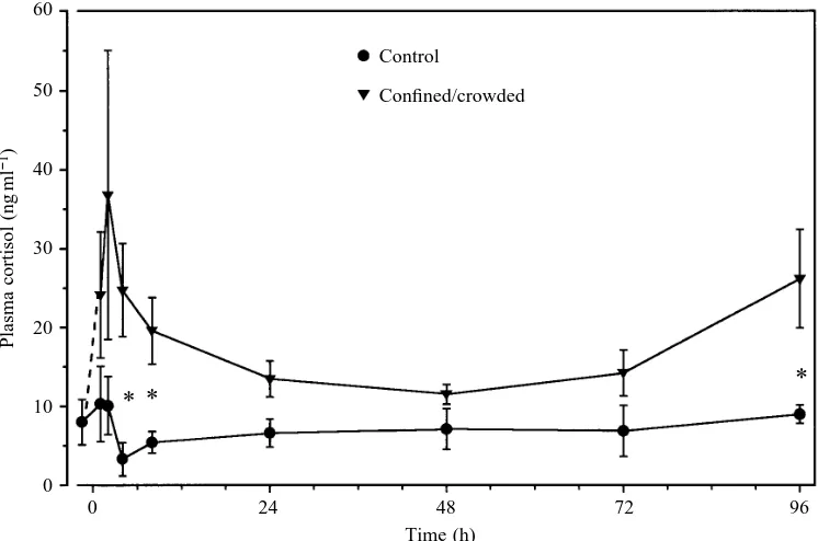
In series I, confinement resulted in a rapid initial two- to fourfold rise in plasma cortisol levels which peaked at 2 h and was rapidly cleared so that by 24 h, levels were not significantly different from controls (Fig. 1). Cortisol levels in confined fish continued to remain low from 24 to 72 h until a second significant increase (Pø0.05) at 96 h. This may represent the start of a chronic increase in plasma cortisol levels. At no time during the experiment were levels of cortisol in control fish significantly different from the values for the initial (20.5 h) sample. Thus, both acute (2 h) and chronic (96 h) elevations of cortisol levels were induced by confinement.
* *
*
60
50
40
30
20
10
0
Plasma cortisol (ng
ml
−
1)
Control
Confined/crowded
0 24 48 72 96
[image:3.609.194.567.493.740.2]Time (h) Fig. 1. Series I. Plasma cortisol
levels in undisturbed gulf toadfish (control, filled circles) and in toadfish subjected to a confinement/crowding stress (stressed, filled triangles). Values are means ± S.E.M., N=8–11,
In series IIa, metyrapone successfully blocked the 2 h acute cortisol peak in confined/crowded fish. Saline-injected confined fish showed a two- to threefold increase in plasma cortisol levels (37.4±2.7 ng ml21, mean ±S.E.M., Pø0.05) after 2 h (very similar to series I fish) compared with metyrapone-injected fish (14.3±1.2 ng ml21, mean ± S.E.M.). In addition, cortisol levels in metyrapone-injected fish were not significantly different from those of our reference group (8.9±1.4 ng ml21, mean ±S.E.M.).
In series IIb, injection of either saline or metyrapone (as above) caused no significant changes for 24 h plasma cortisol levels in toadfish returned to their original aquaria (Fig. 2A). Confinement for 24 h following saline injection elicited a significant fivefold increase (Pø0.05) in plasma cortisol above initial control levels (Fig. 2A). While the plasma cortisol level of confined, saline-injected fish after 24 h in series IIb (26.1±6.2 ng ml21, mean ± S.E.M.) was greater than that of confined toadfish after 24 h in series I (Fig. 1, 24 h, 13.5±2.3 ng ml21, mean ± S.E.M.), it is important to note that in series IIb the plasma cortisol levels of saline-injected fish returned to aquaria were not significantly different from initial-or final-control levels (Fig. 2A). However, confined metyrapone-treated fish showed significantly depressed
cortisol levels (Pø0.05) compared with confined saline-treated fish after 24 h, i.e. they were comparable to values for control
B A
120
80
40
0
Total nitrogen excretion (
m
mol-N
100
g
−
1h
−
1)
0 4 8 12 0 50 100 150 200 250 Plasma cortisol (ng ml−1)
y=3.86x+8.80 r2=0.80 P<0.001
y=−0.03x+63.45 r2=0.001
[image:4.609.317.554.71.431.2]P<0.94
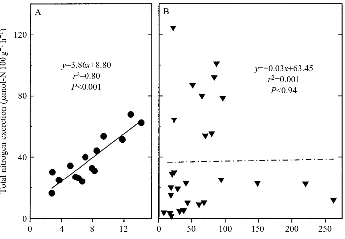
Fig. 3. Series III. Plot of total nitrogen excreted versus plasma cortisol level in (A) undisturbed (control) and (B) crowded/confined (chronically stressed) toadfish. Solid and dashed lines indicate least-squares regressions.
B
*
*
A 30
20
10
0
Cortisol (ng
ml
−
1)
8
6
4
2
0
GNS activity (units
g
−
1)
Control (initial)
Saline Met Saline Met Control (final) Aquaria
Confined
*
*
Control (initial)
Saline Met Saline Met Control (final) Aquaria
[image:4.609.215.562.491.732.2]Confined Fig. 2. Series IIb. Effects of metyrapone (Met; cross-hatching) or
saline injection (diagonal hatching) on (A) plasma cortisol levels and (B) liver glutamine synthetase (GNS) activity in toadfish confined/crowded for 24 h or toadfish returned to aquaria for 24 h. Solid bars represent undisturbed (uninjected) toadfish sampled immediately prior to (control-initial) or immediately following (control-final) the experiment. Liver GNS activity is expressed as units g21(mmol of substrate converted to product per minute per gram
fish and fish injected and returned to aquaria (Fig. 2A). Cortisol levels in fish sampled from aquaria at time zero (control-initial) and at 24 h (control-final) were not significantly different (Fig. 2A). Confinement/crowding with saline injection resulted in a significant doubling of GNS activity over initial levels, while confined metyrapone-treated fish showed no change in GNS activity at 24 h (Fig. 2B). In toadfish injected with metyrapone or saline and returned to aquaria, GNS activity was not significantly elevated above initial levels. GNS activities in fish sampled from aquaria at time zero (initial) and at 24 h (final) were not significantly different.
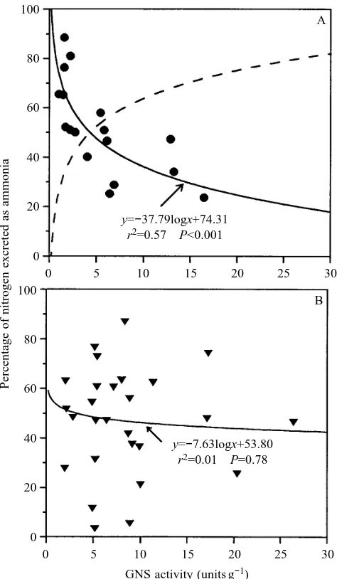
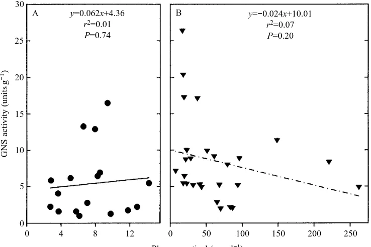
In series III we found a strong linear relationship (r2=0.80) between total nitrogen excretion (the sum of ammonia-N and urea-N) and plasma cortisol levels in unconfined (control post-absorptive) toadfish (Fig. 3A). However, this relationship did not hold for long-term crowded/confined (chronically stressed) fish, and the slope of this regression line was not significantly different from zero (Fig. 3B). The slope of this relationship was also not significantly different from zero in control-fed fish. When the percentage nitrogen excreted as ammonia was plotted against GNS activity for unconfined (control post-absorptive) toadfish, the relationship was not linear. However, by log-transforming hepatic GNS activity, this relationship was much stronger and more than half of the variability (r2=0.57, Pø0.001) in the percentage nitrogen excreted as ammonia was explained by the logarithm of GNS activity (Fig. 4A). For chronically stressed toadfish, the relationship between percentage nitrogen excreted as ammonia and GNS activity was weak to non-existent (Fig. 4B), as was the relationship in control-fed toadfish (data not shown). GNS activity was not correlated with cortisol levels in control post-absorptive (Fig. 5A), chronically stressed toadfish (Fig. 5B) or control-fed toadfish (results not shown). Lastly, a 144–168 h control post-absorptive group of toadfish exhibited cortisol levels of 10.7±1.6 ng ml21 (mean ± S.E.M., N=25), which were not significantly different from values for 72 h control post-absorptive fish.
Discussion
Previous studies with post-absorptive gulf toadfish have shown that short-term exposures to exogenous ammonia, air, confinement and crowding increase the degree of ureogenesis (Walsh et al. 1990, 1994; Walsh and Milligan, 1995). We believe that these short-term treatments can be classified as an acute stress, in that short-term confinement/crowding led to a brief but pronounced rise in plasma cortisol levels which disappeared by 24 h (Fig. 1). Thus, toadfish, like most teleosts (Barton and Iwama, 1991; Brown, 1993), respond to an acute stress such as confinement and crowding with an initial cortisol surge which thereafter declines to near-normal resting levels by 24–48 h (Fig. 1).
Hepatic GNS has been implicated in prior studies with toadfish (Mommsen et al. 1992; Anderson and Walsh, 1995) as one key regulatory or rate-limiting step for ureogenesis. Since the liver is also a major target organ for corticosteroid
action (Henderson and Garland, 1980) and cortisol is the most abundant and most active corticosteroid in fish blood (Idler and Truscott, 1972), Walsh et al. (1994) hypothesized that a classic cortisol stress response may be important in the activation of ureogenesis. In trout and many other animals, metyrapone blocks the synthesis of cortisol by competitively inhibiting
11-b-hydroxylase, which catalyzes the conversion of 11-deoxycortisol to cortisol (Bennett and Rhodes, 1986). Thus, metyrapone treatment results in accumulation of plasma 11-deoxycortisol, which has a low cortisol receptor affinity, is readily metabolized and excreted and is unlikely to have cortisol-like effects (Pottinger, 1990; Pottinger et al. 1992).
B A 100
80
60
40
20
0
Percentage of nitrogen excreted as ammonia
0 5 10 15 20 25 30 100
80
60
40
20
0
0 5 10 15 20 25 30 GNS activity (units g−1)
y=−37.79logx+74.31 r2=0.57 P<0.001
[image:5.609.325.562.73.480.2]y=−7.63logx+53.80 r2=0.01 P=0.78
Fig. 4. Series III. Plot of percentage nitrogen excreted as ammonia
versus liver glutamine synthetase activity (units are as in Fig. 2) in
(A) undisturbed (control) and (B) crowded/confined (chronically stressed) toadfish with best-fitting lines (solid lines) from least-squares regressions. The dashed line in A indicates the percentage of nitrogen excreted as urea, calculated as (1002y). Note that, in control
Metyrapone treatment successfully blocked the initial cortisol surge in toadfish in the present study.
A central finding of this study is that hepatic GNS activity was not elevated in acutely stressed fish previously treated with metyrapone (Fig. 2B), indicating that acute GNS activation probably requires an initial cortisol peak. The lack of a relationship between hepatic GNS activity and baseline cortisol levels (Fig. 5A,B) further supports this view that the peak in cortisol levels is an important factor. However, this finding as well as the observation that GNS activity only accounts for part (at most 57 %) of the percentage of nitrogen excreted as ammonia/urea (Fig. 4), taken together, suggest that cortisol is not the only factor setting GNS activity and degree of ureogenesis.
Recent work by Walsh and Milligan (1995) has demonstrated that the transition to ureogenesis in confined toadfish is mostly due to a reduction in total nitrogen excretion (the bulk of which is as ammonia), whereas the rate of urea excretion remains largely unchanged. Furthermore, they found that it was the cytosolic GNS fraction which increased in confined fish, leading to the hypothesis that GNS acts to turn off ammoniotely by trapping ammonia in the conversion of glutamate to glutamine. Our results demonstrate that a transient surge in cortisol levels is a necessary component of the mechanism which activates GNS (Figs 1, 2).
In addition to these acute dynamics, it is apparent that chronic baseline levels of stress can influence cortisol levels and nitrogen metabolism in toadfish. First, nutritional state appears to alter cortisol dynamics and nitrogen metabolism even in unconfined fish. In control post-absorptive fish, a very strong relationship between total nitrogen excretion rate and plasma cortisol level is evident (Fig. 3A). Interestingly, these levels of cortisol are not very high and even 144–168 h of
starvation does not cause significant elevation of baseline cortisol levels (see Results). The percentage of nitrogen excreted as ammonia is inversely related to liver GNS activity in control post-absorptive fish (Fig. 4A). However, as pointed out above, GNS activity is not correlated to baseline plasma cortisol levels (Fig. 5A). When toadfish are fed, the relationships between nitrogen excretion rate and cortisol levels and between percentage nitrogen excreted as ammonia and GNS are virtually absent. Therefore, the coupling of nitrogen metabolism and baseline cortisol levels in unconfined fish is likely to be related to the degree of body protein catabolism and deamination for gluconeogenesis. Higher levels of long-term baseline stress in unfed toadfish appear to affect nitrogen metabolism differently. The relationships between total nitrogen excretion, cortisol level and the percentage of nitrogen excreted as ammonia and GNS activity are eliminated. These observations indicate a relatively narrow range of plasma cortisol concentration (approximately 0–20 ng ml21) over which nitrogen metabolism is coordinated according to classical models.
Our data indicate that two levels of stress must be distinguished in gulf toadfish: acute stress (physiological), which causes a modest, transient rise in plasma cortisol concentration and plays an important role in the induction of GNS activity, and chronic high-level stress (perhaps aphysiological) caused by long-term confinement or other disturbance, resulting in persistent, higher levels of cortisol and abnormal patterns of nitrogen excretion. Plasma cortisol levels for our undisturbed control fish were always less than 14 ng ml21 (Figs 1, 2A, 3A). Toadfish sampled immediately after capture from Biscayne Bay, Florida, USA, have plasma cortisol levels of 6.9±0.4 ng ml21(mean ±S.E.M., N=384), with a range of 0.4–43.6 ng ml21(T. E. Hopkins and P. J. Walsh,
B A
30
20
15 25
10
5
0
GNS activity (units
g
−
1)
0 4 8 12 0 50 100 150 200 250 Plasma cortisol (ng ml−1)
y=0.062x+4.36 r2=0.01 P=0.74
y=−0.024x+10.01
[image:6.609.193.562.74.320.2]r2=0.07 P=0.20
unpublished data), indicating that the resting undisturbed laboratory values are well within natural limits. Acute stress results in short-lived peaks in plasma cortisol (up to 37 ng ml21), which appear to be involved in the coordination of the transition to ureogenesis. Chronic stress (such as long-term confinement/crowding) produced much higher plasma cortisol values (Fig. 3B). Under other conditions of chronic stress, such as an infection of the ciliate ectoparasite Cryptocaryon iritans (Stoskopf, 1993), the skin and gills of toadfish are covered with trophonts (feeding stage) and excessive mucus production occurs. Toadfish in our laboratory infected with this parasite nearly always die within 3 days, and infected and moribund fish would be classified as chronically stressed with plasma cortisol levels of 64.8±9.4 ng ml21(mean ± S.E.M., N=8, P. J. Walsh, unpublished data). Finally, Wood et al. (1995) found that, in one treatment involving surgery, toadfish which had been initially ureogenic prior to surgery exhibited a large increase in total nitrogen excretion coupled with extremely high cortisol levels (103.0±42.5 ng ml21, mean ± S.E.M., N=7). Under these conditions of chronic, extremely high cortisol levels, the ureotelic pattern is lost and cortisol exerts its well-known proteolytic effects on intermediary metabolism (van der Boon et al. 1991).
Our results show that a transient rise in cortisol levels is a prerequisite for activation of gulf toadfish GNS. We have not shown how it is involved in GNS activation – possibilities include direct cortisol effects on gene transcription, ribosomal translation and allosteric modification of pre-existing enzymes. However, fish GNS does not appear to be allosterically activated (Shankar and Anderson, 1985). We believe that the most fruitful path of investigation should now include studies which examine whether changes in GNS concentration occur and, if so, how cortisol might be involved in regulating GNS concentration (for example, by transcriptional- or translational-level control).
This research was supported by NSF grant IBN-9118819 to P.J.W. and an NSERC operating grant to C.M.W. The authors thank Dr Tom Mommsen for reviewing the manuscript and Jimbo Luznar and Jim ‘Bubba’ Luznar Jr for supplying toadfish.
References
ANDERSON, P. M. ANDWALSH, P. J. (1995). Subcellular localization
and biochemical properties of the enzymes of carbamoyl phosphate and urea synthesis in the batrachoidid fishes Opsanus
beta, Opsanus tau and Porichthys notatus. J. exp. Biol. 198,
755–766.
BARBER, M. L. ANDWALSH, P. J. (1993). Interactions of acid–base status and nitrogen excretion and metabolism in the ureogenic teleost Opsanus beta. J. exp. Biol. 185, 87–105.
BARTON, B. A. ANDIWAMA, G. K. (1991). Physiological changes from
stress in aquaculture with emphasis on the response and effects of corticosteroids. A. Rev. Fish Dis. 1, 3–26.
BENNETT, R. O. AND RHODESIII, C. S. (1986). Evaluation of oral administration of cortisol and metyrapone: the effects on serum
cortisol in rainbow trout (Salmo gairdneri). Comp. Biochem.
Physiol. 83A, 727–730.
BROWN, J. A. (1993). Endocrine responses to environmental
pollutants. In Fish Ecophysiology (ed. J. C. Rankin and F. B. Jensen), pp. 276–296. New York: Chapman & Hall.
HENDERSON, I. W. ANDGARLAND, H. O. (1980). Pisces II. Physiology. In General, Comparative and Clinical Endocrinology of the
Adrenal Cortex, vol. 3 (ed. I. C. Jones and I. W. Henderson), pp.
473–523. London: Academic Press.
IDLER, D. R. ANDTRUSCOTT, B. (1972). Steroids in Nonmammalian
Vertebrates. New York: Academic Press. pp. 127–252.
IVANCIC, I. ANDDEGGOBIS, D. (1984). An optimal manual procedure for ammonia analysis in natural waters by the indophenol blue method. Water Res. 18, 1143–1147.
MOMMSEN, T. P., DANULAT, E. ANDWALSH, P. J. (1992). Metabolic
actions of glucagon and dexamethasone in liver of the ureogenic teleost, Opsanus beta. Gen. comp. Endocr. 85, 316–326.
MOMMSEN, T. P. ANDWALSH, P. J. (1989). Evolution of urea synthesis in vertebrates: the piscine connection. Science 243, 72–75. MOMMSEN, T. P. ANDWALSH, P. J. (1991). Urea synthesis in fishes:
evolutionary and biochemical perspectives. In Biochemistry and
Molecular Biology of Fishes, vol. I (ed. P. W. Hochachka and
T. P. Mommsen), pp. 137–163. New York: Elsevier.
NETER, J., WASSERMAN, W. AND KUTNER, M. H. (1985). Applied
Linear Statistical Models. Illinois: Irwin. 1127pp.
PAGNOTTA, A., BROOKS, L. ANDMILLIGAN, L. (1995). The potential regulatory roles of cortisol in recovery from exhaustive exercise in rainbow trout. Can. J. Zool. (in press).
POTTINGER, T. G. (1990). The effects of stress and exogenous cortisol
on receptor-like binding of cortisol in the liver of the rainbow trout,
Oncorhynchus mykiss. Gen. comp. Endocr. 78, 194–203.
POTTINGER, T. G., MORAN, T. A. ANDCRANWELL, P. A. (1992). The biliary accumulation of corticosteroids in rainbow trout,
Oncorhynchus mykiss during acute and chronic stress. Fish Physiol. Biochem. 10, 55–66.
PRICE, N. M. ANDHARRISON, P. J. (1987). Comparison of methods for the analysis of dissolved urea in seawater. Mar. Biol. 94, 307–317. SHANKAR, R. A. AND ANDERSON, P. M. (1985). Purification and properties of glutamine synthetase from liver of Squalus acanthias.
Archs Biochem. Biophys. 239, 248–252.
STOSKOPF, M. K. (1993). Fish Medicine. Philadelphia: W. B. Saunders
Co. 882pp.
VAN DER BOON, J., GUIDO, E. B., VAN DEN THILLART, J. M. AND
ADDINK, A. D. F. (1991). The effects of cortisol administration on intermediary metabolism in teleost fish. Comp. Biochem. Physiol.
100A, 47–53.
WALSH, P. J., DANULAT, E. ANDMOMMSEN, T. P. (1990). Variation in
urea excretion in the gulf toadfish, Opsanus beta. Mar. Biol. 106, 323–328.
WALSH, P. J. AND MILLIGAN, C. L. (1995). Effects of feeding and confinement on nitrogen metabolism and excretion in the gulf toadfish Opsanus beta. J. exp. Biol. 198, 1559–1566.
WALSH, P. J., TUCKER, B. C. ANDHOPKINS, T. E. (1994). Effect of
confinement/crowding on ureogenesis in the gulf toadfish Opsanus
beta. J. exp. Biol. 191, 195–206.
WOOD, C. M., HOPKINS, T. E., HOGSTRAND, C. AND WALSH, P. J. (1995). Pulsatile urea excretion in the ureagenic gulf toadfish
Opsanus beta: an analysis of rates and routes. J. exp. Biol. 198,
1729–1741.