Printed in Great Britain © The Company of Biologists Limited 1989
WATER UPTAKE IN A CEPHALOPOD AND THE FUNCTION
OF THE SO-CALLED 'PANCREAS'
BY M. J. WELLS AND J. WELLS
Zoology Department, Cambridge, UK and Laboratoire Arago, Banyuls, France
Accepted 30 May 1989
Summary
If the gut of Octopus vulgaris is ligated at both ends, the animal loses weight at about 10% per day at 21 °C, dying after about 48 h. Ligation of the ducts to the midgut gland has the same effect. If one or both ducts are cannulated and led to the exterior with a sufficient length of duct remaining, sea water is taken up by peristalsis and there is little or no weight loss. As body weight falls, blood and urine osmolality remain unchanged, but blood conductivity falls and blood copper concentration rises, indicating a loss of salts as well as water. Muscle dry weight, as a percentage of muscle wet weight, increases as body weight falls. Octopus is hyperosmotic compared with sea water and the rate of ultrafiltration through the branchial heart appendages into the kidney sacs is estimated to be sufficient to account for the observed body weight losses.
Each side of the midgut gland, connected to the gut by a digestive duct, includes two distinct structures. One, the digestive gland, produces enzymes and is also concerned with absorption of the fluid and paniculate products of digestion. The other, the digestive gland appendage (the so-called 'pancreas'), is composed of cells with characteristics that strongly indicate bulk fluid transport; it seems likely that this is the principal site of the fluid uptake. This evidently includes salts as well as water, since sea water taken into the gut (notably by rectal pumping) is not concentrated as fluid is withdrawn.
Introduction
In cephalopods the pressure of the gill hearts is used to drive an ultrafiltrate of the blood through the branchial heart appendages into a pair of pericardial ducts that run into the renal sacs and thence to the exterior (Harrison & Martin, 1965; Schipp & Hevert, 1981). The volume of fluid passed out in this manner has been measured by cannulation of the renal papillae in Octopus dofleini. It averaged 2-6mlkg~1h~1 from animals weighing 10-12kg (Harrison & Martin, 1965). No comparable figures are available for the much smaller Octopus vulgaris but, if one assumes that excretion will scale as metabolic rate (i.e. as mass0'74, Wells et al. 1983) a 0-5 kg animal would be expected to lose around 14 % of its body weight in this manner over 24 h.
216 M . J. W E L L S A N D J. W E L L S
The water lost via the kidneys must be made good. In the account that follows it will be argued that all, or most, of it is taken up through the digestive gland appendages.
Materials and methods
Animals
The experiments were conducted using Octopus vulgaris, collected by trawling or SCUBA diving around Banyuls in August and September 1987 and 1988. The
Octopus weighed 111-1741 g. Each was kept separately and fed on crabs until it
was evident that the individual was undamaged and healthy. Senile animals with large gonads were discarded, since these are physiologically abnormal in ways that affect the excretory system and the water content of body tissues (see Tait, 1986). The experiments were run at 19-23 °C. Animals were starved for 4 days before use.
Anatomy
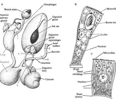
An outline of the anatomy of the gut of Octopus is given in Fig. 1. The digestive gland appendages lie at the entrance to the midgut gland in Octopus and have traditionally been called the 'pancreas', while the digestive gland, also part of the midgut gland, has been termed the 'liver'. Both names are inappropriate, as Bidder (1976) has pointed out. The digestive gland is the site both of enzyme secretion and of digestive uptake and is mainly composed of a single distinctive cell type, the 'boules' cell, which runs through cycles of enzyme production, absorption and waste discharge as digestion proceeds (Boucaud-Camou & Boucher-Rodoni, 1983). The lumen of the digestive gland on each side (it is a bilateral structure, see Wells & Wells, 1989) is confluent with that of the digestive gland appendage, and passes through this into a duct (the 'digestive duct' following the nomenclature in Bidder, 1976) leading to the caecum. The digestive gland appendage is again formed very largely from a single cell type, quite distinctive from the boules cell, with characteristics that strongly indicate bulk fluid transport. (Diagrams illustrating the two dominant cell types are included in Fig. 1. See also Berridge & Oschmann, 1972.)
Surgery
Oesophagus B
Digestive gland
Ink sac
Digestive gland appendages
Anal leaflets Rectum
MicroviUae
Brown body
Caecum
Basal lamina
[image:3.451.15.406.63.397.2]20 /an
Fig. 1. The anatomy of the digestive tract of Octopus vulgaris. (A) The relationship between the principal parts and the points at which surgical interventions were made were as follows. (1) Ligation and section of the oesophagus. (2) Ligation of the rectum. (3) Cannulation of the rectum. The aperture of the cannula faced towards the anus, with the cannula leading out of the animal to a reservoir floating on the surface of the animal's aquarium (as described for oesophageal cannulations in Wells & Wells, 1988). The rectum above the cannula was ligated or exteriorized so as to discharge into the mantle cavity. (4) Ligation of one or both digestive ducts. (5) Cannulation of one or both digestive ducts, close to the digestive gland discharging into the mantle cavity. The ducts on the caecum side were ligated. (6) Cannulation of one or both digestive ducts, leaving the greater part of the lengths of the ducts intact and discharging through the back of the abdomen. The ducts on the caecum side were ligated. (B) The structure of a 'boules' cell from the digestive gland. (After Boucaud-Camou & Boucher-Rodoni, 1983.) (C) A cell from the digestive gland appendage, or 'pancreas'. (From Best, 1981.)
Measurements of the osmolality and conductivity of body fluids and blood copper content
218 M . J. W E L L S AND J. W E L L S
the depression of freezing point of 0-05 ml samples of blood and other body fluids. The machine was calibrated using 400 mosmol kg"1 NaCl and Banyuls sea water as standards; Banyuls laboratory sea water is on open circulation and almost constant at 38 %o and 1130 mosmol kg"1. Conductivity was measured using a Radiometer conductivity meter. Blood copper was assessed using an SP90 absorption spectro-photometer, calibrated from Spectrosol standard copper nitrate solutions.
Correction for scale effects
The animals used ranged in weight over an order of magnitude. Smaller octopuses have a higher specific metabolic rate than large ones; oxygen consump-tion varies as mass0'74 (Wells et al. 1983). It seems reasonable to assume that excretion will scale with metabolic rate, so that the rate of water loss via the kidneys will also scale as mass0'74. In Table 1, the results actually obtained from measurements of weight loss are scaled for a 'standard' animal of 500 g, close to the average size of the octopuses used. So, mass loss observed = am0'74, and mass loss
predicted is a5000'74, where a is a constant and m is mass.
Results
Weight loss after ligation of the anterior oesophagus or the anterior oesophagus and rectum
During and after feeding, octopuses swallow massive quantities of sea water; volumes as large as 50 ml kg"1 h"1 can be collected from floating reservoirs fed by cannulae draining the crop. This is reduced by the end of digestion, but even starving animals continue to drink at around 4 ml kg"1 h"1 (Wells & Wells, 1988). Ligation of the anterior oesophagus (operation type 1, in Fig. 1) did not result in a substantial weight loss. Over the first 24 h six octopuses lost between 2-3 and 7-0 % of their body mass (corrected for a 'standard animal' of 500 g). Some of this appears to have been an immediate result of the operations, possibly attributable to blood loss, but more probably due to a tendency to void fluid from the gut and renal sacs during the operation.
At all events, four of the six actually regained some weight during the second 24 h, so that the total weight loss at 48 h ranged from 0-1 to 8-8%. One would expect a small weight loss simply because the animals were starving; at 20-25°C this is likely to be around 1-1 % of the body weight per day (Mangold & Boletzky, 1967; M. J. Wells, unpublished results).
In sharp contrast were the large and continuing weight losses from octopuses having the rectum ligated as well as the anterior oesophagus. Seven such animals lost between 7-1 and 16-1% of their body weight in the first 24 h, rising to 12-5-20-3 % at 48 h (Table 1). Four of the seven were dead or dying by that time.
contract over the top of the head between the eyes. Similar pale patches develop as pale streaks on the web between the arms, which are held more outstretched than usual, and eventually stiffen at the bases, with the tips flaccid. The animal becomes progressively weaker and less reactive, circulation becomes sluggish and eventu-ally stops, the systemic heart evidently failing first, since freshly dead animals have the auricles distended with blood. Experiments were normally terminated as soon as the external symptoms became obvious.
Rectal cannulation and the collection of ingoing fluid
Direct observation of the anus in lightly anaesthetized animals shows quite clearly that this end of the gut is capable of engulfing water and passing it upstream. The anus expands and surrounds a bolus of water every few seconds. Attempts to quantify this input by cannulating the rectum a few centimetres upstream of the anus only resulted in the collection of substantial volumes in four out of eight experiments. In only one case was the rate of uptake sufficient to balance a 10% loss in body weight over 24 h. The most probable explanation is that the pressure generated in the rectum is minuscule, insufficient to lift the fluid the 3 mm or so needed to push it over the lip in the centre of the floating reservoir.
[image:5.451.35.414.335.646.2]Weight losses in rectal cannulation experiments followed a predictable pattern.
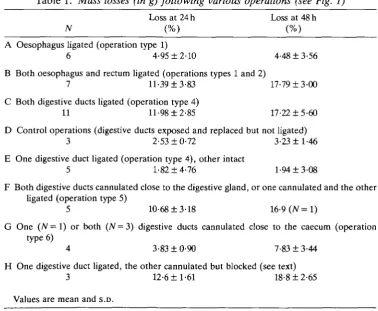
Table 1. Mass losses (in g) following various operations (see Fig. 1)
Loss at 24 h Loss at 48 h N (%) (%) A Oesophagus ligated (operation type 1)
6 4-95 ±2-10 4-48 ±3-56 B Both oesophagus and rectum ligated (operations types 1 and 2)
7 11-39 ±3-83 17-79 ±3-00 C Both digestive ducts ligated (operation type 4)
11 11-98 ±2-85 17-22 ±5-60 D Control operations (digestive ducts exposed and replaced but not ligated)
3 2-53 ±0-72 3-23 ±1-46 E One digestive duct ligated (operation type 4), other intact
5 1-82 ±4-76 1-94 ±3-08
F Both digestive ducts cannulated close to the digestive gland, or one cannulated and the other ligated (operation type 5)
5 10-68 ±3-18 16-9 (N= I)
G One (N = 1) or both (N = 3) digestive ducts cannulated close to the caecum (operation type 6)
4 3-83 ±0-90 7-83 ±3-44 H One digestive duct ligated, the other cannulated but blocked (see text)
220 M . J. W E L L S A N D J. W E L L S
Three animals with the anterior oesophagus open did not lose weight. Four animals with the anterior oesophagus ligated lost 12-4-22-2 % of their body weight over 24-48 h. The remaining animal lost only 6-5% in 48 h, within the 'control' range. This was one of three octopuses with the oesophagus ligated but the intestine open to the mantle cavity upstream of the rectal cannula. It could have pumped water into the gut by dhary or peristaltic action. The other two animals in this class failed to do so and lost weight at rates that implied no fluid uptake.
Ligation of the digestive ducts
The results summarized above show that Octopus must imbibe water through one end of the gut or the other, if it is to maintain its body weight. The following experiments show that the site of the uptake is inside the midgut gland capsule. It will be argued from their structure that the likely site of the uptake is the digestive gland appendages.
Both digestive ducts were ligated and cut (operation type 4) in 11 octopuses. These animals lost between 8-6 and 17-0% of their body weight in the first 24h; five were killed at the end of this period. The remaining six animals had lost 11-9-25-9% of their body weights by the end of 48 h; by then all were dead or dying (Table 1C).
Control animals in which the ducts were exteriorized and replaced without ligation (N = 3) and animals with one duct only ligated (N = 5) lost between 0-4 and 7-3 % of their body weight over 48 h (Table 1D,E)- Most were kept for twice as long, apparently still in good condition.
Cannulation of the digestive ducts close to the midgut gland
The digestive gland of Octopus is concerned not only with the secretion of enzymes, and the absorption of the products of digestion, but also with the discharge of waste materials. Much of the food uptake is by pinocytosis, and the undigested material from the particles taken up in this manner, together with crystalline material (perhaps uric acid) and possibly other wastes, is discharged into the lumen as 'brown bodies' (Boucaud-Camou & Boucher-Rodoni, 1983). Brown bodies accumulate in the intestine and rectum in faecal strings together with solid matter, such as fish scales or fragments of crustacean cuticle, and are periodically discharged through the anus. Starving animals continue to produce brown bodies for a considerable period after the last meal and would still be doing so after the standard 4-day fast imposed on animals before the start of any of the experiments dted above.
Two of the five animals had bilateral cannulations, three had one duct cannulated and the other ligated. It was already known from the five experiments summarized in Table IE that animals can survive a single-sided duct ligation without losing weight.
All five animals with the digestive ducts cannulated lost weight, four of them at rates fully comparable with animals having both ducts ligated. The fifth, at 111 g the smallest animal used in any of the experiments, lost only 5-9 % when scaled to 500 g (8-8% unsealed), overlapping 'control' values. Post-mortem examination of the digestive glands and digestive gland appendages in these animals showed the appendages to be whitish/transparent, and the lumina of the digestive glands to contain only a little brownish fluid on any side with a cannula. Ligated ducts were choked with a thick brownish fluid, filling the lumen of the digestive gland and digestive gland appendages. The latter in these circumstances become brown and spotty. These observations indicate that ducts provided with short cannulae discharge their wastes successfully, so that the weight losses found cannot be attributed to poisons in the system.
Cannulation of the digestive ducts, leaving most of the duct intact
Cannulae inserted into the digestive ducts close to the midgut gland capsule evidently allow discharge of brown bodies and other wastes. But it seems that fluid uptake is prevented. The digestive glands of freshly killed octopuses evidently contain, or are sheathed in, muscles, since they can be seen to contract when dissected out. Contraction would expel fluid from the gland, but cannot suck any in. Fluid intake depends on the digestive ducts, which show active peristalsis, often in the direction from caecum to digestive gland in intact animals. The failure to take up water seen in the animals with cannulae close to the digestive glands might have been due to the extreme shortening of the ducts, rendering them incapable of pumping water inwards.
Seven experiments were made with cannulae to the exterior, leaving the greater part of the digestive ducts intact. The results fall into two categories. Four of the seven maintained their body weights at control levels, with losses of only 2-9-5-0% over 24h and 5-6-11-8% at 48h (N = 3, one killed at 24h). The other three lost weight at 11-3-14-4 % (16-8-21-8 % at 48 h), as fast as animals with both ducts ligated (Table 1G,H).
Careful post-mortem examination showed that one of the three showing substantial weight losses (16-8 %) had the single cannulated duct so twisted that no fluid could pass in or out. The other two (17-8 and 21-8%) had their cannulated ducts choked with thick brown fluid, suggesting that, although no twists or kinks were seen, blockage had nevertheless occurred. Unable to discharge, they were also, presumably, unable to pump in water. The animals that maintained their weights had clear fluid or only a little brown matter in their ducts.
222 M. J. WELLS AND J. WELLS
Blood and urine osmolalities and conductivities; the copper content of the blood
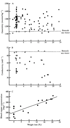
The osmolality of the blood did not vary with the proportion of the body weight lost; nor did the osmolality of the urine. The osmolality of the blood was typically some 15 % higher than that of sea water. The osmolality of the urine was always lower than that of the blood from the same individual, but still typically higher than that of sea water (Fig. 2A).
The smaller number of experiments in which conductivity was measured show blood (always) and urine (usually) having values less than that of sea water. There was no correlation between the conductivity of urine and loss of body weight, but the blood in four samples from animals showing substantial weight losses all proved to be less conductive than blood taken from controls (Fig. 2B). Taken in conjunction with the data showing that osmolality is maintained, this suggested that the non-electrolytes in the blood were being concentrated in animals that lost weight.
The blood pigment in Octopus, as in all cephalopods, is haemocyanin. The copper content of the blood was higher in animals showing substantial weight losses (Fig. 2C). The ultrafutrate includes both salts and water, but blood proteins cannot escape through the branchial heart appendages.
Muscle tissue water content
Samples of muscles from the mantle and second right arm were taken, blotted dry and weighed before drying to a constant weight in an oven at 105 °C. The results are summarized in Fig. 3A,B- Muscle tissue loses water as the body weight falls.
The osmolality of fluid from the rectum
Fluid samples were collected from the rectum or caecum of eight animals showing body weight changes that ranged from a gain of 0-3 % to a loss of 23-2 %. Three of the animals had both ends of the gut ligated, losing 19-2, 20-8 and 23-2 % of their body weights. The fluids collected were all close to the osmolality of Banyuls sea water. There would seem to be no tendency to concentrate the fluid in the gut, even in animals showing large fluid losses. On this evidence, it seems likely that the fluid moving from the gut through the midgut gland into the body is isosmotic with sea water.
Fluid losses from the gut and kidney sacs
1500
f 1400
- A
1300
| 1200 o E
1100
52
a
t
I*!
1
•I
! .•0--0 o ° .
o " o
o
_ o
. Banyuls sea water
12 16 20 24
48
44
•g 40 c o O
36 - B
.o o Banyuls sea water
00
12 16 20 24
c
o
rat
i
c
s
8!_ u- GO
s
73loo
CQ
400
350
300
250
200
150
- C
i i i i i i 8 12 16
Weight loss(%)
[image:9.451.74.355.77.584.2]20 24
224 M. J. WELLS AND J. WELLS 30 28 26 24 22 20 en Q
• rt i
J j 1 1 1 » t
- - - — - " " ' o ' — - " o
! °
o i
o
_ O r
-O o o 0 0
° 8
_—
i i- 5 10 15 20 25
30 28 26 24 22 20
r B
-6 _ | i _ —
1 °
2-o o 1 o o 0 o 1 1 o o n OT^~ 1 - 5 0 5 10 15
Mass loss by animal (%)
[image:10.451.96.362.52.419.2]20 25
Fig. 3. Changes in muscle tissue water content plotted against body weight loss. (A) Mantle muscle samples, y = Q'12x+22-7, r2 = 0-22; (B) Arm muscle samples, >> = 0-09x+23-l,r2 = 0-21.
operation or the beginning of a control run. Animals that have recently fed typically have much larger volumes of fluid visible in the gut.
Control of the activities of the digestive ducts
The ducts carry nerves from the gastric ganglion into the capsule that surrounds the digestive gland and the digestive gland appendages. These nerves appear to be the only connection between the midgut gland and the rest of the nervous system (Young, 1967). The gastric ganghon was removed from two animals of 150 and 240 g. This did not stop the animals from feeding. One took two crabs, and the other three, over the next 3 days. They killed and dismembered the crabs normally, ate all the flesh and discarded the hard parts, including the epiphragmal skeleton, perfectly cleaned out.
because chips of flesh passed through the digestive system almost unchanged, emerging as one would normally find them in the crop. In one instance, the first such remains were discharged through the funnel, along with mucous strings, while the octopus was still hunched up over, and apparently still eating, the crab. Further fragments of flesh appeared in the tanks over the next several hours following each daily meal. The animals were still apparently fit at the end of day 3, when they were killed.
If the gastric ganglia were controlling water uptake as well as digestion, these small octopuses would have died within the first 1 or 2 days. These results therefore confirm the findings from the experiments in which the ducts were disconnected and exteriorized through the back of the abdomen (see above). Digestive duct peristalsis and water uptake can proceed without central nervous input.
Discussion
The experiments described above show that octopuses lose weight if sea water is prevented from reaching the midgut gland. Sea water normally enters the gut by the mouth and/or the rectum and is passed up into the gland by peristalsis of the digestive ducts. This last process, at least, can proceed in the absence of central nervous control. Fluid uptake is apparently isosmotic, with salts as well as water being absorbed, since sea water taken into the gut is not concentrated there. The rate of weight loss (about 10 % in 24 h) that follows when uptake via the midgut gland is blocked is somewhat less than the estimated rate of urine production in
Octopus vulgaris (about 14 % of the body weight in 24 h). Since the animal is
considerably hyperosmotic compared with the sea water in which it lives, osmotic uptake through the gills and/or over the general body surface is likely.
226 M . J. W E L L S A N D J. W E L L S
capsule. There is an additional epithelial layer, not present in octopods, separated from the inner epithelium by a blood space; the outer epithelium faces into the renal sacs. The deep infoldings of the inner epithelial cells are typical of cells concerned in bulk fluid transport (Berridge & Oschmann, 1972).
On the basis of this experimental and anatomical evidence, we suggest that the digestive gland appendages are the principal site of fluid uptake in Octopus and in other cephalopods.
References
BERRIDGE, M. & OSCHMANN, J. L. (1972). Transporting Epithelia. New York: Academic Press. BEST, E. M. H. (1981). Aspects of the digestive system and its control in Octopus vulgaris. PhD
thesis, University of Cambridge, UK.
BIDDER, A. M. (1976). New names for old: the cephalopod 'mid-gut gland'. /. Zooi, Lond. 180, 441-443.
BOUCAUD-CAMOU, E. & BOUCHER-RODONI, R. (1983). Feeding and digestion in cephalopods. In The Mollusca, vol. 5 (ed. A. S. M. Saleuddin & K. M. Wilbur), pp. 149-187. New York, London: Academic Press.
HARRISON, F. M. & MARTIN, A. W. (1965). Excretion in the cephalopod, Octopus dofleini. J. exp. Biol. 42, 71-92.
MANGOLD, K. & BOLETZKY, S. V. (1967). New data on reproductive biology and growth of Octopus vulgaris. Mar. Biol. 19, 7-12.
SCHIPP, R. & BOLETZKY, S. (1976). The pancreatic appendages of dibranchiate cephalopods. I. The fine structure of the "pancreas" in Sepioidea. Zoomorphologie 86, 81-98.
SCHIPP, R. & HEVERT, F. (1981). Ultrafiltration in the branchial heart appendages of dibranchiate cephalopods: a comparative ultrastructural and physiological study. /. exp. Biol. 92, 23-35.
TAIT, R. W. (1986). Aspects physiologiques de la senescence post reproductive chez Octopus vulgaris. PhD thesis, University of Paris.
WELLS, M. J . , O ' D O R , R. K., MANGOLD, K. & WELLS, J. (1983). Diurnal changes in activity and metabolic rate in Octopus vulgaris. Mar. Behav. Physiol. 9, 275-287.
WELLS, M. J. & WELLS, J. (1988). Fluid flow into the gut of Octopus. Vie Milieu 38, 221-226.
WELLS, M. J. & WELLS, J. (1989). The control of enzyme secretion by the digestive gland of Octopus. Vie Milieu (in press).