/. exp. Biol. 155, 103-125 (1991) 1 0 3
Printed in Great Britain © The Company of Biologists Limited 1991
ADHESION AND DETACHMENT OF THE TOE PADS OF
TREE FROGS
BY GAVIN HANNA AND W. JON P. BARNES*
Department of Zoology, University of Glasgow, Glasgow G12 8QQ, Scotland
Accepted 7 August 1990
Summary
The mechanisms by which the toe pads of tree frogs adhere to and detach from surfaces during climbing have been studied in Osteopilus septentrionalis and other tree frogs using a variety of techniques.
The experiments on attachment lend general support to the theory that toe pads stick by wet adhesion. First, the presence of a meniscus surrounding the area of contact shows that pad and surface are connected by a fluid-filled joint. Second, experiments on single toe pads of anaesthetised frogs demonstrate that the pads exhibit the velocity-dependent resistance to shear forces expected of any system employing a fluid as an adhesive mechanism. Third, the largest adhesive forces that toe pads can generate (approx. 1.2mNmm~2, calculated from data on sticking ability) are within the range that can be produced by wet adhesion. Simple measurements of the forces needed to separate a pair of metal discs joined by mucus demonstrate that both viscous forces (Stefan adhesion) and surface tension (the two components of wet adhesion) are likely to play significant roles in the tree frog's adhesive mechanism.
The experiments on detachment demonstrate that toe pads are detached from surfaces by peeling, the pads being removed from the rear forwards during forward locomotion up a vertical surface. When the frogs were induced to walk backwards down this vertical slope, peeling occurred from the front of the pad rearwards. Use of a force platform to measure directly the forces exerted by the feet during climbing shows that, during forward locomotion up a vertical slope, this peeling is not accompanied by any detectable detachment forces. Such forces of detachment are seen, however, during backward walking down the slope and when belly skin comes into contact with the platform. That peeling occurs automatically during forward locomotion is supported both by observations of peeling in single toe pads of anaesthetised frogs and by the inability of frogs to adhere to vertical surfaces in a head-down orientation. Indeed, frogs on a rotating vertical surface were observed to adjust their orientations back towards the vertical whenever their deviation from the vertical reached 85.1 ±21.5°. During forward locomotion peeling seems to occur as a natural consequence of the way in which the toes are lifted off surfaces from the rear forwards, while during
*To whom requests for offprints should be sent.
104 G. HANNA AND W. J. P. BARNES
backward locomotion it is an active process involving the distal tendons of the toes.
Introduction
Frogs from several families have independently adopted an arboreal way of life and are known as tree frogs (Duellman and Trueb, 1985). They possess large disc-like pads at the tip of each toe to provide adhesion during climbing. These pads have several interesting morphological features, being characterised by a special-ised epidermal layer made up of several layers of cells (Ernst, 1973a). The outermost cell layer consists of flat-topped hexagonal or cuboidal columnar cells with free apices, separated from each other by canal-like spaces (Welsch etal. 1974; Green, 1979; Emerson and Diehl, 1980; McAllister and Channing, 1983; Green and Simon, 1986). Although flat, the outer surfaces of these cells appear rough under the scanning electron microscope, and transmission electron mi-croscopy shows the surface to be completely covered by minute peg-like hernidesmosomes (Ernst, 1973a). Between the cells are channels into which mucous glands open (Ernst, 19736). They supply the fluid that forms an essential part of the adhesive mechanism of both toe pads and normal skin. Additionally, in members of the families Rhacophoridae and Hylidae, there is an intercalary cartilage present between the two most distal phalanges (Noble and Jaeckle, 1928).
The physical mechanisms by which toe pads enable tree frogs to adhere to a variety of surfaces have been the subject of much debate. Both friction (Noble and Jaeckle, 1928) and suction (Peters, 1964; Porter, 1972) have been suggested as well as wet adhesion (Nachtigall, 1974). Wet adhesion, the means by which a piece of wet paper sticks to glass, involves a fluid-filled joint which resists cavitation by viscous and surface tension forces. Both Emerson and Diehl (1980) and Green (1981) favoured surface tension as the main component of the adhesive force based on the following experimental observations. A meniscus can be observed at the toe pad-substratum interface (Green, 1981), sticking forces are directly proportional to toe-pad area (Emerson and Diehl, 1980), and adhesion is abolished when the surface tension is lowered by the presence of detergent (Green, 1981) or when the air-water interface is disrupted by immersing the toe pads in water (Emerson and Diehl, 1980). A role for other mechanisms cannot, however, be excluded. For instance, on rough surfaces, the hemidesmosome pegs on the epidermis could interdigitate with irregularities of the substratum, so providing adhesion by mechanical interlocking (Emerson and Diehl, 1980).
Toe-pad adhesion in tree frogs 105
their pads to detach them each time they take a step? Or is there a mechanism of detachment which allows the frogs to move about freely, but grip when required? The investigation was based around observations of the movements of the feet, using video equipment, and analyses of the forces produced by the feet in various positions, using a force platform.
Materials and methods
Experimental animals
Preliminary experiments were carried out on three species of. tree frog, Hyla versicolor Le Conte, a native of North America, Polypedates leucomystax Gravenhorst from Asia and Osteopilus septentrionalis Dumeril and Bibron from the West Indies. O. septentrionalis was found to be the most actively climbing and jumping species and subsequent experiments, including all those described here with the exception of that illustrated in Fig. 7A, were carried out on five adults of this species obtained from Glasgow Zoo. The frogs were maintained in a vivarium at approximately 32°C and fed on live crickets at weekly intervals. Their weight increased slightly over the 18-month period during which experiments were carried out.
Scanning electron microscopy
Feet from freshly pithed frogs were prepared for scanning electron microscopy as follows: the material was fixed in buffered 2.5 % glutaraldehyde for 1.5 h and rinsed in phosphate-buffered sucrose (2 % w/v) for several days; this was followed by postfixation in buffered 1 % osmium tetroxide for 1 h, washing in distilled water, dehydration in an acetone series and critical point drying from CO2. The samples were then mounted on stubs and gold-coated (300 nm thickness) before being viewed under a Philips SEM 500 scanning electron microscope.
Measurement of sticking ability
106 G. HANNA AND W. J. P. BARNES
Video analysis
Movements of the limbs of the tree frogs during different aspects of their climbing behaviour, including forward and backward locomotion, jumping and repositioning, were filmed using Philips and Panasonic WV-1550/B video cameras equipped with both a 16 mm wide-angle lens and a 50 mm macro lens to which 20 mm or 40 mm extension tubes were added for close-up work. Filming was done through the wall of a clear Perspex tank, with a synchronised stroboscope. Use of a Panasonic NV-8050 time-lapse video recorder gave a filming speed of 50 pic-tures s"1 and allowed frame-by-frame analysis of the recordings. A video timer (For A, model VTG-33F) was used to determine the exact time course of all events.
To study the preferred orientation of frogs in the vertical plane, animals were filmed on a slowly rotating vertical surface. The base of a Perspex tank was cemented to the end of a horizontally mounted kymograph spindle. A frog was placed in the middle of this surface and the kymograph set in motion at an angular velocity of 4.8°s"1.
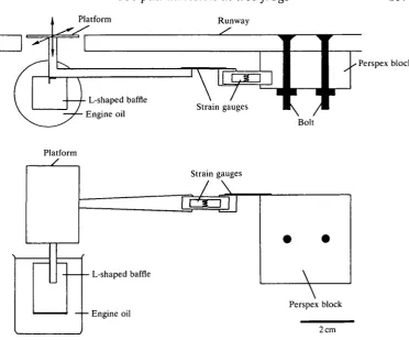
Measurement of forces
Toe-pad adhesion in tree frogs
Platform Runway
107
Perspex block
Bolt
Platform /
Strain gauges
L-shaped baffle
Engine oil
T
Perspex block [image:5.451.39.412.61.370.2]2 cm
Fig. 1. The force platform used to measure forces generated by climbing tree frogs. The upper drawing shows the force platform, which was constructed of light plastic materials viewed from above, the lower drawing the platform viewed from in front with the runway omitted for clarity.
To measure the ability of individual toe pads to withstand shearing forces applied in different directions or at different velocities, frogs were anaesthetised in a solution of Benzocaine (200 mg I"1). Individual toes (third toe of right manus) were either loosely supported or firmly clamped using a mounted pair of forceps and a rubber sleeve around the digit so that the toe pad made contact with a horizontally mounted force platform which could be moved forwards or back-wards at a range of velocities. This was achieved by mounting the force platform on a micromanipulator whose coarse control was directly linked to the spindle of a horizontally mounted kymograph. Forces at which slippage or detachment of the toe pads occurred were measured in each case for three frogs.
108 G. H A N N A A N D W. J. P. BARNES
sufficient quantities for these tests, mucin from a pig's stomach obtained from a commercial source (Sigma Chemical Company, catalogue no. M-2378) was used instead. The discs were subjected to a steady pull by means of the micromanipula-tor/kymograph device described in the previous paragraph, while a strain gauge (Tinsley Telcon, type 2/120/EC11) was used to measure the maximum adhesive force generated before the plates separated.
Results
Surface morphology
Osteopilus septentrionalis is one of the larger tree frog species (one of the frogs used in this study weighed 28 g) and has well-developed toe pads, located on the expanded tips of each digit. Four occur on each forelimb, five on each hindlimb. Each toe pad is ovoid in surface view (Fig. 2A), and is raised above the digit's ventral surface (Fig. 2D). The thickness of the pad, combined with its flexibility, means that small movements of the toes, such as those that occur when a frog changes its weight distribution, need not result in movement of the pad epithelium with respect to the substratum. Each toe pad is completely surrounded by a circumferential groove, as previously described by Green (1979), and lateral grooves continue down the margin of the ventral surface of each digit (Fig. 2A,C). Proximal to each toe pad are located subarticular tubercles (Fig. 2A), whose epithelial surface appears identical to that of the toe pads (Fig. 2B).
The surface morphology of the toe pads of O. septentrionalis is typical of that of other tree frog species (Welsch et al. 1974; Green, 1979; McAllister and Channing, 1983; Green and Simon, 1986). It is important to note that no anterior/posterior asymmetry in the toe-pad epithelium (e.g. backwardly pointing spines) is visible in surface view that could explain why toe pads resist backward, but not forward, pulls (see Fig. 10A where the term 'forward pull' refers to the direction of movement of the force platform with respect to the frog and is thus equivalent to a backward pull on the animal).
Sticking ability
The adhesive ability of the tree frog species used in this study was directly tested using the method of Emerson and Diehl (1980). Frogs were placed in turn on a platform whose angle with respect to the horizontal could be smoothly increased from 0° (horizontal) through 90° (vertical) to 180°, and the angle at which each frog lost contact with the surface was noted.
Fig. 2. Scanning electron micrographs of toe pads and subarticular tubercles of
Osteopilus septentrionalis. (A) Overview of two toe pads and three subarticular
Toe-pad adhesion in tree frogs
110
G. HANNA AND W. J. P. BARNESo
o ^
"oh a) 180 150-90 60 30-j 0 Head-up
• H
-Head-down
10 12 14 16 18
180- 150- 120-90 60H 30 0 Perspex Sandpaper
[image:8.451.33.427.56.191.2]10 12 14 16 18 Body mass (g)
Fig. 3. Means and standard deviations of maximum angles of stick plotted against body mass for five Osteopilus septentrionalis. (A) Animals tested up and head-down on Perspex. (B) Animals tested head-up on Perspex and sandpaper. Each frog was given five trials in each situation. The graph shows that, in the head-up situation, the 8.6g tree frog had a maximum angle of stick on Perspex of 151±10.7°. Adhesive force [force component perpendicular to surface, which equals cos(180°—maximum angle of stick) xbody mass], thus has a mean value of 7.7 g (75.5 mN).
At angles of up to between 90° and 100°, the adhesion of toe pads and subarticular tubercles was aided by that of belly and thigh skin pressed against the substratum (see Fig. 5). Directly after belly and thigh skin lost contact with the platform, head-down frogs usually fell from the platform. There is no evidence from these experiments that toe pads facing down a slope significantly aid the frogs in maintaining a grip. This is a point we will return to later. In contrast, frogs rotated head-up on the platform maintained a grip with their toe pads long after belly and thigh skin had lost contact with the platform (Fig. 3A). The nature of the surface also affects sticking ability; replacement of the smooth Perspex surface of the platform with sandpaper (English Abrasives Glass Paper, grade 1.5, grit no. 120, approx. 45 sand particles per mm2) reduced the maximum angle of stick by an average of 35.7° (Fig. 3B).
The mean maximum angle of stick of the 8.6 g frog in the head-up situation on Perspex was 151° (Fig. 3). From this value, the adhesive force, the force component perpendicular to the surface, can be calculated by simple trigonometry as 7.7g or 75.5mN. Since the toe-pad area of this frog, measured from a photograph, was 63mm2, this represents an adhesive force of U r a N r a m '1.
Toe-pad adhesion in tree frogs 111
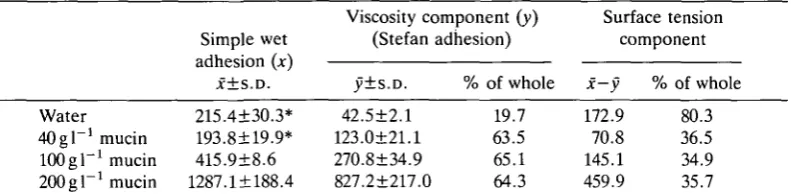
Table 1. Forces (mN) required to separate two metal discs joined by a film of water or mucous solutions of different concentrations
Water 40 g I"1 mucin 100 g P1 mucin 200 el"1 mucin
Simple wet adhesion (x) X±S.D. 215.4±30.3* 193.8±19.9* 415.9±8.6 1287.1 + 188.4
Viscosity component (y) (Stefan y±s.D. 42.5±2.1 123.0±21.1 270.8±34.9 827.2±217.0 adhesion)
% of whole
19.7 63.5 65.1 64.3 Surface tension component
x—y % of whole
172.9 80.3 70.8 36.5 145.1 34.9 459.9 35.7
* Values for simple wet adhesion with water and with 40 mg 1 l mucin between the discs are not significantly different from each other (r-test).
The figures in the left-hand column represent wet adhesion where the liquid/solution is restricted to the area between the discs. The figures in the centre columns represent Stefan adhesion where the liquid/solution surrounds the discs as well. The figures in the right-hand columns (surface tension component) are obtained by subtraction.
N=5 in all cases.
a factor of 5 (Table 1), though low mucin concentrations do not produce this effect since they reduce the surface tension component of adhesion. It can also be seen that the proportion of the total force that is due to surface tension forces is reduced from about 80 % when water lies between the discs to about 36 % when water is replaced by pig stomach mucin.
Because of the viscoelastic properties of the sealing fluid, any attachments utilising wet adhesion should be able to withstand appreciable fast transient shear forces, which, if applied slowly and steadily, would cause slippage of the surfaces relative to each other. To test whether the forces exhibited by tree frog toe pads were velocity dependent, single toe pads of anaesthetised frogs were held against a force platform which was then moved in the shear plane at different velocities. As predicted, low resistive forces were generated during low-velocity movements, the toe pad simply slipping over the surface of the platform (Fig. 4). Indeed, the toe-pads of tree frogs on smooth vertical surfaces are continuously slipping and being repositioned on the surface, so long as adhesion is not being aided by belly and thigh skin. In contrast, rapid movements of the platform led to the generation of large resistive forces (up to 177 mN in one instance) before significant slippage of the pad occurred.
Video analysis
112 G. HANNA AND W. J. P. BARNES
140i
z.
-a
CJ
o
it
sm
j
o 120
100
80
60
40
20
0.0 0.5 1.0 1.5 2.0
Velocity of platform (mms~')
[image:10.451.102.351.77.226.2]2.5 3.0
Fig. 4. Means and standard deviations of the maximum resistive forces exhibited by a single toe pad of an 8.7 g Osteopilus septentrionalis to movement in the shear plane (three trials at each of five velocities). Third digits of the right manus of anaesthetised frogs were supported in their normal orientation so that the toes exerted a downward pressure on the horizontally mounted force platform of approx. 0.25 g. Horizontal force was then applied in a forward direction, mimicking the forces that would be applied to the toe pads of a frog tending to slide backwards down a vertical slope. The graph shows that resistive forces of single toe pads to movement in the shear plane are dependent on the velocity of the applied movement.
the forelimbs, they remain so pressed until the end of the stance phase of locomotion. This is presumably a reflection of the outward pull acting on the forelimbs and the inward force acting on the hindlimbs. As might be predicted from the mechanics of this situation, the uppermost fore- and hindlimbs of frogs moving across a slope show the behaviour just described for the forelimbs, while the lower two feet remain pressed against the substratum as described above for the hindlimbs.
In all recorded examples (over 50), toe-pad removal occurred by peeling, contact with the substratum being progressively broken from the rear forwards when the frog was climbing up the slope (Fig. 5B). The area of contact between toe pad and plastic is progressively reduced as the line representing the posterior limit of the area of contact moves upwards towards the tip of the toe. In contrast, when the frogs walked backwards down the slope, the pads were again peeled from the surface, but this time from the front edge of the pad backwards (Fig. 5C).
During forward locomotion, the toes had an angle of about 15° to the substratum when the pads were placed on a surface. The proximal portions of the
114 G. H A N N A AND W. J. P . B A R N E S
toes were then pivoted forwards through an angle of up to 65° before any peeling took place. From this observation, we suspect that one of the main functions of the intercalary cartilage in the toes of tree frogs (illustrated in Fig. 9A) is to increase the angle through which the toes can move without initiating peeling of the pads. In this way, adequate adhesion is provided until the swing phase of locomotion is initiated.
Forces exerted by the limbs during locomotion
The force platform illustrated in Fig. 1 was used to measure directly the forces exerted by the feet of tree frogs as they climbed up and down a vertical Perspex surface (Fig. 6A). During forward walking up the slope, the first detectable force component is a transient inward force (Fig. 6Ci-iii, lower traces). This is presumably associated with the pressing of the toe pads onto the surface at the start of the locomotor stance phase. During the rest of the stance phase of both fore- and hindlimbs, the vertical force component acts downwards, resisting the force of gravity (Fig. 6Ci-iii, upper traces), while the horizontal force components normal to the surface act outwards for forelimbs and inwards for hindlimbs as predicted (Fig. 6Ci-iii, lower traces).
Were the frog to need to pull its feet off the substratum at the end of each stance phase, there would be a substantial upward and outward force component present at the end of each stance phase during forward walking up the runway (Fig. 6B). Were the frog instead to have an effective mechanism of toe-pad detachment, such forces would be absent, as seen in the lower set of theoretical traces. The actual data for frogs walking forwards up the slope (Fig. 6Ci-iii) clearly correspond to the lower set of theoretical traces. It can therefore be concluded that the peeling mechanism used by the frogs to detach their pads during forward locomotion is an effective one, and removes the need for the frogs to overcome directly the substantial sticking forces exerted by their pads every time they take a step. As Fig. 6Civ shows, such detachment forces are also absent when a back foot jumps off the platform.
In Up
Down -Out
Toe-pad adhesion in tree frogs
B
Front foot Back foot
115
If no detachment mechanism were present
Front foot Back foot
If detachment mechanism were present
Ci Cii Ciii Civ
Front foot walk Back foot walk Front and back foot walk Back foot jump
Down
20 mN
20 mN
L
I n
Is
Fig. 6. Sample force platform recordings of the vertical (upward and downward) and horizontal (inward and outward) forces exerted by the front and back feet of
Osteopilus septentrionalis during uninterrupted walking up a vertical slope (Ci-iii), and
by a back foot being removed from the platform during a jump (Civ). The traces correspond to the lower set of theoretical traces (B) in that no upward and outward forces are detectable when the toe pads are removed from the platform, the result that would be expected if the frog had an effective mechanism of toe-pad removal (see text for explanation). The positioning of a foot on the vertically mounted force platform and the directions of force components recorded by it are illustrated in A. The recordings illustrated in Ci-iii were from a frog weighing 8.7 g, while Civ came from a frog weighing 8.5 g.
116
G. HANNA AND W. J. P. BARNESA Bi Bii Biii Biv Bv
Back foot walk. Front foot. Back foot. Front foot, Front foot walk, Belly skin abnormal step backwards walk backwards walk jump belly skin
-A- VVU -JU
[image:14.451.59.392.56.226.2]Is Is
Fig. 7. Sample force platform recordings of the vertical (upward and downward) and horizontal (inward and outward) forces exerted by Hyla versicolor (A) and Osteopilus
septentrionalis (B) in situations where observable detachment forces were recorded.
Experimental arrangement as in Fig. 6A. A shows the only instance during forward walking where such detachment forces were seen. This occurred in a preliminary experiment (note undamped force platform) during a visibly abnormal step. Bi and Bii illustrate removal of feet from the force platform during backward walking down the vertically orientated runway, and Biii removal of a front foot from the platform during a jump. In Biv belly skin made contact with the platform during forward walking up the runway, while in Bv a frog at rest with its belly skin touching the platform resumed forward walking. The recordings illustrated were from frogs weighing 7.1 g (A), 8.9g (Bi), 8.8g (Bii), 9.5 g (Biii), 28g (Biv) and 8.7g (Bv).
are detectable as the foot detaches from the platform during backward walking. These detachment forces are shown as open blocks on the histograms.
Mechanisms of toe-pad removal
Two large tendons run along the dorsal surface of each toe of O. septentrionalis and a single large tendon runs along the toe's plantar surface, dividing into two near its distal end (Fig. 9). The former are the tendons of the extensor brevis profundus, while the latter is the tendines superficialis, which attaches either to the aponeurosis plantaris or to the flexor digitorum brevis superficialis, or to both, depending on the digit (Dunlap, 1960). These tendons are secured at their distal ends to the terminal phalange. When the two dorsal tendons were pulled, the terminal phalange moved in the same way as it did when the pads were being peeled off from the front during backward walking. We therefore suspect that, during backward walking, peeling occurs as a direct result of the contraction of the muscles attached to these dorsal tendons.
Toe-pad adhesion in tree frogs
111
20 n O
if
0
SERIES A Force up Force in
o z
60J
201
11 10 11 10 20
SERIES B Force up Force in
14 9 14 9
JZ
^ s
u
CQ
Force down Force out Force up Force in
3 3
60J
2 0 -i
Force down Force up
60 Force down Force out 60-^
N
Force out Force in
6 2 N
Force down Force out
l::A Front foot HH Back foot
•
[image:15.451.91.359.47.399.2]Forces generated by removal of a foot
Fig. 8. Means and standard deviations of peak values of major force components generated by Osteopilus septentrionalis walking forwards up or backwards down over a force platform located in the middle of a vertically oriented Perspex runway. Measurements of initial pad attachment forces (see Fig. 6) are excluded from the figure. During backward locomotion (but not forward locomotion), forces associated with the removal of a foot were clearly seen. Such forces are indicated by open blocks on the figure. Series A: data obtained from a frog weighing 7.2g. Series B: data obtained from two frogs weighing 8.6g and 8.7 g. N, number of observations.
118
G. HANNA AND W. J. P. BARNES A BIntercalary cartilage Tendines superficial Terminal phalange
lmm
D
Ligament
Tendons of the extensor brevis profundus
Fig. 9. Bones and tendons of the third digit of the left manus of Osteopilus septentrionalis. A illustrates the presence of an intercalary cartilage between the two terminal phalanges, while B, C and D illustrate, respectively, ventral, lateral and dorsal views of the ligaments and tendons present in the digits.
(equivalent to a forward movement of the toe), the toe pad simply turned over with negligible generation of force (Fig. 10A). In both parts of Fig. 10A, the toe-pad forces are compared to the situation that arises when the force platform is moved against a stop. They show that, even when substantial resistive forces are generated, some slippage of the pad occurs. This experiment confirms that toe pads peel automatically when pulled forwards, but does not exclude the possibility that there is, in addition, some inherent asymmetry in the structure of the pads that allows them to resist shear forces applied in one direction but not the other. This possibility was examined by securely clamping the same toe pads and pressing them firmly against the force platform with the aim of preventing, as far as possible, the toe pads from turning over. Fig. 10B illustrates 10 superimposed trials of both forward and backward pulls and a direct comparison of two of these traces {a and b). They show that, although forward pulls result in the generation of larger resistive forces, since it is not possible to prevent the toes from turning over eventually, the time course of the force generation during the period before this occurred was often similar in the two situations. This result, combined with the lack of anatomical evidence for any structural asymmetry in the toe-pad surface (Fig. 2C), convinces us that the toe pads themselves are inherently able to resist shear forces applied in any direction.
A Toe pad free Forward pull
Toe-pad adhesion in tree frogs
Backward pull
119
100 mN 10 mN
B Toe pad fixed
[image:17.451.47.399.65.295.2]Forward pulls Backward pulls10 s
Fig. 10. Sample recordings of the resistive forces exhibited by a single toe pad of
Osteopilus septentrionalis to movement in the shear plane at a velocity of 1.3 mm s"1. In A, the third digit of the right manus of an anaesthetised frog was supported in its normal orientation so that the toe exerted a downward pressure on the horizontally mounted platform of approx. 0.2 g. Horizontal force was then applied in a forward or backward direction. In both traces (and in B below), the toe-pad forces are compared to the forces that result from the platform being moved against a stop. In B, the same toe was firmly clamped so that the toe exerted a downward pressure on the platform of approx. 3.3 g with the aim of preventing, as far as possible, the toe pad from turning over. The figure illustrates 10 superimposed trials of both forward and backward pulls and a direct comparison of two of these traces (a and b). The mass of the frog in this experiment was 8.8 g.
Fig. 3, head-down frogs fell from the platform directly its angle was increased beyond the vertical. If a frog is placed head-up on a vertical surface which is then rotated (Fig. 11), the frog adjusts its position back towards the vertical whenever the deviation of its body from the head-up position reaches approximately 90° Thus, it never allows itself to reach the head-down position. Also, even though frogs may often rest on a vertical surface facing across rather than up the slope (Fig. 5 A), the distal portions of most of the toes are aligned with the vertical rather than the anterior-posterior axis of the frog. This occurs both as a result of the feet being placed facing up the slope and because of the ability of the toes themselves to bend at their joints to face towards the vertical (Fig. 5A).
Discussion
120
G. HANNA AND W. J. P. BARNES40
•a
m 120
10 20 30
Time (s)
40 50 60
Fig. 11. The effect of substratum rotation on positioning of Osteopilus septentrionalis on a vertical surface. The frog was placed head-up on a vertical Perspex surface, which was rotated about its horizontal axis at a velocity of 4.8°s~'. Throughout the experiment, the frog repositioned itself whenever its deviation from the head-up position reached 85.1±21.5° (N=29).
locomotion. The experiments on detachment mechanisms show for the first time that tree frogs possess mechanisms for detaching their feet during locomotion that obviate the need to overcome directly the sticking forces exerted by their pads. It should, however, be stressed that, as toe pads have arisen independently several times in tree frogs, the following observations apply particularly to one species, Osteopilus septentrionalis. In spite of considerable convergence in toe-pad structure (Green, 1979), members of other families of tree frogs may well utilise mechanisms of attachment and detachment that differ in detail from those we discuss below.
Mechanisms of toe-pad adhesion
Nachtigall's proposal that wet adhesion is the prime mechanism of adhesion to smooth surfaces has been supported by experimental data from Emerson and Diehl (1980), Green (1981) and Green and Carson (1988). The results described in this study lend further support to this argument.
First, as previously described by these authors, our video analysis has clearly shown the presence of a meniscus surrounding the area of contact between pad and surface (Fig. 5), so that pad and surface are connected by a fluid-filled joint, a central requirement of wet adhesion.
Second, our experiments on single toe pads of anaesthetised frogs (Fig. 4) show that the pads exhibit the velocity-dependent resistance to shear forces expected of any system employing a fluid as an adhesive mechanism. That these results on anaesthetised animals are representative of the properties of pads during normal behaviour is shown by the observation that frogs on smooth vertical surfaces constantly slip unless they increase their area of contact with the surface by the use of thigh and belly skin, as illustrated in Fig. 5A.
Toe-pad adhesion in tree frogs 121
_ 2
(1.2mNmm ) is well within the range that can be produced by wet adhesion (7 mN mm"2, Nachtigall, 1974). This is also true for the data of Emerson and Diehl (1980) for Hyla cinerea, whose method was used in this study, from which a value of 1.4mNmm~2 can be calculated.
The use of a fluid to provide adhesion between pad and substratum provides the frog with distinct advantages. Since the sealant does not harden, as would a glue, the joint is instantly fully functional and requires less force to break it. The underlying physical mechanism is, however, quite complex, and involves both surface tension and viscous forces, since both will help resist cavitation, the entry of air into the joint and its subsequent fracture. Stefan (1874), on both theoretical and experimental grounds, showed how the time of separation (t) of two discs each of radius R is influenced by their initial separation (D), the viscosity (rf) of the fluid in which they are submerged, and the normal force applied (f):
. (1) Since longer separation times represent better adhesion, adhesion is increased by increasing the viscosity, increasing the size of the discs or decreasing their separation distance. Since force per unit area (/) equals F/JTR2, equation 1 can be rewritten as follows (Bikerman, 1968):
This analysis neglects surface tension (capillarity) and is thus strictly applicable only to discs that are completely immersed in the liquid. Indeed, if the liquid is restricted to the area between the discs, there is additionally a surface tension component which, if the two surfaces are fully wettable, is 2A/D, where A is the surface tension of the liquid in air (Bikerman, 1968). The tree frog toe-pad situation is thus best described by the following equation, in which the first and second expressions represent the viscosity, or Stefan adhesion, component and the surface tension component of wet adhesion, respectively (Emerson and Diehl, 1980):
fi-
3
-A+-- «
J
4 D2 D
Emerson and Diehl (1980), Green (1981) and Green and Carson (1988) favour surface tension as the main adhesive force on the grounds that adhesion fails if surface tension is lowered by a detergent (Green, 1981) or abolished by immersing the whole of the pad in water (Emerson and Diehl, 1980). These experiments do, of course, strongly suggest that surface tension is involved, but do not exclude a role for viscosity as well. Indeed, all these experiments have actually shown is that with the surface tension component reduced (Green) or absent (Emerson and Diehl), the remaining force components are insufficient to support the frogs.
122 G. H A N N A AND W. J. P. B A R N E S
carried out a few simple tests using a pair of polished cupronickel discs as previously described. The mean maximum force obtained in these tests for simple wet adhesion with water between the discs was 0.68mNmm"2. Since this is not only less than the theoretical value (which is not unexpected, and can be explained by the plastic and viscous flow that precedes fracture and the existence of flaws and dislocations in the surface of the discs, Davies and Rideal, 1961), but an order of magnitude less than the experimental value of 7 mN mm"2 given by Nachtigall (1974), our conclusions must necessarily be tentative. The fact remains, however, that high concentrations of mucin can increase the adhesive force by a factor of 5, and that, while low mucin concentrations do not have this effect (the values for simple wet adhesion with water and with "Wmgl"1 mucin between the discs are not significantly different from each other), the proportion of the total force that is due to the surface tension component is reduced from about 80% when water lies between the discs to about 36% when water is replaced by mucin (Table 1).
Thus, while it would be reasonable to agree with the statements of Emerson and Diehl (1980) and Green (1981) that tree frogs adhered by surface tension forces (capillarity) were the fluid secreted by the toe pads to be water, the extent to which it is a mucous solution will make this conclusion invalid. Unless further work shows that the mucus secreted by the frogs has very different physical properties to those of the pig stomach mucus used in the above tests, it is safer to state that toe pads adhere to smooth surfaces by wet adhesion, and make no assumptions as to whether the surface tension or the viscosity component is of greater significance. Indeed, frog mucus has already been shown to be a good wet adhesive, since it is a viscous, long-chain, high molecular weight polymer and has many functional groups capable of providing good cohesive strength (Lipson and Silbert, 1968; Dapson, 1970).
Previous authors, notably Emerson and Diehl (1980), consider the possibility that the hemidesmosome pegs on the epidermis could aid adhesion by mechanical interlocking on rough surfaces. Since adhesion to rough surfaces is relatively poor (Fig. 3B), this suggestion is not supported by our data. Indeed, the hemidesmo-some pegs are so small that it is difficult to see them playing such a role. More likely, as suggested by Green (1981), they are the non-functional remains of the desmosomes that are important in keeping the different layers of the epidermis of the toe pads firmly attached to each other.
Mechanisms of toe-pad detachment
Toe-pad adhesion in tree frogs 123
pads are removed by peeling, the pads being removed from the rear forwards during forward locomotion up a vertical slope, and from the front of the pad rearwards when frogs are induced to move backwards down a vertical slope (Fig. 5B,C). Use of a force platform to measure directly the forces exerted by the feet during locomotion shows that, during forward locomotion up a vertical slope, this peeling is not accompanied by detectable detachment forces, although such forces were seen when the frogs backed down the slope, when the frogs jumped off the platform (forelimbs only), during a single abnormal step (in another species, Hyla versicolor) or when belly skin came into contact with the platform (Fig. 7). Thus, we can be sure that such detachment forces would have been seen had they been within the range detectable by our force platform.
In theory, such a detachment mechanism could have its basis in the physical structure of the pads. For instance, ski mountaineers used to put seal skins on their skis when they wished to go uphill, which allowed smooth movement of the skis in a forward direction up the slope, but did not slip backwards readily because all the hairs pointed backwards. We have, however, failed to find any structural asymmetries in the surface of the toe pads (Fig. 2), nor are any such asymmetries visible from published scanning electron micrographs (Welsch et al. 1974; Green, 1979; Emerson and Diehl, 1980; Green and Simon, 1986). When single toe pads of anaesthetised frogs were gently placed on a force platform which was then moved in the shear plane (Fig. 10A), substantial forces were generated in response to fast movements when the platform was moved forwards with respect to the frog (equivalent to a frog sliding backwards down a vertical slope), but negligible force when the platform was moved in the other direction since the pads simply turned over (peeled off). However, when the pads were held as firmly as possible and pushed against the platform (Fig. 10B), no directionality was observable in the forces generated during the initial phase of the response (though, as it was impossible to prevent the pads turning over eventually, differences appeared with time). We thus conclude on both anatomical and physiological grounds that there are no asymmetries in pad structure that could underlie the frog's mechanism of detaching its toe pads.
124 G. H A N N A AND W. J. P. B A R N E S
applied in a backward direction, they were completely powerless to resist being lifted if the pull was forwards and upwards.
During backward walking down a slope, peeling no longer occurred automati-cally, but was mimicked, in a dissected foot, by pulling on the tendons that run along the dorsal surface of each toe. We thus suspect that, during backward walking, the peeling of the toe pads from the front backwards is an active process, occurring by contraction of the extensor brevis profundus in the hindlimb and the equivalent muscle in the forelimb. That this method is not as efficient as the automatic peeling occurring during forward locomotion is shown by the detectable peeling forces observed during backward locomotion (Fig. 7).
Such a mechanism of toe-pad detachment by peeling has important conse-quences for the lives of the frogs, for it means that the pads can never be positioned so that they face down a slope, or the weight of the frog's body acting on them would automatically initiate peeling. Indeed, we can see from video stills (e.g. Fig. 5A), that even when an animal positions itself across a slope, most of the toes are positioned so that they face up the slope. Also, as clearly demonstrated by the results illustrated in Fig. 11, a frog on a vertical surface slowly rotated from its head-up position adjusted its position back towards the vertical whenever the deviation of the body from the vertical reached 85.1±21.5°. Tree frogs are thus clearly unable to walk forwards down steep slopes but, since they jump so efficiently, this does not appear to be a significant disadvantage. The greatest asset of the detachment mechanism is its effectiveness, in that frogs do not need to waste energy overcoming the substantial sticking forces that their toe pads can generate whenever they wish to take a step.
We would like to thank Dr Roger Downie for his helpful comments on the manuscript.
References
BIKERMAN, J. J. (1968). The Science of Adhesive Joints. New York: Academic Press.
DAPSON, R. (1970). Histochemistry of mucus in the skin of the frog, Rana pipiens. Anat. Rec.
166, 615-626.
DAVIES, J. T. AND RIDEAL, E. K. (1961). Interfacial Phenomena. New York: Academic Press. DUELLMAN, W. E. AND TRUEB, L. (1985). Biology of Amphibians. New York: McGraw-Hill. DUNLAP, D. G. (1960). The comparative myology of the pelvic appendage in the Salentia.
J. Morph. 106, 1-76.
EMERSON, S. B. AND DIEHL, D. (1980). Toe pad morphology and mechanisms of sticking in frogs. Biol. J. Linn. Soc. 13, 199-216.
ERNST, V. (1973a). The digital pads of the tree frog Hyla cinerea. I. The epidermis. Tissue & Cell 5, 83-96.
ERNST, V. (19736). The digital pads of the tree frog Hyla cinerea. II. The mucous glands. Tissue
& Cell 5, 97-104.
GREEN, D. M. (1979). Tree frog toe pads: comparative surface morphology using scanning electron microscopy. Can. J. Zool. 57, 2033-2046.
GREEN, D. M. (1981). Adhesion and the toe pads of tree frogs. Copeia 1981, 790-796. GREEN, D. M. AND CARSON, J. (1988). The adhesion of treefrog toe-pads to glass: cryogenic
Toe-pad adhesion in tree frogs 125
GREEN, D. M. AND SIMON, M. P. (1986). Digital microstructure in ecologically diverse sympatric microhylid frogs, genera Cophixalus and Sphenophryne (Amphibia: Anura), from Papua New Guinea. Aust. J. Zool. 34, 135-145.
LIPSON, M. AND SILBERT, J. (1968). Glycosaminoglycans of adult frog skin. Biochim. biophys.
Ada 158, 344-350.
MCALLISTER, W. AND CHANNING, A. (1983). Comparison of toe-pads of some southern African climbing frogs. S. Afr. J. Zool. 18, 110-114.
NACHTIGALL, W. (1974). Biological Mechanisms of Attachment. Berlin, Heidelberg, New York: Springer-Verlag.
NOBLE, G. K. AND JAECKLE, M. E. (1928). The digital pads of the tree frogs. A study of the phylogenesis of an adaptive structure. /. Morph. 45, 259-292.
PETERS, J. A. (1964). Dictionary of Herpetology. New York: Hafner Publ. Co. PORTER, K. R. (1972). Herpetology. Philadelphia: W. B. Saunders Co.
STEFAN, J. (1874). Versuche iiber die scheinbare Adhasion. Sitzber. Akad. Wiss. Wien (Abt.
II. Math.-Phys.) 69, 713-735.