THE ROLE OF V-ATPase IN NEURONAL AND ENDOCRINE
SYSTEMS
BY YOSHINORI M0R1YAMA*, MASATOMO MAEDA AND MASAMITSU FUTAI
Department of Organic Chemistry and Biochemistry, Institute of Scientific and Industrial Research, Osaka University, Ibaraki Osaka 567, Japan
Summary
Synaptic vesicles have important roles in the neural transmission at nerve terminals: the storage and the controlled exocytosis of neurotransmitters. At least two different factors are responsible for the concentration process: the vacuolar-type H+-ATPase (V-ATPase), establishing an electrochemical gradient of protons, and specific transport systems for transmitters. We will discuss our recent progress on the energy-transducing systems in synaptic vesicles: (1) structural aspects of V-ATPase; (2) energy coupling of transport of transmitters; (3) reconstitution of transporters; (4) effects of neurotoxins and neuron blocking agents; (5) function of synaptic-vesicle-like microvesicles from endocrine tissues.
Introduction
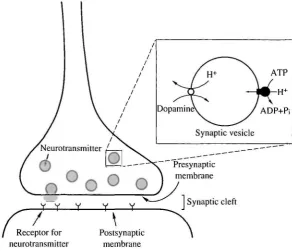
Synaptic vesicles play an important role in neuronal transmission in nerve termini. They accumulate high concentrations of neurotransmitters and, upon receiving the proper signal, secrete them by exocytosis. Similarly, secretory granules in endocrine cells accumulate biogenic amines and secrete them by exocytosis (Ueda, 1986; Johnson, 1988; Njus et al. 1987). We have been interested in the bioenergetics of the accumulation process, which is an initial step in neuronal transmission or biogenic amine secretion. Our results are consistent with a chemiosmotic energy coupling mechanism for the accumulation: a V-ATPase forms an electrochemical gradient of protons across vesicular membranes and transmitters are taken up through specific transporters coupled with this gradient (Fig. 1). Here, we discuss our recent results supporting this model.
Structure of vacuolar-type H+-ATPase
V-ATPases purified from adrenal chromaffin granules (Moriyama and Nelson, 1987a) and brain clathrin-coated vesicles (Xie and Stone, 1986) have essentially the same subunit structures (115, 72, 57, 41, 39, 34, 33, 20 and l6XKfiMr subunits with a
stoichiometry of 1, 3, 3, 1, 1, 1, 1, 1 and 5-6) (Arai et al. 1988). Cold treatment in the presence of MgATP and appropriate salts (e.g. 0.1 moll"1 NaNCb) caused rapid inactivation accompanied by release of a group of hydrophilic subunits (subunit A, B, C,
*To whom correspondence should be addressed.
Y y Y Y Y
ATP
Dopamine\ / ADP+Pi
Synaptic vesicle
Presynaptic membrane
1 Synaptic cleft
[image:2.451.84.377.73.324.2]Receptor for Postsynaptic neurotransmitter membrane
Fig. 1. Energy coupling of uptake of transmitter by synaptic vesicles.
D and E of 72, 57,41, 34 and 33X 103 Mr, respectively) but four polypeptides (115, 39,20 and 1 6 x l 03Mr) remained in the membrane (Moriyama and Nelson, 1989a). Subunit A contains an ATP binding site (Moriyama and Nelson, 1987ft) and is homologous to the subunit of Fi-ATPase (Zimniak et al. 1988). Subunit c (16X103Mr) binds N,N'-dicyclohexylcarbodiimide (DCCD) and is homologous to the c subunit of the Fo portion of F-ATPase (Mandel et al. 1988; Sutton and Apps, 1981). These findings indicate that the enzyme is composed of two different sectors (catalytic and hydrophobic membrane sectors). All V-ATPases from plants and animals show similar properties and essentially the same subunit structures (Moriyama and Nelson, \9%9b,c). This conclusion was confirmed by analyses of the primary structures of the subunits (for reviews, see Nelson,
1989;Forgac, 1989; Futai etal. 1990).
Immunoelectron microscopy, using proteoliposomes reconstituted with purified chromaffin granule V-ATPase, demonstrated the presence of particles of lOnm diameter on the membrane: these particles could be removed by cold treatment and were recognized by antibodies against the hydrophilic sector of chromaffin granule ATPase (Moriyama et al. 1991c). Thus, this particle is the hydrophilic sector of V-ATPase and is similar to F| of F-ATPase.
Synaptic vesicles
1
Synaptic vesicles
1
I
\Q-3xM r
72>
57>
Ann 72
Anti57
34 33
Anti 41
2A
B
Fig. 2. Release of the peripheral portion of synaptic vesicle V-ATPase by alkaline treatment. Synaptic vesicles (1 mgml~') were treated with sodium carbonate and hydrophilic sectors released from the membranes were obtained. See experimental details in Moriyama and Futai (1990a). (A) SDS-polyacrylamide gel electrophoresis. Lane 1, V-ATPase purified from chromaffin granules (5 fig); lane 2, synaptic vesicles (20 jig protein); lane 3, depleted membranes after alkaline treatment (20/xg); lane 4, supernatant (hydrophilic sector) after alkaline treatment (4/xg). Proteins were stained with Coomassie Brilliant Blue. (B) Immunological identification of subunits of V-ATPase. Proteins in the gel (see A) were transferred onto a nitrocellulose sheet and their cross-reactivity with antibodies against subunits A, B and D of chromaffin granule V-ATPase was examined with 125I-labelled protein
A. Autoradiograms are shown. The contents of lanes are the same as in A.
mRNAs encoding subunits of V-ATPase were higher in the brain than in other tissues (Nelson et al. 1990; Wang et al. 1988; Hirsch et al. 1988). Is the V-ATPase of synaptic vesicles structurally similar to those in other organelles, or are different isoforms expressed in different organelles? To answer this question, we cloned a cerebellar cDNA encoding a 16X103 A/
r subunit, a major intrinsic membrane protein of V-ATPase (Hanada
et al. 1991). The deduced amino acid sequence showed that this brain polypeptide (155
lobe, Drosophila and yeast. Glu-139, thought to be essential for proton transport, was also conserved as a potential DCCD binding site.
We then cloned four different 16X lCPAfr genes including pseudogenes from a human genomic bank using the cDNA as a probe (Hasebe et al. 1992). One gene corresponded to the gene encoding the ^XlCPMr subunit expressed in HeLa cells and had a coding sequence separated by two introns. The second intron was located in the DNA segment coding for a loop region between the second and third transmembrane helices, suggesting that the \f>Y.\0PMT subunit with four transmembrane helices had evolved by gene
duplication. We are analyzing whether other genes are actually transcribed. The primary sequence deduced from the second clone had a termination codon behind the third transmembrane helix. Possible translation products from other clones had no putative acidic residues essential for proton transport. We do not yet have evidence for organelle-specific differences in V-ATPases. However, detailed studies on genes from different sources may eventually answer this question.
Uptake of neurotransmitter into synaptic vesicles
Energy coupling
Synaptic vesicles contain transporters for monoamines, acetylcholine, anionic glutamate and y-aminobutyrate (GABA) as well as zwitterionic glycine (Ueda, 1986; Johnson, 1988; Njus et al. 1987; Hell et al. 1988; Kish et al. 1989). A monoamine transporter that is sensitive to reserpine and tetrabenzine is also present in various secretory granules. We concluded that V-ATPase couples the uptakes of these transmitters to ATP hydrolysis, because selective inhibition of the ATPase by bafilomycin Ai and cold treatment or treatment with sodium carbonate diminished the uptakes (Moriyama and Futai, 1990a).
The electrochemical gradient of protons established by V-ATPase consists of a proton gradient and a membrane potential. The uptakes of monoamines and GABA are sensitive to nigericin plus K+ or ammonium (a dissipator of the proton gradient) but are not affected by valinomycin plus K+ (a dissipator of the membrane potential), indicating that these uptakes are coupled to a proton gradient. In contrast, glutamate uptake is coupled to a membrane potential (inside positive), because it is sensitive to valinomycin plus K+ or a high concentration of Cl~ but insensitive to nigericin plus K+ and slightly accelerated by ammonium (Ueda, 1986; Moriyama et al. 1990).
Solubilization and reconstitution of the neurotransmitter transporter with F-ATPase
Escherichia coli cells (Moriyama et al. 19916). The monoamine transporter was solubilized from chromaffin membranes after removal of V-ATPase by cold treatment. It was mixed with purified FiF0 and azolectine liposomes, freeze-thawed and diluted with an appropriate buffer. The resulting proteoliposomes exhibited reserpine-sensitive, ATP-dependent monoamine uptake, which was sensitive to sodium azide (an inhibitor of F-ATPase) but resistant to bafilomycin Ai and N-ethylmaleimide (inhibitors of V-F-ATPase), indicating that the driving force for the uptake was supplied by the F-ATPase. The glutamate transporter could be reconstituted by a similar method (H.-L. Tsi, Y. Moriyama and M. Futai, unpublished observation). It should be noted that limited numbers of proteins derived from chromaffin vesicles were incorporated into liposomes, suggesting that the transporters were partially purified. Thus, the reconstitution systems using F1F0 will be useful for identifying and purifying the transporters.
Effects of neuron blocking reagents and neurotoxin on the energy transducing systems in secretory vesicles
Effects of neuron blocking reagents
Neuronal transmission is perturbed by reagents that abolish the accumulation of transmitters in vesicles. These reagents can be classified as inhibitors of various steps in the formation and utilization of the electrochemical gradient in the vesicles. One class of such compounds interacts directly with the transporters and inhibits their activities. Reserpine, an example of a specific inhibitor of vesicular transport, binds to the monoamine transporter from inside the vesicles and blocks adrenergic neurons (Shore and Norn, 1971). Another class of inhibitors dissipates the electrochemical gradient of protons (e.g. CCCP and ammonium), resulting in indirect inhibition of the uptake (Johnson, 1988). We found that some neuron blocking reagents show both types of inhibition.
Uptake of 4-methylphenylpyridinium (MPP+), a neurotoxin causing Parkinsonism, into synaptic vesicles
False transmitters, another type of reagent interacting with the transporters, are recognized as substrates by the transporters and accumulate in the vesicles. MPP1", a highly bioactive metabolic product of MPTP, is a neurotoxin that causes degradation of dopaminergic neurons in man and experimental animals (artificial Parkinsonism) (Langston etal. 1983; Heikkila etal. 1984). We found that this neurotoxin is the substrate of the vesicular monoamine transporter.
Upon addition of ATP, MPP+ was actively accumulated in chromaffin membrane vesicles and synaptic vesicles, leading to a concentration gradient of about 100-fold (Y. Moriyama, K. Amakatsu and M. Futai, in preparation). The uptake was sensitive to bafilomycin Ai and reserpine, indicating that it was mediated by V-ATPase and the monoamine transporter. These results suggest that MPP"1" accumulates in these organelles in vivo by this mechanism. Furthermore, addition of MPP+ inhibited dopamine uptake but did not affect the uptake of glutamate or GABA. These results are consistent with the hypothesis that MPP+ is toxic to dopaminergic neurons but has little effect on other neurons. The compartmentation of MPP+ may also be important for understanding the mechanism of long-term toxicity of the compound and will provide one molecular basis for development of a Parkinsonian syndrome.
Conclusions
In this article, we have described the structure and function of V-ATPase in synaptic vesicles. The electrochemical gradient of protons established by the ATPase is used as a
c '5
s
T
00
E
CO
a. o Q
[image:6.451.135.318.405.574.2]0 5 10 15 Time (min)
driving force for the concentration of transmitters inside the organelles. Various drugs (e.g. neuron blockers, local anesthetics) or toxins (e.g. MPP+) may affect the uptake into the synaptic vesicles and so perturb neuronal transmission. Thus, V-ATPase has key physiological and pharmacological roles in the nervous and endocrine systems.
This work was supported by grants from the Ministry of Education, Science and Culture of Japan, the Human Frontier Science Program and Naito Fundation.
References
ARAI, H., TERRS, G., PINK, S. AND FORGAC, M. (1988). Topography and subunit stoichiometry of the coated vesicle proton pump. J. biol. Chem. 263, 8792-8802.
CIDON, S. AND SHIRA, T. S. (1989). Characterization of a H+-ATPase in rat brain synaptic vesicles: coupling to L-glutamate transport. J. biol. Chem. 264, 8281-8288.
FORGAC, M. (1989). Structure and function of vacuolar class of ATP-driven proton pumps. Physiol. Rev. 69,765-796.
FUTAI, M., HANADA, H., MORIYAMA, Y. AND MAEDA, M. (1991). Proton translocating ATPase (FoFi): understanding its molecular structure and function. In New Era of Bioenergetics (ed. Y. Mukohota), pp. 7 3 - 108. Tokyo: Academic Press.
FUXE, K., FREDHOLM, B. B., OGREN, S.O., AGNATI, L. F., HOKFELT, T. AND GUSTAFSSON, J. (1978). Ergot
drugs and central monoaminergic mechanism: a histochemical, biochemical and behavioral analysis.
Fedn Proc. Fedn Am. Socs exp. Biol. 37, 2181 - 2 1 9 1 .
HANADA, H., HASEBE, M , MORIYAMA, Y., MAEDA, M. AND FUTAI, M. (1991). Molecular cloning of
cDNA encoding the 16kDa subunit of vacuolar H+-ATPase from mouse cerebellum. Biochem.
biophys. Res. Commun. 176, 1062- 1067.
HASEBE, M., HANADA, H., MORIYAMA, Y., MAEDA, M. AND FUTAI, M. (1992). Vacuolar type H+-ATPase
genes: presence of four genes including pseudogenes for the 16-kDa proteolipid subunit in the human genome. Biochem, biophys. Res. Commun. 183, 856—863.
HEIKKILA, R. E., HESS, A. AND DUVOSIN, R. C. (1984). Dopaminergic neurotoxicity of l-methyl-4-phenyl-l,2,5,6-tetrahydropyridine in mice. Science 224, 1451 — 1453.
HELL, J. W., MAYCOX, P. R., STADLER, H. AND JAHN, R. (1988). Uptake of GABA by rat brain synaptic vesicles isolated by a new procedure. EM BO J. 7, 3023-3029.
HIRSCH, S., STRAUS, A., MASOOD, K., LEE, S., SUKHATME, V. AND GLUCK, S. (1988). Isolation and
sequence of a cDNA clone encoding the 31-kDa subunit of bovine kidney vacuolar H+-ATPase. Proc.
natn. Acad. Sci. U.S.A. 85, 3004-3008.
JOHNSON, R. G. (1988). Accumulation of biological amines into chromaffin granules: a model for hormone and neurotransmitter transport. Physiol. Rev. 681, 232 — 307.
KISH, P. E., FISCHER-BORENKERK, C. AND UEDA, T. (1989). Active transport of GABA and glycine into synaptic vesicles. Proc. natn. Acad. Sci. U.S.A. 86, 3877-3881.
LANGSTON, J. W., BALLARD, P., TETRUD, J. W. AND IRWIN, I. (1983). Chronic Parkinsonism in human due to a product of meperidine-analog synthesis. Science 219, 574—578.
MANDEL, M., MORIYAMA, Y., HULMES, J. D., PAN, Y. C. E., NELSON, H. AND NELSON, N. (1988). cDNA
sequence encoding the 16-kDa proteolipid of chromaffin granules implies gene duplication in the evolution of H+-ATPase. Proc. natn. Acad. Sci. U.S.A. 85, 5521 -5524.
MARSHALL, I. G. AND PARSONS, S. M. (1987). The vesicular acetylcholine transport system. Trends
NeurosciAO, 174-177.
MORIYAMA, Y., AMAKATSU, K., YAMADA, H., PARK, M.-Y. AND FUTAI, M. (1991a). Inhibition of
neurotransmitter and hormone transport into secretory vesicles by 2-(4-phenylpiperadino)cyclohexanol and 2-bromoergocryptine: both compounds act as uncouplers and dissipate the elecrochemical gradient of protons. Archs Biochem. Biophys. 290, 233—238.
MORIYAMA, Y. AND FUTAI, M. (1990a). H+-ATPase, a primary pump for accumulation of neurotransmitters, is a major constituent of brain synaptic vesicles. Biochem. biophys. Res. Commun. 173,443-446.
Escherichia coli H+-ATPase (F0F1) and its reconstitution into liposomes with neurotransmitter transporters. J. biol. Chem, 266, 22141-22146.
MORIYAMA, Y., MAEDA, M. AND FUTAI, M. (1990). Energy coupling of L-glutamate transport and vacuolar H+-ATPase in brain synaptic vesicles. J. Biochem,, Tokyo 108, 689-693.
MORIYAMA, Y. AND NELSON, N. (1987a). The purified ATPase from chromaffin granule membranes is an anion-dependent proton pump. J. biol. Chem. 262, 9175-9180.
MORIYAMA, Y. AND NELSON, N. (1987b). Nucleotide binding sites and chemical modification of the chromaffin granule proton ATPase. J. biol. Chem. 262, 14723-14729.
MORIYAMA, Y. AND NELSON, N. (1989a). Cold inactivation of vacuolar proton-ATPases. J. biol. Chem. 264,3577-3582.
MORIYAMA, Y. AND NELSON, N. (19896). H+-translocating ATPase in Golgi apparatus: characterisation as vacuolar H+ ATPase and its subunit structures. J. biol. Chem. 264, 18445 -18450.
MORIYAMA, Y. AND NELSON, N. (1989C). Lysosomal H+-translocating ATPase has a similar subunit structure to chromaffin granule H+-ATPase complex. Biochim. biophys. Acta 980, 241 —247. MORIYAMA, Y. AND NELSON, N. (1990). Proton translocating ATPase from chromaffin granules and
synaptic vesicles. In Molecular Structure, Function and Assembly of the ATP Synthase (ed. S. Marzuki), pp. 129-139. New York: Plenum Press.
MORIYAMA, Y., YAMAMOTO, A., TASHIRO, Y. AND FUTAI, M. (1991C). Chromaffin granule H+-ATPase has Fi-like structure. FEBS Lett. 291, 92-96.
NELSON, H., MANDIYAN, S., NOUMI, T., MORIYAMA, Y., MIEDEL, M. C. AND NELSON, N. (1990).
Molecular cloning of cDNA encoding the C subunit of H+-ATPase from bovine chromaffin granules.
J. biol. Chem, 265,20390-20393.
NELSON, N. (1989). Structure, molecular genetics, and evolution of vacuolar H+-ATPases. J. Bioenerg.
Biomembr. 21, 553-572.
NJUS, P., KELLY, P. M. AND HARNADEK, G. J. (1987). Bioenergetics of secretory vesicles. Biochim.
biophys. Acta 853, 237-265.
SHORE, P. A. AND NORN, S. (1971). Failure to affect tissue reserpine concentrations by alteration of adrenergic nerve activity. Biochem. Pharmac. 20, 2133—2135.
SUTTON, R. AND APPS, D. K. (1981). Isolation of a DCCD-binding protein from bovine chromaffin-granule membranes. FEBS Lett. 130, 103-106.
UEDA, T. (1986). Glutamate transport in the synaptic vesicles. In Excitatory Amino Acids (ed. P. J. Robert, J. Storm-Mathisen and H. Bradford), pp. 173-195. London: Macmillan Press.
WANG, S.-Y., MORIYAMA, Y., MANDEL, M., HULMES, J. D., PAN, Y. C. E., DANHO, W., NELSON, H. AND
NELSON, N. (1988). Cloning of cDNA encoding a 32-kDa protein: an accessory polypeptide of the H+ -ATPase from chromaffin granules. J. biol. Chem. 263, 17638-17642.
XIE, X. S. AND STONE, D. K. (1986). Isolation and reconstitution of the clathrin-coated vesicles proton translocating ATPase complex. J. biol. Chem. 261, 2492-2495.
ZIMNIAK, L., DITTRICH, L., GOGARTEN, J. P., KIBAK, H. AND T A E , L. (1988). The cDNA sequence of the
69-kDa subunit of the carrot vacuolar H+-ATPase: homology to the /3-chain of FoFi-ATPase. J. bioL
PAGE
GRINSTEIN, S., NANDA, A., LUKACS, G. AND ROTSTEIN, O. V-ATPases in phagocytic cells 179
CHATTERJEE, D., CHAKRABORTY, M., LEIT, M., NEFF, L., JAMSA-KELLOKUMPU, S., FUCHS, R.,