David M. Yousem, Rena J. Geckle, Warren B. Bilker, Donald A. McKeown, and Richard L. Doty
PURPOSE: To evaluate the sites of injury in patients with posttraumatic olfactory deficits and to compare damage with findings on clinical olfactory tests. METHODS: Twenty-five patients with posttraumatic olfactory dysfunction were examined by means of olfactory testing, endoscopy, and MR imaging. MR surface-coil scans through the olfactory bulbs and tracts and head-coil scans of the temporal lobes were evaluated. Quantitative and qualitative gradings of damage to the olfac-tory bulbs, tracts, subfrontal region, hippocampus, and temporal lobes were compared with results on tests of odor identification, detection, memory, and discrimination. RESULTS: Twelve patients were anosmic, eight had severe impairment, and five were mildly impaired. Injuries to the olfactory bulbs and tracts (88% of patients), subfrontal region (60%), and temporal lobes (32%) were found, but these did not correlate well with individual olfactory test scores. Volumetric analysis showed that patients without smell function had greater volume loss in olfactory bulbs and tracts than did those posttraumatic patients who retained some sense of smell. Qualitative and quantitative assessments of damage showed few significant correlations with olfactory tests, probably because of multifocal injuries, primary olfactory nerve damage, and the constraints of a small sample size on the detection of clinically significant differences. CONCLUSION: MR imaging shows abnormal-ities in patients with posttraumatic olfactory dysfunction at a very high rate (88%), predominantly in the olfactory bulbs and tracts and the inferior frontal lobes.
Index terms: Brain, magnetic resonance; Head, injuries; Nerves, olfactory (I)
AJNR Am J Neuroradiol17:1171–1179, June 1996
Posttraumatic anosmia occurs in approxi-mately 31% of patients who have sustained ma-jor closed head injuries, as defined by posttrau-matic amnesia for more than 24 hours (1). However, even in persons whose head injuries are not accompanied by loss of consciousness or in whom amnesia lasts less than 1 hour, the frequency of anosmia ranges from 3% to 8% (1). The exact cause of olfactory dysfunction is un-clear, although many mechanisms have been
proposed. Shearing injuries at the cribriform plate that lacerate the primary olfactory nerves extending from the nasal cavity to the olfactory bulb are believed to be one of the common mechanisms involved in posttraumatic smell loss (1–5)(N. D. Zasler, R. M. Costanzo, P. G. Heywood, “Neuroimaging Correlates of Olfac-tory Dysfunction after Traumatic Brain Injury,”
Arch Phys Med Rehabil 1990;71:814,
ab-stract). Alternatively, direct injury to the olfac-tory bulbs or tracts, intracerebral hematomas compressing these structures, injury to the sep-tal nuclei in the inferior fronsep-tal region, orbito-frontal cortex injuries, or medial temporal lobe lacerations have been postulated as events that can precipitate posttraumatic olfactory deficits (1–5)(Zasler et al, “Neuroimaging Correlates”). Several postmortem neuropathologic studies have been performed in this regard (1, 3, 5).
As part of a broader study of smell and taste disorders, we examined 25 patients with olfac-tory dysfunction resulting from head trauma by using both surface-coil and head-coil magnetic resonance (MR) imaging to analyze injuries to
Received September 8, 1995; accepted after revision December 21. Supported by research grant 5 PO1 DC 00161–15 from the National Institute on Deafness and Other Communication Disorders, National Insti-tutes of Health.
From the Department of Radiology (D.M.Y., R.J.G.), the Smell and Taste Center (D.M.Y., R.J.G., D.A.M., R.L.D.), and the Department of Biostatistics and Epidemiology and Center for Clinical Epidemiology and Biostatistics (W.B.B.), University of Pennsylvania Medical Center, Philadel-phia.
Address reprint requests to David M. Yousem, MD, Department of Radiology, University of Pennsylvania Medical Center, 3400 Spruce St, Philadelphia, PA 19104.
AJNR 17:1171–1179, Jun 1996 0195-6108/96/1706 –1171
qAmerican Society of Neuroradiology
odor detection, identification, discrimination, and memory.
Subjects and Methods
Twenty-five patients with olfactory dysfunction after head injuries were referred for smell testing to the Smell and Taste Center of the University of Pennsylvania. An extensive review of these patients’ clinical histories was carried out to ascertain the origin of their olfactory dys-function. All patients noticed the olfactory decline within 7 days of the head injury. Because some patients had altered consciousness at the time of their injury, the report of smell dysfunction was not immediate in some cases. These find-ings are in keeping with a report by Schecter and Henkin (6) in which 83% of patients experiencing posttraumatic smell loss did so immediately after the trauma, while the remainder noticed the deficits within 3 weeks to 4 months after the injury. No patient in our series reported olfactory deficits that predated the head trauma.
The study group included 14 men and 11 women who were 16 to 51 years old (mean, 36 years; SD, 9.8). The traumatic event occurred 3 to 183 months before the MR examination, with a mean of 23 months (SD, 37 months) and a median of 12 months. No patient was examined in the acute phase of the injury. All patients signed approved consent forms for participation in this study.
The patients underwent a battery of psychophysical olfactory tests designed to ascertain the specific nature of their olfactory deficits. All tests were administered to each side of the nose separately and included the University of Pennsylvania Smell Identification Test (UPSIT) (20 items to each side); a 16-item odor discrimination test; a 12-item odor memory test (with retention intervals of 0, 30, and 60 seconds); and a single-staircase odor detection threshold test using the odorant phenylethyl alcohol (7–10). The results of these smell tests were used to determine the severity of the chemosensory deficit. The UPSIT is the most widely used test of odor identification in the world and is the standard for determining whether a patient can identify smells. The other tests determine whether a pa-tient can distinguish one odor from another (odor discrim-ination), remember an odor and recognize it later (odor
the MR study.
MR images of the olfactory bulbs and tracts were ob-tained with a 12.7-cm, round, general-purpose surface coil centered on the nasion. After a sagittal localizing scan, coronal images with parameters of 500/15/2 (repetition time/echo time/excitations) were obtained with 3-mm in-terleaved scans and a 2563256 matrix with a 12-cm field of view (Fig 1). These coronal scans were followed by 3-mm interleaved coronal fast spin-echo T2-weighted im-ages through the same anatomy with scan parameters of 2000/84/2 and a matrix of 256 3 192. With the use of high-resolution surface-coil imaging, one can be quite confident of the location of the olfactory bulbs and tracts and can trace the tracts to the entrance to the brain near the septal nuclei.
inferior frontal/septal regions, the right and left temporal lobes, and the right and left hippocampal/parahippocam-pal/amygdaloid regions. Spearman correlations were used to evaluate the relationship among these grades, the quan-titative assessments of volume, and the olfactory tests for the patients who could smell. We used Wilcoxon’s scores to determine whether there was a difference in the volumes of the temporal lobes and the olfactory bulbs and tracts between anosmic and nonanosmic patients.
A control population (six women and two men, with a slightly older age range of 43 to 70 years) with no history of head trauma, no reported olfactory deficits, and olfac-tory test results in the normal range was also examined. A Mann-Whitney test, which is a nonparametric alternative to the two-sample t test for independent samples, was used to compare UPSIT scores and mean volumes of the olfactory bulb/tract system and the temporal lobes among the control subjects, the posttraumatic anosmic patients, and the posttraumatic nonanosmic patients.
Results
Cognitive and Endoscopic Testing
Twenty-four of 25 patients had perfect scores on the 40-item PIT, with one patient getting one picture incorrect. Only one patient scored below 28 on the MMSE. These data suggest that the patients had sufficient cognitive skills to follow the directions adequately on the specific olfac-tory tests. No patient had findings at endoscopy that were sufficient to account for severe smell deficits. Therefore, for the purposes of this study, all 25 patients were included in the anal-ysis of the relationship of olfactory system dam-age to olfactory test scores.
Olfactory Testing
Twenty-four patients had olfactory dysfunc-tion as measured by the UPSIT. Twelve were anosmic (UPSIT score , 18), 8 were severely impaired (UPSIT score$18,#25), and 4 were mildly impaired (UPSIT score $27,#34). De-spite subjective claims of a distortion in the sense of smell, one patient had normal smell function on the basis of standardized UPSIT norms (her UPSIT score was 35, which is the lower limit of normal) (7, 8). This patient did have deficits in odor memory and in phenethyl alcohol odor-detection threshold, so she was included in the data analysis. Another patient with mild impairment had an UPSIT score that was 5 items better on the right than the left; otherwise the deficits were seen to be bilateral and symmetric.
None of the olfactory test scores showed any relationship to the time interval between the traumatic event and the MR study. The interval from event to MR imaging for the patients with mild to unimpaired olfactory ability ranged from 3 to 62 months (mean, 27 months); for the anosmic group, the interval ranged from 5 to 183 months (mean, 29 months).
The mean normal value for persons without olfactory impairment on the bilateral 24-item odor memory test is 16 (SD, 2.8). Only 6 of our 25 patients scored above 10 on the 24-item odor memory test, and none of these patients was anosmic by UPSIT criteria. Three patients were mildly impaired and 3 were severely
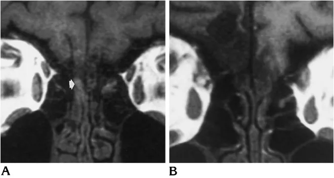
im-Fig 1. Grade 0 (no volume loss) in three patients through various parts of the anatomy. A, Coronal T1-weighted MR image shows normal-sized olfactory bulbs (arrows) bilaterally.
[image:3.612.58.557.87.267.2]paired on the basis of UPSIT scores. The patient with “normal” smell function correctly remem-bered 9 of 24 odors presented (a score of 6 on the left and 3 on the right).
The mean score for persons with no olfactory impairment on the bilateral 32-item odor dis-crimination test is 21.8 (SD, 2.8). Three of our patients scored 20 or higher on the combined 32-item odor discrimination test. These 3 pa-tients had the 3 highest UPSIT scores (35, 33, and 28, respectively). Two of these 3 also scored above 10 on the odor memory test.
Odor detection thresholds for phenethyl alco-hol are measured in logarithmic dilutions, with 22.0 the strongest odor concentration and 210.0 the weakest concentration. On average, persons with normal smell function can detect a 26.3 dilution of phenethyl alcohol (SD, 1.6). Seventeen of the 25 patients studied scored a 22.0 on at least one side of the nose. The22.0 value is the maximum concentration and the value expected of anosmic persons.
Imaging Findings
Because the earliest MR study was performed 3 months after the traumatic event, the imaging findings in our population represent the late changes one finds in patients with posttrau-matic olfactory deficits. Twenty-two of the 25 patients had damage (ie, volume loss) to the olfactory system (olfactory bulbs, olfactory tracts, inferior frontal region, hippocampi, or temporal lobes). All 22 had injury to the olfac-tory bulbs or tracts, and in all but 2 the injury was bilateral (Fig 2). The damage to the olfac-tory bulbs or tracts was moderate to severe in
15 of the 22 patients, and mild in the other 7. Frontal lobe injury occurred in 15 patients (moderate to severe in 12) and 13 had bilateral inferior frontal damage. Temporal lobe injury occurred unilaterally in 5 patients and bilater-ally in 3 patients; 1 patient had isolated right hippocampal trauma. Four patients had moder-ate to severe temporal lobe injury and 4 had mild injury (Fig 3).
Of the anosmic patients, only 1 did not have olfactory bulb or tract damage. The single pa-tient with no smell impairment by UPSIT had mild bulb damage on the right side but no other areas of volume loss by inspection. The olfac-tory bulbs and tracts had minimal to no damage in 4 of the 5 patients who could smell but in just 3 of the 12 anosmic patients. Of the 4 patients who showed a differential between right and left UPSIT, odor memory, or odor discrimination scores who could definitely smell (UPSIT score . 27), only 1 had asymmetric damage (more disease was present ipsilateral to the nostril with the larger deficit). Only 1 patient in whom tem-poral lobe injury was evident on MR images could smell: his odor memory and odor discrim-ination deficits were the worst among the mildly impaired or unimpaired patients.
[image:4.612.225.555.87.263.2]77.1 mm3. The mean volumes of the right and left temporal lobes of the 25 patients were 70 094 mm3 and 69 635 mm3, respectively (SD, 8216 mm3 and 10 516 mm3, respective-ly). Ranges from 45 028 mm3 to 89 035 mm3 were present for the temporal lobes. The aver-age of the right and left temporal lobes was 69 864.5 mm3.
Because the olfactory tracts are typically hy-perintense on long-repetition-time pulse se-quences obtained with a surface coil, the value of assessing signal-intensity abnormality on T2-weighted images was reduced; in fact, no areas of frankly abnormal signal intensity were iden-tified in a bulb or tract that had normal volume.
Statistical Analysis: Reliability Data
Intraclass correlation coefficients were per-formed to assess the reproducibility of the quantitative measurements of the right and left olfactory bulbs and tracts and the temporal lobes for each examiner (each examiner having performed the quantitative volumetric analysis twice to determine intraobserver reliability). The intraclass correlation coefficients ranged from .90 to .96, signifying an outstanding degree of reproducibility for the two examiners (Table). The intraclass correlation coefficients are graded on the same scale as thekstatistic, with values above .80 regarded as “almost perfect” (13). When the valuesbetweenexaminers were analyzed, the intraclass correlation coefficients
were in the .90 to .95 range, again signifying “almost perfect” reliability between examiners. We then assessed the percentage of differ-ence between examiners’ values by dividing the absolute difference between examiners’ values by the most experienced examiner’s values. This yielded a mean percentage of absolute dif-ference between interpretations of 7.0% for the left temporal lobes, 7.5% for the right temporal lobes, 12.2% for the left olfactory bulbs and tracts, and 18.6% for the right olfactory bulbs and tracts. The degree of difference between the two interpretations of the same structure by the same person averaged 3.6% for the temporal lobes and 16.7% for the olfactory bulbs.
Anosmic (UPSIT ,18) versus Nonanosmic
(UPSIT $ 18) Patients
There was a statistically significant difference between the volume of the left olfactory bulbs and tracts in the patients who were anosmic
Intraclass correlation coefficients within and between observers
Anatomic Region
Correlation Coefficient Observer 1:
Two Interpretations
Observer 2: Two Interpretations
Between Observers
L temporal lobe .96 .95 .93
R temporal lobe .90 .91 .93
L olfactory bulb/tract .92 .92 .95
[image:5.612.57.555.87.251.2]R olfactory bulb/tract .94 .94 .90
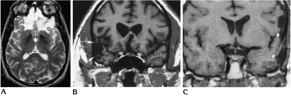
Fig 3. Cortical damage in three patients.
A, Axial T2-weighted MR image shows bifrontal encephalomalacic change, which was symmetric in severity. The patient was anosmic and also had olfactory bulb and tract injury.
B, Bilateral temporal lobe injury was present in a different patient, more severe on the right side (arrows). No asymmetry in smell test scores was noted.
68 273 mm3, respectively). No differences in qualitative grades of damage were discovered between patients with and without residual ol-faction.
Anosmic and Hyposmic Posttraumatic Patients versus Control Subjects
The UPSIT scores of the control subjects ranged from 35 to 40, with a mean of 37. These values were significantly different from those of patients with head trauma (P,.0001), whether they did (P 5 .0006) or did not (P 5 .0002) have residual olfactory function. The mean vol-ume of the olfactory bulbs and tracts for the 8 control subjects was 126.6 mm3 (SD, 38.4), and the mean volume of the temporal lobe was 70 319 mm3(SD, 11 139). The volumes of the olfactory bulbs and tracts differed statistically from those of all posttraumatic patients (P 5
.029) and from the posttraumatic anosmic pa-tients (P 5 .008), but not from those of post-traumatic hyposmic patients (P5.307). There was no statistically significant difference in tem-poral lobe volumes between control subjects and patients with posttraumatic olfactory defi-cits (Pvalues ranged from .839 to .901).
Qualitative versus Qualitative Ratings
We found statistically significant correlations between the qualitative degree of damage to the olfactory bulbs and that to the ipsilateral tracts (P ,.001) and frontal lobe (P, .001), as well as that to the contralateral bulb (P , .001), tracts (P ,.001), and frontal lobe (P ,.001). These data suggest that the injuries sustained are often multifocal and will usually affect both bulbs, tracts, and frontal lobes. Only two pa-tients had damage that was isolated to one site. Damage scores for frontal lobe injuries and
the qualitative assessment of volume loss, the quantitative assessment of the volume of the olfactory bulbs and tracts and temporal lobes, and the assessment of individual olfactory tests. We used a significance level of .05 to search for differences between groups. Only the 13 pa-tients who could smell as determined by UPSIT scores were included to help establish the rela-tionship between smell test scores and location of injury. A multiple comparisons adjustment devised by Hotchberg (14), which is a modified Bonferroni procedure that achieves the required type 1 error while achieving a lower type 2 error, was applied to the significance tests. With this modified Bonferroni correction applied, no cor-relations between smell tests and qualitative or quantitative gradings of olfactory-eloquent structures were noted. Because the smell tests differ in reliability, however, and because such tests are generally correlated with one another (15), we developed a composite olfactory test score based on a weighted average of the four tests administered so as to simplify data presen-tation and decrease the probability of a type 1 error. The test-retest reliability coefficients of these tests, used as weights within this compos-ite, were proportioned to equal 1.0 (total UPSIT score, 0.316; phenethyl alcohol score, 0.302; odor memory, 0.234; and odor discrimination, 0.148) (15, 16). Again, there was no significant correlation between composite scores and the volume of the olfactory bulbs and tracts and temporal lobes.
Discussion
tract, fibers pass in the olfactory stria to septal nuclei at the base of the brain just inferior and anterior to the rostrum of the corpus callosum. From the medial and lateral septal nuclei, fibers extend to the limbic system with branches to the uncus, hippocampus, parahippocampal region, septum pellucidum, fornices, amygdala, and gyrus rectus regions.
Thus far, it is unclear which olfactory func-tions correspond to the various anatomic sites. The location of the source of olfactory dysfunc-tion has been surmised from animal and human models. The nasociliary olfactory nerves, olfac-tory bulbs, and olfacolfac-tory tracts are necessary for odor detection. Sectioning these nerves re-sults in anosmia. With orbitofrontal or medial thalamic lesions, odor discrimination and odor quality recognition are affected, though in some instances odor detection may be unaffected or even more sensitive than that in control subjects (4). The ability to recognize, interpret, and re-member odors is located more classically in the uncus and hippocampus, whereas the emo-tional response to smell is tied into the entire limbic system (B. E. Wexler, R. K. Fulbright, C. Greer, et al, “An fMRI Study of Human Brain Response to Attractant and Aversive Odors,” Sarasota, Fla: Association for Chemoreception Sciences, April 22, 1995, abstract 219). We had hoped that this study of posttraumatic injuries of olfactory-eloquent regions would lead to a clearer understanding of anatomic-functional relationships as measured by smell tests.
The prevalence of posttraumatic anosmia ranges from 24% to 30% among patients who have sustained severe head injuries, 15% to 19% among those with moderate head injuries, and 0% to 16% among patients with mild head injuries (17). This disorder is commonly asso-ciated with blows to the frontal region or the occiput. Sumner (1) found that a blow to the occiput has five times the chance of inducing anosmia than does a blow to the forehead if posttraumatic amnesia is present (indicating a severe head injury). This may be due to contre-coup shearing effects at the cribriform plate and inferior frontal lobe region. Because frontal in-juries are more common than occipital blows, posttraumatic anosmia is most often seen in the setting of a frontal contact injury (1, 18, 19). Fractures of the skull or face are seen in 45% to 68% of patients with bilateral posttraumatic anosmia (5, 19).
Most patients reporting olfactory dysfunction
after head trauma are totally anosmic, but ap-proximately one fourth may have hyposmia or parosmia (distortion of smells) (5, 18, 20). Re-tention of the sense of smell in one nostril is uncommon, occurring in fewer than 11% of posttraumatic patients examined for chemo-sensory abnormalities (3, 17–19). In patients who have partial or incomplete loss of olfactory function, the deficit may go completely unno-ticed.
Recovery of olfactory function after head trauma is variable. Most large series report a return of olfactory function in 14% to 39% of patients who were initially anosmic (1, 5, 21), especially if the interval of posttraumatic amne-sia is less than 24 hours. While 74% of patients recovering olfactory function do so within 12 weeks, one study reported that an additional 22% will regain function by the second year after the injury (1). However, reports of return of olfactory function as long as 7 years after injury have been published, although few studies have used quantitative tests of olfactory function (1, 5, 18). Olfactory neurons have the capacity for neurogenesis, allowing new receptor growth, so it is surmised that the late return of function may be related to a peripheral (olfactory nerves/ bulbs/tracts) mechanism rather than a more central one (22). In hamsters, recovery of odor detection after unilateral olfactory nerve transection occurs in over half the cases (22). In humans, though, it is believed that there may be fibrotic scarring that occurs at the cribriform plate that may prevent regenerating axons from connecting to the secondary neurons of the ol-factory bulb (23). In our study group, the rate of recovery of function was less than 10%, possibly because the mean time from evaluation to trau-matic event was 23 months. However, we have not performed serial testing in this group.
beam-harden-noid bone.
We have shown in this MR study that the most common sites of injury in patients with posttrau-matic olfactory dysfunction are the olfactory bulbs and tracts followed by the inferior frontal lobes. The prevalence of temporal lobe and hip-pocampal injury is low. The finding that the volume of the olfactory bulbs and tracts in pa-tients with anosmia is less than that of papa-tients with residual smell function or control subjects suggests that the source of the olfactory deficit may be at the level of the olfactory bulbs and tracts or proximally in the olfactory neurons. The lack of a statistical difference between anosmic patients and persons with normal smell function (both control subjects and post-traumatic patients) in the volume of their tem-poral lobes and in the qualitative grade of injury at multiple intracerebral sites (frontal, temporal, or hippocampal regions) supports this hypoth-esis.
Why were no significant relationships found between individual olfactory tests and qualita-tive and quantitaqualita-tive measures of damage (as seen on MR images) to olfactory-eloquent re-gions of the brain? We believe that several fac-tors account for this phenomenon. 1) Olfactory deficits may result from extraparenchymal in-jury at the ciliary nerve or olfactory epithelium level, which MR imaging cannot detect. 2) Injury occurs at multiple sites; only two of our patients had an injury that was isolated to one olfactory site (ie, usually the frontal lobes and olfactory bulbs were damaged together), making it diffi-cult to correlate findings on smell tests with single anatomic sites. 3) Few (n 5 13) of our patients could still smell, and those with severe deficits (n 5 8) had unreliable scores on odor memory, discrimination, and threshold tests. 4) Recent work by Doty et al (15, 16) suggests that many olfactory tests are unreliable and may
patients who had posttraumatic olfactory dys-function with that of posttraumatic patients with normal olfactory function. However, the volume difference between anosmic and hyposmic pa-tients, between anosmic persons and control subjects with normal smell function, and be-tween the combined anosmic-hyposmic group and subjects with normal smell function sug-gests that the volume loss seen in the olfactory bulbs and tracts may reflect a propensity for smell loss. One cannot presume cause and ef-fect from our study. Most of the patients we saw were referred to the Smell and Taste Center; one would have to assess all head trauma patients with surface-coil examinations of the olfactory bulbs and tracts to produce a control population with documented damage to this system but without smell dysfunction. The prevalence of damage to olfactory bulbs and tracts and the frontal lobe would be expected to be greatest in patients with acceleration-deceleration injuries, in which the plane of impact was in an antero-posterior direction.
be-tween intracranial sites of injury and specific results on smell tests.
References
1. Sumner D. Post-traumatic anosmia.Brain1964; 87:107–120 2. Costanzo RM, Heywood PG, Ward JD, Young HF. Neurosurgical
applications of clinical olfactory assessment.N Y Acad Sci1987; 510:242–244
3. Levin HS, High WM, Eisenberg HM. Impairment of olfactory rec-ognition after closed head injury.Brain1985;108:579 –591 4. Potter H, Butters N. An assessment of olfactory deficits in patients
with damage to prefrontal cortex. Neuropsychologia 1980;18: 621– 628
5. Zusho H. Posttraumatic anosmia. Arch Otolaryngol 1982;108: 90 –92
6. Schecter PJ, Henkin RI. Abnormalities of taste and smell after head trauma.J Neurol Neurosurg Psychiatry1974;37:802– 810 7. Doty RL, Shaman P, Dann M. Development of the University of
Pennsylvania Smell Identification Test: A standardized microen-capsulated test of olfactory function. Physiol Behav 1984;32: 489 –502 (Monograph)
8. Doty RL, Ugrawal U, Frye RE. Evaluation of the internal consis-tency reliability of the fractionated and whole University of Penn-sylvania Smell Identification Test (UPSIT).Percept Psychophys 1989;45:381–384
9. Smith RS, Doty RL, Burlingame GK, McKeown DA. Smell and taste function in the visually impaired.Percept Psychophys1993; 54:649 – 655
10. Bromley SM, Doty RL. Odor recognition memory is better under bilateral than unilateral test conditions.Cortex1995;31:25– 40 11. Vollmecke TA, Doty RL. Development of the Picture Identification
Test (PIT): A research companion to the University of Pennsylva-nia Smell Identification Test.Chem Senses1985;10:413– 414 12. Folstein MF, Folstein SE, McHugh PR. Mini-mental state.J
Psy-chiatr Res1975;12:189 –198
13. Landis JR, Koch GG. The measurement of observer agreement for categorical data.Biometrics1977;33:159 –174
14. Hotchberg Y. A sharper Bonferroni procedure for multiple tests of significance.Biometrika1988;75:800 – 803
15. Doty RL, McKeown D, Lee WW, Shaman P. Test-retest reliability of 10 olfactory tests.Chem Senses1995;20:645– 656
16. Doty RL, Newhouse MG, Azalina JD. Internal consistency and short-term test-retest reliability of the University of Pennsylvania Smell Identification Test.Chem Senses1985;10:297–300 17. Costanzo RM, Zasler ND. Head trauma. In: Getchell TV, Doty RL,
Bartoshuk LM, Snow JB Jr, eds.Smell and Taste in Health and Disease.New York, NY: Raven Press; 1991:711–730
18. Leigh AD. Defects of smell after head injury. Lancet1943;1: 38 – 40
19. Symonds CP. Discussion on differential diagnosis and treatment of post-contusional states.Proc Royal Soc Med1942;35:601– 614 20. Mott AE, Leopold DA. Disorders in taste and smell. Med Clin
North Am1991;75:1321–1353
21. Varney NR. Prognostic significance of anosmia in patients with closed-head trauma.J Clin Exp Neuropsychol1988;10:250 –254 22. Constanzo RM. Neural regeneration and functional reconnection following olfactory nerve transection in hamster.Brain Res1985; 361:258 –266
23. Jafek BW, Eller PM, Esses BA, Moran DT. Post-traumatic anos-mia: ultrastructural correlates.Arch Neurol1989;46:300 –304 24. Truwitt CL, Barkovich AJ, Grumbach MM, Martini JJ. MR imaging
of Kallmann syndrome: a genetic disorder of neuronal migration affecting the olfactory and genital systems.AJNR Am J Neurora-diol1993;14:827– 838
25. Bick DP, Ballabio A. Bringing Kallmann syndrome into focus. AJNR Am J Neuroradiol1993;14:852– 854
26. Iida Y, Naito M, Asahina N, et al. Magnetic resonance imaging of the olfactory apparatus.Arch Otolaryngol Head Neck Surg1994; 120:869 – 872
27. Knorr JR, Ragland RL, Brown RS, Gelber N. Kallmann syndrome: MR findings.AJNR Am J Neuroradiol1993;14:845– 851 28. Li C, Yousem DM, Doty RL, Kennedy DW. Imaging evaluation of
olfactory deficits.AJR Am J Roentgenol1994;162:411– 418 29. Vogl TJ, Stemmler J, Heye B, et al. Kallman syndrome versus
idiopathic hypogonadotrophic hypogonadism at MR imaging. Ra-diology1994;191:53–57
30. Yousem DM, Turner WJD, Li C, Snyder PJ, Doty RL. Kallmann’s syndrome: MR evaluation of olfactory system.AJNR Am J Neu-roradiol1993;14:839 – 843