In many amphibian genera, including Bufo, Rana and Xenopus, tadpoles hatch at an early ‘tailbud’ stage of development before the mouth has opened and when the nervous system is in a very simple state of differentiation. Despite this simplicity, tadpoles can usually swim and would be expected to have sensory systems that allow them to detect stimulation and responses that help them to survive. For example, well before hatching, the skin is excitable so that stimuli can be detected anywhere on the body and initiate movements even before the sensory innervation of the skin is complete (Roberts and Smyth, 1974). Observations on hatchling Xenopus laevis tadpoles lying
unattached on the bottom of a small dish in 1 cm of water (Boothby and Roberts, 1995) have shown that when touched on one side of the tail they flex slightly to the opposite side and, in 89 % of responses, swim forwards away from the stimulated side. When touched on the head skin, the initial flexion is stronger, only 31 % swim forwards, and the direction of swimming is unpredictable. In all these swimming responses, the tadpoles tended to swim belly-down, dorsal-up. We suggested that swimming away or showing an unpredictable response could help to avoid predation. However, this behaviour was limited by the shallow water.
Printed in Great Britain © The Company of Biologists Limited 2000 JEB2629
Many amphibian tadpoles hatch and swim before their inner ears and sense of spatial orientation differentiate. We describe upward and downward swimming responses in hatchling Xenopus laevis tadpoles from stages 32 to 37/38 in which the body rotates about its longitudinal axis.
Tadpoles are heavier than water and, if touched while lying on the substratum, they reliably swim upwards, often in a tight spiral. This response has been observed using stroboscopic photography and high-speed video recordings. The sense of the spiral is not fixed for individual tadpoles. In ‘more horizontal swimming’ (i.e. in directions within ±30 ° of the horizontal), the tadpoles usually swim belly-down, but this position is not a prerequisite for subsequent upward spiral swimming.
Newly hatched tadpoles spend 99 % of their time hanging tail-down from mucus secreted by a cement gland on the head. When suspended in mid-water by a mucus strand, tadpoles from stage 31 to 37/38 tend to swim spirally down when touched on the head and up when touched on the tail. The three-dimensional swimming paths of stage 33/34 tadpoles were plotted using simultaneous video images recorded from the side and from above. Tadpoles spiralled for 70 % of the swimming time, and the probability of spiralling increased to 1 as swim path angles became more vertical.
Tadpoles were neutrally buoyant in Percoll/water mixtures at 1.05 g cm−3, in which anaesthetised tadpoles floated belly-down and head-up at 30 °. In water, their centre of mass was ventral to the muscles in the yolk mass. A simple mathematical model suggests that the orientation of tadpoles during swimming is governed by the action of two torques, one of which raises the head (i.e. increases the pitch) and the other rotates (rolls) the body. Consequently, tadpoles (i) swim belly-down when the body is approximately horizontal because the body is ballasted by dense yolk, and (ii) swim spirally at more vertical orientations when the ballasting no longer stabilises orientation.
Measurements in tethered tadpoles show that dorsal body flexion, which could produce a dorsal pitch torque, is present during swimming and increases with tailbeat frequency.
We discuss how much of the tadpole’s behaviour can be explained by our mathematical model and suggest that, at this stage of development, oriented swimming responses may depend on simple touch reflexes, the organisation of the muscles and physical features of the body, rather than on vestibular reflexes.
Key words: Xenopus laevis, tadpole, swimming, orientation, torque, pitch, mathematical model.
Summary
Introduction
SIMPLE MECHANISMS ORGANISE ORIENTATION OF ESCAPE SWIMMING IN
EMBRYOS AND HATCHLING TADPOLES OF XENOPUS LAEVIS
ALAN ROBERTS1,*, N. A. HILL2,‡ANDROBIN HICKS1
1School of Biological Sciences, University of Bristol, Bristol BS8 1UG, UK and 2Department of Applied
Mathematics, University of Leeds, Leeds LS2 9JT, UK
*e-mail: A.Roberts@bristol.ac.uk ‡e-mail: N.A.Hill@leeds.ac.uk
Like other tadpoles, free-living Xenopus laevis tadpoles occur naturally in ponds. For a day or so after hatching, they spend almost all their time immobile and attached to solid objects such as plants or to the surface meniscus (Jamieson and Roberts, 2000). During this period, their bodies hang in a head-up, tail-down orientation from a strand of mucus secreted by a cement gland on the head. If they become detached, they sink, and so can reach the substratum. Their swimming behaviour can therefore influence their position in the water and their site of attachment (Bles, 1905), as well as their likelihood of capture by predators such as aquatic insect larvae. In deeper water, we found that tadpoles lying on the substratum very often swam nearly vertically upwards along a corkscrew path when touched. However, if touched on the head while attached to the surface, they often swam spirally downwards. In this paper, we describe these unusual spiral swimming responses that occur around the time of hatching and suggest that they depend initially not on vestibular reflexes controlling the orientation of swimming but on simple touch reflexes and features of the physical organisation of the embryo and young tadpole. As development continues, the control of swimming becomes altogether more sophisticated, and the body shape changes to the fat head and thin tail form whose swimming has been studied in great detail (Liu et al., 1997).
Materials and methods Observations and lesions
Tadpoles were staged according to Nieuwkoop and Faber (1956) and produced by induced mating using a colony of adult
Xenopus laevis (Daudin). Most observations were made in
glass containers with a specific depth of dechlorinated Bristol tapwater. When necessary, tadpoles were anaesthetised in 0.1 % MS222 in saline (composition in mmol l−1: NaCl, 115; KCl, 3; CaCl2, 2; MgCl2, 1; NaHCO3, 2.4; Hepes, 10, at
pH 7.4). Operations were performed in this saline. To record the movements of ‘tethered’ tadpoles, an enlarged shadow of the tadpole’s body was cast horizontally onto a flat photocell 4 mm×10 mm using a 5 mm diameter, clear 6 V light bulb with a simple filament. This was powered by a battery and placed close to the tadpole so that the shadow of part of the dorsal edge of the tail lay across the photocell (see Fig. 14A). The voltage generated by the photocell was amplified and displayed on a Graphtec 7600 thermal arraycorder.
Video recordings of swimming
Tadpoles were manipulated using a Pasteur pipette or a mounted needle tangled in the mucus secreted by the cement gland on the head. They were stimulated by touching them with a fine needle or a mounted gerbil hair. Swimming paths were recorded through the side of a 100 mm×100 mm×100 mm glass container using a Sony Handycam Video 8 CCD-TR303E or a VHS Panasonic NVM7B. A front-silvered mirror angled at 45 ° was used to obtain simultaneous top and side views. When recording swimming paths, the camera was at least 1 m away so that parallax error in the recording tank was only 2 % and
could be ignored. High-speed video recordings were made using a NAC 400 camera with stroboscopic illumination to record tadpoles at 200 frames s−1. Recordings were played back, and tadpole positions or outlines every 0.1 s were then traced manually from the video screen to allow measurement of successive positions or to assemble montages of the initial phases of the response to touch. Tadpole positions during swimming were plotted in three dimensions using Move3D software written by Professor J. M. V. Rayner (Applied Biology, University of Leeds, UK).
Stroboscopic flash photography
A Nikon 35 mm camera with a Macro lens was used to take photographs onto Technical Pan film through the front of a 100 mm×100 mm×100 mm glass tank. A tadpole was placed on the bottom of the tank in front of a vertical glass partition 20 mm from the front of the tank. This region was illuminated from the left side by a stroboscope flashing at 10 Hz, the camera shutter was opened, the tadpole was stimulated by touch with a fine needle, and the shutter was closed when the response was finished.
Observations in Percoll/water mixtures
Percoll consists of colloidal silica particles 15–30 nm in diameter coated with polyvinylpyrrolidone (registered trademark of Pharmacia Inc; from Sigma; density 1.130±0.005 g cm−3, viscosity 10×10−2±5×10⁻²Pa s at 20 °C, osmolality <20 mosmol kg−1H2O). We have used it because it
is sufficiently dense but has low osmolality and viscosity. Tadpoles were anaesthetised and transferred in a close-fitting pipette with as little water as possible. In initial observations, they were transferred to a glass chamber made from microscope slides containing approximately 20 ml of a mixture of Percoll and water in known proportions. For later observations, linear density gradients 65–80 mm high were prepared in a glass tube 25 mm in diameter by controlled mixing of two Percoll/water solutions, one with a higher and one with a lower density (Leif, 1968). These gradients ranged in density from 1.0973 g cm−3at the bottom to 1.0315 g cm−3 at the top.
Results
Responses of tadpoles lying on the substratum
recordings of tadpoles at stages 33/34 and 35/36 (Fig. 1A,B) show that some paths are nearly vertical, while others are more tortuous with irregular periods of more horizontal swimming (i.e. in directions within ±30 ° of the horizontal). In deeper water (200 mm), periods of horizontal swimming became more common.
Since observations from one side can only give information about the angle of swimming normal to the direction of observation, upward swimming was compared at different developmental stages by taking video recordings from above a tank with a water depth of 30 mm. Animals were placed on the bottom and stimulated to swim by touching the tail. When the swimming paths were traced, periods of spiralling and
normal swimming could be distinguished (Fig. 1C–F). The maximum horizontal width of the swimming path was measured (see small arrows in Fig. 1E,F) to define the diameter (in mm) of the minimal vertical cylinder that contains each swimming path (small numbers by each animal in Fig. 1E,F). At each stage, 15 swims to the surface were recorded using a fresh animal for each trial. The minimum cylinder diameter and the number of failures to reach the surface were plotted for stages 31–41 (Fig. 2).
These video recordings showed (Figs 1, 2) that, when swimming nearly vertically, tadpoles from stages 31–37/38 rotated about their own longitudinal axis and moved upwards following a tight, spiral path that could be either clockwise or
A
B
0 mm 30 mm
50 mm Stage 35/36
Stage 33/34
C
Stage 33/34D
Stage 35/36E
F
Stage 37/38 Stage 3210 mm
[image:3.609.211.563.71.568.2]18 17 12 9.3 9 9.3 12 22
anticlockwise. In the tracings of swimming paths shown in Fig. 1C–F, spiralling is indicated by solid lines and normal or non-spiral swimming by dotted lines. Such non-spiralling was not seen after stage 37/38. From stage 39 onwards, tadpoles still swam with a strong upward trend, but there was an increase in the speed of swimming, in the number of tadpoles failing to reach the surface and in the diameter of the upward swimming cylinders (Fig. 2). From stages 33/34 to 37/38 (45–54 h of development), tadpoles often swim for a short period more horizontally (±30 ° from horizontal, dotted paths in Fig. 1C–F) before turning to spiral vertically towards the surface (solid paths in Fig. 1C–F). During this developmental period, upward swimming is the most vertical and occurs in the smallest diameter cylinders (Fig. 2). When swimming is first becoming effective at stage 31, it is rather unreliable, weak and slow, but the tadpoles already spiral and can reach the surface in more than 50 % of trials.
More detailed observations on upward swimming
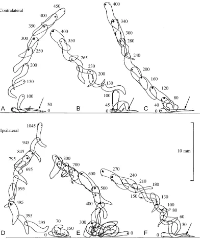
Upward swimming was recorded using high-speed video at 200 frames s−1and stroboscopic photography at 10 flashes s−1. The initial stage of upward swimming in response to a touch on the tail was seen most clearly in high-speed video recordings of 41 swimming responses in stage 37/38 tadpoles. The usual response to touch is to flex first on the opposite side (Boothby and Roberts, 1995). When the tadpole is lying on one side on the substratum, the upper side will be stimulated. If the first contraction is on the opposite side, it will bend into the substratum, but it will rise from the substratum on the second flexion and can quickly attain an upward swimming trajectory (Fig. 3A–C). When the first flexion is away from the
substratum, upward swimming can start at once (Fig. 3F), but it can also be preceded by complex struggling movements (Fig. 3D,E; Soffe, 1991). It is clear from these examples that, following stimulation, an upward swimming path can emerge from a wide range of movements: sometimes the body is dorsal up (Fig. 3C at 120 ms), sometimes ventral up (Fig. 3D at 295 ms). An almost vertical path is therefore not the result of a fixed sequence of manoeuvres. In the sequences shown in Fig. 3, spiralling started immediately and was clockwise (Fig. 3A,E,F) or anticlockwise (Fig. 3B–D) with full rotation times of between 0.4 and 0.7 s.
Flash photography of 84 tadpoles from stage 32 to stage 37/38 at 10 Hz allowed longer periods (up to 2 s) of upward swimming to be recorded at better resolution. A detailed examination was made of 10 swimming sequences in water 85 mm deep. In half of these, the swimming was entirely upwards with spiralling (e.g. Fig. 4C–E), while the rest included turns from or to more-horizontal swimming (e.g. Fig. 4A,B). In six cases, upward swimming started straight after stimulation (e.g. Fig. 4C–E). In the other four cases, the tadpole was swimming at 45 ° or less to the horizontal before it turned to spiral upwards, but in three of these cases it was already spiralling and in the fourth (Fig. 4B) it was upside-down before the turn. This suggests that the dorsal-up position is not particularly stable during more vigorous horizontal swimming. Vertical spiral lengths were 9.6–18 mm with rotation times of 0.3–0.7 s.
Individual tadpoles could change their direction of spiralling from clockwise to anticlockwise (or vice versa) during a single ascent to the surface, particularly in deeper water. This suggested that the direction of spiralling is not fixed for an individual. Thirty-two stage 37/38 tadpoles were therefore tested by laying them alternately on their left or right sides on the bottom of a container in 55 mm of water. They were then stimulated to swim 12 times by touch or by dimming the light, and the initial direction of spiralling was noted. Only one of the 32 tadpoles always spiralled in the same direction. Analysis of variance showed that spiralling direction was not significantly correlated with the individual tadpole, with the side that was in contact with the bottom or with the type of stimulus.
Responses of tadpoles hanging from a mucus strand
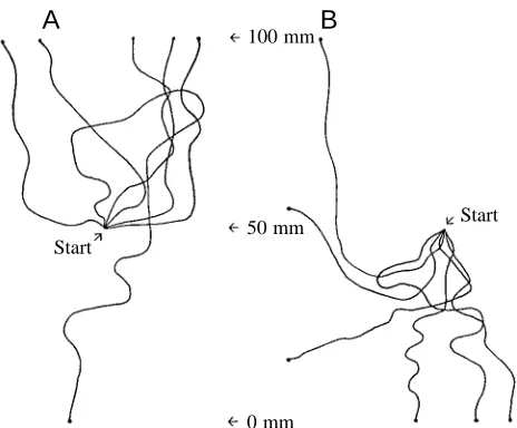
Since tadpoles spend most of their time attached by mucus secreted from their cement gland, they were observed as they hung by mucus at the end of a wire in the centre of a water-filled 250 ml beaker with a horizontal line marked around the middle. Forty tadpoles at stage 37/38 were each tested once by touching them on the head or tail with a gerbil hair while they hung tail-down. Tail stimulation led mainly to upward spiral swims (13/20 upwards, 12 of these spirals), whereas head stimulation led to mainly downward spiral swims (15/20 downwards, 11 of these spirals). These tests showed that tadpoles can swim downwards and that, when they do this, they tend to spiral in just the same way as during upward swimming (Fig. 5).
0 5 10 15 20 25 30
Cylinder diameter (mm)
Failures
0 5 10
3840 45 50 54 57 66 76
Time (h)
3132 33 35 37 39 40 41
Stage
[image:4.609.47.290.77.258.2]The upward and downward swimming responses to head and tail stimulation are clear in tracings from video recordings taken from the side (Fig. 5). The vertical direction of swimming responses was tested by observing the initial swimming direction (up, down, unclear or none) in larger samples of tadpoles from stage 31, when swimming is first effective, to stage 37/38. At each stage, 300 tadpoles were each
tested once by touching the head, trunk or tail (Table 1). The direction of swimming was significantly different in response to head and tail stimuli at each stage of development (P<0.05, by χ2tests). Touching the head led to downward swimming
and touching the tail led to upward swimming, but there was no clear vertical direction in responses to touching the trunk. Since the numbers of responses categorised as none or unclear
A
B
C
Contralateral
D
E
F
Ipsilateral
10 mm 100
150 200
250 300
350
350
265
230
200
130
100
945
0 0
150 845
795
300
0 500
270 240
210 180
130
100 80
60
30 600
700 800
400 695
595
495
395 295
150 70
45 0
40 400
340
300
280
240
200
160
120
80
1045
0 50
0 400
[image:5.609.115.507.71.539.2]400 450
Fig. 3. Initial stages of swimming showing rapid attainment of an upward direction and rotation about the longitudinal axis. Tracings from video recordings taken through the side of a glass tank at 200 frames s−1of stage 37/38 tadpoles stimulated to swim by a touch with a fine hair
dropped markedly after stage 32, we used stage 33/34 tadpoles in subsequent tests on directional swimming responses.
Since spiralling can occur when the tadpole swims either up or down, we made simultaneous video recordings from the side and above (using a mirror angled at 45 °) of stage 33/34 tadpoles swimming in response to head or tail stimulation. Our aim was to determine how swimming behaviour changed with the angle of the swimming path. Twenty swimming responses were analysed from 100 recorded examples, and the x,y,z coordinates of the swimming paths were measured every 0.1 s to the nearest 1 mm. This allowed us to plot three-dimensional swimming paths (Fig. 6) and to calculate
[image:6.609.244.557.72.578.2]swimming speeds and angles (where 0 ° is up, 90 ° is horizontal and 180 ° is down). As expected, touching the tail usually led to an initial upward swimming angle after 0.1 s (mean 63.9±37.0 °, N=8) while touching the head led to downward angles (mean 122.1±32.0 °, N=12, P<0.01, t-test; means ± S.D.). The swimming episodes lasted 1–2.6 s (mean 1.5±1.3 s) because they were limited by the size of the tank and usually ended when the tadpole hit the surface of the water (Fig. 6A–C,F), the bottom (Fig. 6D,E) or the side of the tank. In 18 out of 20 cases, the tadpole swam broadly in the same direction (either up or down) and for one part of the episode showed spiral rather than simple belly-down Fig. 4. Examples of upward swimming at different
stages of development (A and B at stage 32, C at stage 33/34, D at stage 35/36, E at stage 37/38) traced from multiple flash photographs at 10 Hz. The cement gland on the rostral ventral surface of the head is shown as a solid dark spot. The eyes on either side of the head are shown as crescents with the open side ventral. In the ribbon diagrams, which interpret the spiralling, the right side is shaded, the dorsal midline is the thick line and the ventral midline is the thin line. In A and B, the sequence starts on the bottom; in C–E, the sequence starts within 5 mm of the bottom.
A
C
D
E
B
swimming (e.g. Fig. 6 A,B,D,E, solid lines). In two cases, the swimming paths were more complex with changes in vertical direction (Fig. 6C,F). During swimming, 70 % of time was spent spiralling and there appeared to be a bias, since 66 % of this was spiralling upwards and 34 % was spiralling downwards despite the fact that in 12 out of 20 cases the initial path angle was horizontal or downwards (⭓90 °). Furthermore, the duration of individual upward spiral swimming sequences was longer (1.5±0.3 s, N=10) than for downward sequences (0.6±0.3 s, N=11; means ± S.D.) (compare Fig. 6A–C,F with Fig. 6D,E).
During both upward and downward swimming, spiralling seems to occur more often at more vertical angles. This was clear when the probability of spiralling was plotted against swimming path angle for all 20 swimming paths (Fig. 7A). However, there is no sudden cut-off angle, and belly-down swimming can occur from horizontal to quite vertical path angles. Swimming speed ranged from 10 to 113 mm s−1 but with no clear dependence on the path angle (Fig. 7B).
Another way to compare upward and downward swimming is to measure the minimum diameter of a vertical cylinder (i.e. maximal extent in the horizontal x,y plane) that will contain the swimming paths video-recorded from above (Fig. 2). Tadpoles hanging from a wire at a depth of 100 mm in water 200 mm deep were stimulated to swim up or down. The minimum cylinder diameters were significantly smaller for upward swims (66.03±24.77 mm, N=40) than for downward swims (115.75±31.9 mm, N=40, P<0.001, t-test; means ± S.E.M.).
When stimulated while hanging from mucus in mid-water, tadpoles can swim spirally up or down, but upward spiral swimming is more stable.
Basic properties of the tadpole
Our analysis of the properties of the tadpoles has concentrated on stages 33/34–37/38 when the spiral swimming response is most clear (Table 1). At these stages, body lengths range from 4.5 to 6.5 mm (Nieuwkoop and Faber, 1956) and body masses were determined after careful blotting of surrounding water under a dissecting microscope. Tadpole volumes were measured after the tadpole had been sucked into a glass tube that tapered to an inside diameter of 0.75 mm. This compressed the tadpole to a cylinder of circular cross section whose length was measured. We conclude that tadpoles at these stages have a mass close to 2 mg and a volume close to 2 mm3(Table 2).
[image:7.609.58.290.69.261.2]All tadpoles tested from stage 31 to stage 41 sank in Bristol tapwater (at least five tadpoles were tested at each stage). In 14 tadpoles from stages 32–35/36, the mean sinking rate was 12.3±1.1 mm s−1. Two methods were used to examine the density of tadpoles and their orientation near neutral density. First, solutions of different densities were prepared by mixing distilled water and Percoll, which consists of colloidal silica particles coated with polyvinylpyrrolidone and has a density of 1.13±0.005 g cm−3. Anaesthetised tadpoles (six at stage 32, 11 at stage 33/34, three at stage 35/36 and 13 at stage 37/38, N=33) floated in a 1:1 Percoll/water mixture (density 1.064 g cm−3), whereas 23 out of 27 sank in a 1:1.8 mixture (density 1.045 g cm−3). Second, anaesthetised tadpoles were placed at the top of a linear Percoll/water density gradient to measure the level to which they sank and, from this, to calculate their densities (Table 2). The two methods suggest that tadpole densities lie between 1.035 and 1.060 g cm−3. In Percoll/water mixtures in which they were close to neutral density, all tadpoles at all the stages examined were oriented dorsal side up and floated either nearly horizontally or head-up tail-down with the body at an angle of head-up to 35 ° to the horizontal.
Fig. 5. Tracings of swimming paths from video recordings of stage 33/34 embryos taken from the side (as in Fig. 1A,B). The tadpole began the sequence suspended in the middle of the water. The tracings show that when touched on the tail (A) the initial swimming path is usually upwards, and when touched on the head (B) it is usually downwards.
A
B
0 mm
Start
Start
50 mm
100 mm Table 1. Directional responses to local skin touch at different
developmental stages of tadpoles of Xenopus laevis
Stage Response Head Trunk Tail
31 Swim up 5 42 46
Swim down 43 14 1
None or unclear 52 44 53
32 Swim up 14 55 60
Swim down 38 26 5
None or unclear 48 19 35
33/34 Swim up 2 58 77
Swim down 81 29 5
None or unclear 17 13 18
35/36 Swim up 8 57 66
Swim down 74 20 2
None or unclear 18 23 32
37/38 Swim up 8 60 73
Swim down 80 22 10
[image:7.609.317.568.97.303.2]These observations suggest that tadpoles at early stages of development may swim dorsal side up because the body is
[image:8.609.39.292.96.164.2]ballasted by denser material in the ventral yolk mass. We therefore determined the position of the centre of mass in 10 anaesthetised tadpoles by recording the position of the body when it was suspended in water from a single micropin inserted through the fin near its periphery (Fig. 8). By repeating this process three times using different suspension points, the centre of mass was found to lie within the yolk mass, ventral to the muscles, approximately 2 mm from the front of the head and 0.5 mm from the ventral surface of the belly (see the average tadpole in Fig. 8). The centre of mass was assumed to lie on the sagittal midline. We then dissected off the yolk mass with its overlying skin (by cuts across the tail just caudal to the anus, along the ventral edge of the myotomes, and across the Table 2. Mean body mass, volume and density of tadpoles at
different stages of development
Mass Volume Density
Stage (mg) (mm3) (g cm−3)
33/34 1.97±0.16 2.14±0.25 1.0437±0.0033 (N=5) 35/36 1.92±0.20 1.94±0.13 1.0416±0.0022 (N=5) 37/38 2.11±0.26 2.32±0.20 1.0353±0.004
Values are means ±S.E.M.; N=10 unless otherwise specified.
A
D
B
E
C
F
[image:8.609.211.565.251.733.2]body just caudal to the gills) and compared its density with that of the whole body in anaesthetised and fixed stage 37/38 tadpoles using the density gradient method. In glutaraldehyde-fixed tadpoles, densities were increased for the dissected out belly compared with the intact animal (whole body 1.0586±0.0057 g cm−3, N=10; belly 1.0801±0.0068 g cm−3, N=11; means ± S.D.), but dissection of the yolk mass was cleaner than in the live anaesthetised tadpoles (belly 1.0657±0.0081 g cm−3, N=10). In both cases, the belly region was significantly more dense than the whole body (P<0.001, t-test).
Are special senses necessary for upward swimming? Since light usually comes from above, the upward orientation of swimming in tadpoles lying on the bottom could depend on input from the pineal eye (Roberts, 1978; Foster and Roberts, 1982). To determine whether upward swimming
depends on the light direction, two groups of 40 stage 37/38 tadpoles were each tested once. They were placed individually at the centre of the bottom of a 70 mm deep dish with black
0 20 40 60 80 100
Speed (mm s
-1)
0 20 40 60 80 100 120 140 160 180
Swimming path angle (degrees)
Up Down
0 0.2 0.4 0.6 0.8 1
Spiral probability
0 20 40 60 80 100 120 140 160 180
Swimming path angle (degrees)
A
[image:9.609.319.562.65.523.2]B
Fig. 7. Effect of path angle for each 0.1 s segment of swimming path on the probability of spiralling. (A) The probability of spiralling increases as the swimming path angle becomes more vertical (0 ° is up, 90 ° is horizontal and 180 ° is down). Open columns indicate upward spirals; filled columns indicate downward spirals. (B) The swimming speed as a function of path angle. Speed is calculated as distance/time for each 0.1 s swimming segment in 20 swimming responses. Points show means, and error bars are standard deviations. The data for both parts of this figure came from 20 swimming paths recorded as shown in Fig. 6.
1 mm Stage 32
Stage 33/34
Stage 35/36
Stage 37/38
Average of 10 (stages 32–39) a
b c
[image:9.609.48.298.73.400.2]sides illuminated equally either from above or below (at 926µmol m−2s−1≈50 000 lx). They were stimulated to swim and their final location recorded. The number reaching the surface was 39 with top light and 35 with bottom light. This difference was not significant (χ2=0.3 and 1.4, P>0.05 for both
groups), suggesting that the direction of light does not determine the direction of swimming at this stage.
If postural reflexes controlled the direction of swimming, one would expect the inner ears to be involved. However, when the otic capsules were removed, four out of five stage 32 embryos and nine out of nine stage 35/36 embryos were still able to respond with upward spiral swimming when touched.
These observations suggest that special senses are not necessary at embryonic stages of development for upward spiral swimming.
The mathematical model
A mathematical model was constructed to demonstrate that mechanical torques generated by the swimming tadpole are sufficient to reproduce and explain the horizontal belly-down and the upward spiral (more correctly ‘helical’) modes of swimming that characterise the tadpole’s motion. The model does not include the details of the motion of the tadpole’s body during swimming or the associated motion of the fluid, but rather seeks to demonstrate that spiral swimming does not require active control by the animal.
[image:10.609.321.549.391.677.2]In the model, the tadpole’s body is taken to be a rigid ellipsoid that swims forwards with a constant speed and is subject to two constant torques. One torque, G, acts about the tadpole’s long (k) axis and tends to cause the tadpole to roll (Fig. 9). The model’s centre of mass C is displaced ventrally towards its belly away from its geometric centre (centroid, O), through which the buoyancy forces act, because of the greater relative density of the yolk mass. This ballasts the model and prevents G from rolling the tadpole when it is swimming horizontally. The other torque, L, acts about the sagittal (j) axis and tends to alter the pitch of the animal by raising its head.
In essence, if the tadpole starts off swimming horizontally and belly-down, L raises the tadpole’s head towards the vertical until the ballast can no longer prevent G from rolling
the tadpole as it swims forwards. The model shows that, for certain realistic ranges of parameter values, this motion can settle into a stable upward spiral regardless of the initial orientation. Further details of the model are given in the Appendix.
Results and trajectories corresponding to different starting orientations for fixed values of G and L lying within the range giving realistic behaviour are shown in Figs 10, 11 and 12. The model’s orientation is described by three Euler angles (θ, φ, ψ) (see, for example, the textbook by Synge and Griffith, 1970, pp. 259–261) that vary with time t. θ (0 °⭐θ⭐180 °) is the pitch, i.e. the angle that the long (k) axis makes with the upward vertical laboratory (K) axis. θ=0 ° corresponds to swimming vertically upwards, θ=90 ° to horizontal swimming and θ=180 ° to swimming vertically downwards. The yaw, φ (0 °⭐φ<360 °), measures the rotation of the tadpole’s long (k) axis around the upward vertical laboratory (K) axis, and ψ (−180 °⭐ψ<180 °) gives the angle of rotation (roll) of the tadpole’s body about its own long (k) axis. When swimming horizontally (θ=90 °), the tadpole is the right way up (i.e. dorsal up) if ψ= 0 ° and upside down if ψ=−180 °. Only the rate of change of the yaw with time (i.e. the angular velocity about the vertical axis) is relevant, not the absolute value of φ.
A trajectory with the initial orientation (θ, ψ)=(98 °, 164 °) is depicted in Fig. 10. Thus, the trajectory begins with the
j
i w
k
G
L
O
C l
h
[image:10.609.42.292.587.667.2]Fig. 9. The conceptual tadpole that was used to construct the mathematical model. i, j and k are unit vectors along the axes of the model centred on the geometric centre (centroid) O. G and L are torques acting to rotate the tadpole’s body (in roll and pitch, respectively). h and l are the offsets of the centre of mass, C, through which the weight w acts.
Fig. 10. A typical escape trajectory calculated from the mathematical model. The tadpole’s initial trajectory for this example is belly-up and slightly head-down. The trajectory begins by spiralling downwards while the tadpole rights itself and finally settles into a steady upward spiral with pitch θof 33 ° and roll ψof 24 °.
0
x (cm) y
(cm) 1
-1 -2 -3 4
3 2
1
z
(cm)
0 60
40
20
0
-20
tadpole lying slightly head-down and almost belly-up. This position corresponds to a possible orientation immediately after an escape response. Viewed from above, the tadpole first spirals clockwise and slightly downwards, reorients itself and settles into an anticlockwise spiral. Further details are shown in Fig. 11.
This trajectory is the most complicated escape trajectory produced by the model because of the belly-up starting condition. Starting orientations that are belly-down settle very rapidly into the same upward spiral. This is illustrated in Fig. 12, which shows θ(t) and ψ(t) for a number of initial orientations, all of which approach the steady values of θ=33 ° and ψ=24 ° corresponding to the upward spiral after 4 or 5 s.
A mechanism for dorsal torque generation?
Experiments in larger volumes of water have shown that dimming the illumination reliably causes tadpoles that are swimming horizontally to turn upwards and start to spiral (Jamieson and Roberts, 2000). This turning response provides an opportunity to see how the tadpole turns upwards and to look for the basis for an upwards torque, but was difficult to resolve in video recordings of freely swimming tadpoles. It was therefore investigated in ‘tethered’ tadpoles. They were
anaesthetised and then held with two pins pressing against either side of the neck region positioned so the tadpole’s body and tail protruded over the edge of a Sylgard block (see Fig. 14A) (see Kahn et al., 1982). In this way, they were held in a dorsal-up position by the part of the body that moves least during swimming, while the head, trunk and tail were free to move. After recovery from the anaesthesia, tadpoles were video-recorded from the side and stimulated to swim by touching so that the effect of dimming the light during swimming could be recorded.
[image:11.609.47.301.363.645.2]In 13 out of 16 tadpoles between stages 33/34 and 37/38 that were tested, dimming the light from a light pipe illuminating the top of the head from 2000 to 4 mmol m−2s−1led to a dorsal flexion of the body and raising of the tail (e.g. Fig. 13A). In one tadpole, the tail was always held in a raised position when it swam, and dimming made no difference. To measure the dorsal flexion of the body in tracings such as those of Fig. 13A, a line was drawn between the dorsal edge of the body at the neck, where it was held by the pins, and a point on the dorsal edge of the fin at a fixed distance from the pins. (An example is shown by the arrow at 8 s in Fig. 13A). The angles of these lines relative to the vertical (vertically up = 0 °) are plotted in Fig. 13B and show a clear and rapid decrease after dimming and a slower increase when the light is restored.
During these observations, it was noticed that the body was dorsally flexed and the tail raised at the start of a swimming episode. As swimming continued, this flexion decreased and the tail was lowered. Following a sensory stimulus, swimming usually started at a high frequency that then decreased slowly (Kahn et al., 1982). To monitor tailbeat frequency and the dorso-ventral position of the tail simultaneously, a shadow of the dorsal edge of the tethered tadpole’s tail in lateral view was
Fig. 11. Analysis of the trajectory shown in Fig. 10. (A,B) Plots of θ(t) and ψ(t), the changes in pitch θand roll ψwith time t, showing that the tadpole settles into the upward spiral after approximately 5 s. (C) A plot of the (θ, ψ) phase plane; the arrowheads show how θand ψvary with t. (D) A plot of the rotation rate dφ/dt versus time, where φis yaw. The tadpole reverses its sense of direction when dφ/dt=0 at t=2.83 s. (E,F) Projections of the tadpole’s trajectory in Fig. 10 onto the horizontal x,y plane and the vertical z axis.
0 90 180
θ
(degrees)
0 5 10 15 20
Time (s)
A
-180 -90 90 0 180 ψ (degrees)0 5 10 15 20
Time (s)
B
-8 -6 -4 -2 2 0 4 d φ /d t (rev s -1)0 5 10 15 20
Time (s)
D
-2 -1 0 1 3 2 4 y (cm)-3 -2 -1 0 1
x (cm)
E
-20 0 40 20 60 z (cm)0 5 10 15 20
Time (s)
F
-180 -90 90 0 180 ψ (degrees)0 90 180
[image:11.609.330.556.484.698.2]θ(degrees)
C
Fig. 12. Plots of pitch θ(A) and roll (B) ψversus time for several escape trajectories with different starting orientations. All the trajectories tend to an upward spiral with θ→33 ° and ψ→24 °.
A
B
-180 -90 0 90 1800 90 180 ψ (degrees) θ (degrees)0 2 4 6 8 10
cast horizontally onto a photocell (Fig. 14A). The voltage from the photocell showed oscillations due to the lateral movements of the tail during swimming about a mean value indicating the dorso-ventral position of the tail (Fig. 14B,C). In seven tadpoles from stage 33/34 to stage 37/38, the tail was flexed dorsally at the start of swimming. From an initial maximum of 22–26 Hz, the tailbeat frequency fell slowly and this dorsal flexion decreased. When swimming finally stopped, the tail fell back to its resting position.
Discussion
We have examined the swimming behaviour of developing
Xenopus laevis near the time of hatching. At the earlier
developmental stages (31–35/36), these would be considered to be embryos, and at the later stages (37/38–41) tadpoles. In tadpoles at these stages it seems likely that the inner ear becomes functional because labyrinthectomy leads to
continuous rolling about the longitudinal axis during horizontal swimming (Horn and Rayer, 1978). Furthermore, from stage 42, there are compensatory eye movements during imposed roll movements of the body (vestibular-ocular reflex; Horn et al., 1986). In the embryos, it is unlikely that the inner ear is functional because the main responses we describe from stages 32 to 35/36 are not affected by labyrinthectomy. This means that the direction of the embryo’s swimming in three dimensions is controlled by simpler features of its organisation such as the location of the centre of mass, the state of flexion of the body and simple flexion reflexes. We now give a preliminary interpretation of our observations partly based on the behaviour of our mathematical model, and then consider how the embryo’s swimming responses may help it to survive.
A
B
C
Pin Photocell
26–20 Hz
22–14 Hz
Sylgard
1 s Swim
[image:12.609.312.566.70.387.2]Rest
[image:12.609.55.281.81.368.2]Fig. 14. The influence of swimming frequency on dorsal flexion of the tail. (A) A diagram of a tadpole held at the neck with two pins inserted into a Sylgard block so that the body at rest hangs over the side of the block (solid outline). During swimming, dorsal flexion raises the tail (dashed outline). A shadow of part of the dorsal edge of the tail is cast horizontally onto a photocell (rectangular outline), and raising the tail reduces the amount of light falling on the photocell. (B,C) Photocell output during the final part of two swimming episodes in stage 35/36 (B) and stage 37/38 (C) tadpoles as frequency decreases before swimming stops (frequency range indicated below each trace). The trace moves up as dorsal flexion raises the tail (and reduces light to the photocell). Frequency and dorsal flexion are both higher initially and decrease gradually. When swimming stops, the tail falls back to its rest position (dotted line). Fig. 13. Light dimming during swimming leads to dorsal flexion.
(A) Tracings of single frames from a video recording of a tadpole viewed from the side during a single swimming episode (the time is indicated in seconds. The light was dimmed between 8 and 12 s. The tail is raised when the light is dimmed. Tadpoles were held gently by pins around the neck region (vertical line) so their bodies were dorsal-up and hung over the edge of a Sylgard block (see Fig. 14A). An example of the line drawn to measure tail angle is shown in the outline at 8.0 s. (B) A graph of tail angle versus time (90 ° is horizontal, 0 ° is up) for the response in A. This shows that the angle decreases after dimming and returns slowly to a more horizontal value when light is restored.
A
B
75 80 85 90 95 100 5.8
6.0
6.4
7.0
7.8
12.6
13
13.6
14
15 8.0
9.0
10
11
12
Tail angle (degrees)
5 7 9 11 13 15
Time (s) Light dimmed
Light Dim Light
Interpretation of the swimming movements in hatchling
Xenopus laevis
Tadpoles spend 99 % of their time hanging from mucus secreted by a cement gland on the front of the head (Jamieson and Roberts, 2000). Since they are heavier than water, they hang from the mucus in a highly predictable vertical spatial orientation with their head pointing up. They also sink to the bottom if the mucus strand breaks.
When swimming near the horizontal, tadpoles swim dorsal-up because the body is ballasted by a large volume of dense yolk in the undifferentiated belly region and, as a result, the centre of mass lies in the ventral half of the body.
While swimming, we assume that a torque is generated by slight asymmetries in the movements of the body and tail. This torque acts to roll the body about its longitudinal axis but it is not constant so, in different swimming episodes, the tadpole can rotate clockwise or counterclockwise. Near the horizontal, the ballast can counter this torque, even in the absence of any stabilisers such as fins. If swimming becomes more vertical (up or down), tadpoles spiral about their own longitudinal axis because the moment of the ballast is no longer sufficient to stabilize their orientation. The sense of the spiral is not fixed even in an individual tadpole.
The mathematical model suggests that stable upward spiral swimming depends on the presence of another torque that acts to lift the head in the pitch plane. We suggest that this torque results from a dorsal flexion of the body that occurs during swimming and is more marked at higher swimming frequencies. Dorsal flexion is stronger following sensory stimulation at the start of swimming episodes or when excitatory stimulation given during swimming leads to an increase in tailbeat frequency. This could explain why tadpoles turn upwards when light dimming excites the pineal eye during horizontal swimming (Jamieson and Roberts, 2000). In this case, it has been shown that excitation of the pineal eye leads to an increase in swimming frequency. If the dorsal torque does depend on dorsal flexion of the body, our observations suggest that dorsal torque will vary as a function of tailbeat frequency (Fig. 14).
During swimming, tadpoles change direction frequently. At more vertical angles, they spend more time spiralling (70 %) than swimming belly-down. The model predicts that upward spiral swimming is a stable condition and that upward spiralling is more common than downward spiralling.
When hanging from its mucus strand, the tadpole is in a very predictable orientation in space. The result of this is that simple responses to touch that have been described previously (Boothby and Roberts, 1995) can lead to predictable swimming paths in three-dimensional space. When touched on the tail, the tadpole swims up because there is little body flexion so it swims straight ahead when swimming starts. When a hanging tadpole is touched on the head, it swims down because there is a strong body flexion when swimming starts that directs the head downwards. When stimulated while lying on the bottom, tadpoles show a strong tendency to swim up because they cannot swim down (edge effect) and upward spiral swimming is a stable condition.
Observations on the model tadpole suggest that upward spiral swimming is more stable than downward swimming. In tadpoles, three-dimensional plots of swimming paths show that upward spiral swimming is more common and that, during upward swimming, the minimum cylinder containing the swimming path is narrower. Second, the model shows the importance of the initial orientation of the tadpole on subsequent swimming paths. Since the hatched tadpole spends nearly all its time hanging head-up, this would be expected to increase the probability of an upward swimming path. In a large volume of water, the model tadpoles will all end up at the water surface, although we can only guess at the possible advantages that this distribution may have (Jamieson and Roberts, 2000). Although our model is very basic and omits many known features of the tadpoles’ behaviour, for example turns during swimming, these parallels with tadpoles are encouraging.
Biological relevance of swimming orientation
Many studies have shown that amphibian tadpoles are heavily predated by aquatic larvae of, for example, water beetles and Odonata (Chovanec, 1992; Skelly and Werner, 1990). Odonate larvae are certainly common in South African inland waters (Chutter, 1961), and preliminary observations show that such larvae are common in ponds in South Africa where young Xenopus laevis tadpoles were found (A. Roberts and L. Minter, unpublished observations). Odonate larvae are fairly sedentary, but also walk about on vegetation or the substratum to search for prey (Pritchard, 1965). Since hatched
Xenopus laevis tadpoles spend 99 % of their time immobile and
hanging from a mucus strand (Jamieson and Roberts, 2000), they would be expected to be found mainly attached to vegetation, other solid objects or the surface meniscus. Once their mucus strand breaks, they will sink and could reach the bottom or start swimming. If disturbed and touched by a potential insect predator, the responses that we have described appear to be appropriate for escape. When approached from above and touched on the head, the tadpole swims down. When approached from below and touched on the tail, it swims up. If touched while lying on the substratum, it swims up.
We had previously been interested in the unpredictable sidedness of responses to head stimulation and suggested that this might have survival value as it were to lead to unpredictable swimming paths after stimulation (Boothby and Roberts, 1995). However, if the main function of responses to head stimulation is to direct swimming downwards in tadpoles that are hanging head-up from a mucus strand, then the sidedness is not important. What is needed is a strong flexion to either side that will point the head downwards. This response leads to a predictable initial downward swimming path that removes the tadpole from the source of stimulation.
mathematical model indicates that upward spiral swimming is a stable state. What are the advantages of swimming towards the surface? Does this behaviour remove tadpoles from less favourable physical or chemical condictions near the bottom? Is it to avoid bottom-living predators such as adult Xenopus
laevis? Our own observations and those of Bles (1905) suggest
that, in holding tanks, tadpoles accumulate near the surface. In a companion study (Jamieson and Roberts, 2000), we have shown that tadpoles swimming into shadow will turn upwards and attach to objects that cast shadows. This response is mediated by the pineal eye, which is excited by light dimming. We also discuss other ways that changes in light levels and a tendency to swim upwards may affect the vertical distribution of the tadpoles and their ability to find stable solid attachment sites at this non-feeding stage of their early development.
It is clear that, shortly after hatching, the Xenopus laevis tadpole inner ear becomes functional (Horn and Rayer, 1978; Horn et al., 1986) and its behaviour becomes more complex and directed (Tunstall and Sillar, 1993). A similar developmental picture has been drawn for other anuran tadpoles by Herter (1921), who studied the effects of labyrinthectomy on swimming and other behaviour in Rana
esculenta, Bufo vulgaris, Pelobates fuscus and Hyla arborea.
It is likely that the ballasting of the body by yolk can contribute to dorsal-up swimming in these tadpoles as well as Xenopus
laevis, but at present there is no evidence that these other
anuran tadpoles show the stable spiral swimming behaviour that we have described in hatchling Xenopus laevis.
Appendix The mathematical model
We begin by assuming that the tadpole swims with a constant speed and so only the torques that orient the animal need to be considered. Furthermore, we suppose that the tadpole swims in such a way that it generates a torque (or moment of force) s, where:
s= Gk−Lj , (A1) where G and L are constants and L>0. The unit vectors i, j and k are fixed in the tadpole’s body at its centre of buoyancy and form a right-handed triad (Fig. 9). i is dorsal to ventral, k is posterior to anterior, and j is sagittal. Thus, the component of the ‘swimming’ torque Gk acts to rotate the tadpole’s body about its long axis from the tail to the head and −Lj acts to raise the tadpole’s head. The centre of mass of the tadpole does not coincide with its centre of buoyancy and lies at the point (hi+lk) (see Fig. 9).
It is also convenient to define a second right-handed triad of unit vectors (I, J, K) fixed in a stationary laboratory frame of reference, in which K is vertical. The weight of the tadpole can now be written as −wK. The two sets of unit vectors are related by Euler angles (θ, φ, ψ) (Synge and Griffith, 1970), which represent three rotations needed to map (I, J, K) into (i, j, k). Specifically, the three rotations are: ᑨ1=φK, which rotates (I, J, K) into a first intermediate triad (I′, J′, K); ᑨ2=θJ′, which
rotates (I′, J′, K) into a second intermediate triad (I′′, J′, k); and ᑨ3=ψk, which rotates (I′′, J′, k) into the body triad (i, j, k).
The tadpole’s weight can now be written in the body frame as
−wK = w(sinθcosψi−sinθsinψj−cosθk) , (A2) and the gravitational torque about the hydrodynamic centre is:
g= (hi + lk) ×(−wK) (A3)
= w[l sinθsinψi + (h cosθ+ lsinθcosψ)j−h sinθsinψk] . (A4) The Reynolds number for the forward motion of the tadpole is approximately 250 estimated on a swimming speed of 50 mm s−1and a body length of 5 mm. However, we need to estimate the torques required to rotate the body rather than the thrust needed for forward motion. The Reynolds numbers associated with the rotational motion (⭐12) are an order of magnitude smaller than those needed to rotate the body. As a first approximation, we shall assume that the Reynolds number for rotational motion is sufficiently small that inertia can be neglected in the rotational motion.
As the tadpole rotates, the fluid exerts a viscous torque on the surface of the tadpole that opposes the rotation. At low Reynolds number, the viscous torque, v, is proportional to the angular velocity, ⍀, of the body. Relative to the body triad, ⍀ can be written in terms of the Euler angles and their time derivatives, denoted by overhead dots, as
⍀= (θ˙ sinψ − φ˙ sinθcosψ)i
+ (θ˙ cosψ+ φ˙ sinθsinψ)j + (φ˙ cosθ+ ψ˙ )k , (A5) so that the viscous torque is given by
v= −κ1(θ˙ sinψ − φ˙ sinθcosψ)i
− κ2(θ˙ cosψ+ φ˙ sinθsinψ)j− κ3(φ˙ cosθ+ ψ˙ )k , (A6) where κ1, κ2and κ3are the resistance coefficients and depend on the shape of the tadpole’s body. (Strictly, we have made some additional assumptions about the symmetry of the body shape so that a rotation about one of the body axes only produces a torque about that same axis.) In this case, the equations of rotational motion for the tadpole reduce to an overall torque balance:
[image:14.609.310.554.653.742.2]s+ g+ v= 0 . (A7) This last equation can be solved in principle to find the Euler
Table 3. Tadpole parameter values used in mathematical model
Length l 5 mm
Depth b 1.2 mm
Width c 0–0.3 mm
Density ρ 1.05 g ml−1
angles (θ, φ, ψ) as functions of the time t. The torque balance equation can be written in components as
αl˜sinθsinψ= θ′sinψ − φ′sinθcosψ, (A8)
−L˜ + h˜ cosθ+ l˜sinθcosψ= θ′cosψ+ φ′sinθsinψ, (A9) 1 − βh˜ sinθcosψ= φ′cosθ+ ψ′, (A10)
where α=κ2/κ1, β=κ2/κ3, h˜=whκ3/Gκ2, l˜=wlκ3/Gκ2, L˜=Lκ3/
Gκ2, (.)′=d(.)/dt˜ and t˜=Gt/κ3 is a scaled time. Alternatively, in
a form more suitable for numerical integration
θ′= l˜sinθ(αsin2ψ+ cos2ψ) + (h˜ cosθ −L˜)cosψ, (A11) ψ′= 1 − βh˜sinθsinψ − φ′cosθ, (A12)
φ′= sinψ[(h˜cosθ −L˜)cosecθ+ (1 − α)l˜cosψ] . (A13) The right-hand side of equation A13 is independent of the angle φ, which gives the tadpole’s angle of rotation about the vertical laboratory axis. Only the angular velocity dφ/dt is of interest, not the absolute horizontal angle φ, so the problem reduces to solving the two coupled, nonlinear, ordinary differential equations A11 and A12 for θand ψ, on substituting for φ′from equation A13 into equation A12.
In general, no simple analytical solution has been found for these equations; instead, they have been solved numerically for realistic parameter values based on experimental data. We want to find ‘steady’ solutions in which the model tadpole swims vertically up a spiral trajectory of constant pitch as seen in experiments. Mathematically, these are solutions in which θ and φ are fixed, i.e. θ′=ψ′=0, 0⭐θ<π/2 and φ′ is constant.
A special case
One special case, for which an analytical solution has been found, is given below. It is useful because it serves as a check on the modelling and on the numerical solutions. Suppose that α=1 and h˜=L˜⇔wh=L, in other words the gravitational
torque due to the tadpole being ‘head-heavy’ is exactly equal to the torque due to swimming that acts to raise the head when the tadpole is swimming vertically. There is then a solution in which θ=0 and φ′+ψ′=1. This solution corresponds to the animal swimming vertically upwards rotating about the vertical axis with an angular velocity of magnitude equal to
G/κ3. This is most easily seen from equations A8, A9 and
A10.
A
C
E
B
D
F
090 180 270 360
ψ
(degrees)
ψ
(degrees)
ψ
(degrees)
0 90 180
θ(degrees)
0 90 180 270 360 0
90 180 270 360
0 90 180 270 360 0
90 180 270 360
0 90 180 270 360
0 90 180
[image:15.609.195.563.357.740.2]θ(degrees) Limit cycle Fig. 15. Phase portraits of solutions
Experimental data
The parameter values used in the model, given in Table 3, are estimated from the experimental data given in the Results and Table 2.
To estimate the values of the resistance coefficients κ1, κ2
and κ3, the tadpole’s body is modelled as a rigid ellipsoid, this
being one of the shapes for which full analytical results are known. If the ellipsoid has semi-major axes of lengths a, b and
c (a>b>c), the resistance coefficient for rotation about its
longest axis is:
κ1= 16πµ(b2+ c2)/3(b2β0+ c2γ0) , (A14)
where
and µis the viscosity of the fluid (Edwardes, 1892). Similar expressions for κ2and κ3can be derived by permuting a, b and
c. The integrals in the above expressions have to be evaluated
by quadrature. Since the lateral width of the tadpole’s body tapers along its length, the values of the resistance coefficients were calculated for values of c=0.01, 0.02 and 0.03 mm, and for a=2.5 mm and b=0.6 mm.
These values are κ1=7.0×10⁻⁴to 7.3×10⁻⁴, κ2=5.4×10⁻⁴to
6.5×10⁻⁴ and κ3=0.8×10⁻⁴ to 1.0×10⁻⁴. Given the relatively
small variations in the coefficients due to different choices of
c, the following mean values are used hereafter:κ1=7.2×10⁻⁴, κ2=6.1×10⁻⁴ and κ3=0.9×10⁻⁴. Consequently, α=0.85 and β=6.8.
Given the convex shape of the tadpole’s body, its centre of mass must lie within its body, and given that the tadpole floats head-up at 30 ° to the horizontal, we assume that h=√3 mm and
l=−1 mm in the absence of further data, which in turn implies that l˜=−h˜/√3.
To determine an upper bound on G, we appeal to the observation that the tadpole does not rotate about its long axis when it is horizontal but only as its head is raised. This means that when θ=π/2, G must be less than the maximum gravitational torque about the k axis, which occurs when
ψ=±π/2. Thus, G<wh. It follows that h˜>β−1=0.15.
Numerical results
On the basis of the experimental data and on the assumptions detailed in the previous section, we are left with two undetermined quantities, the torques G and L, which correspond to varying h˜ and L˜ in the system of scaled equations A11 and A12 with φ′determined by equation A13. These equations have been solved numerically for various values of h˜ and L˜, seeking steady solutions in which θ′=0 and ψ′=0, and θ≈25 ° and
φ˙ =12.5 s−1, i.e. in which the tadpole swims upwards along a
vertical spiral as observed in experiments. We have to search through the possible values of h˜ and L˜ in the model to find the particular values that yield the desired values of θand φ˙ . The results of these numerical trials are described in this section.
The numerical procedure
Using the public domain software package Dstool 1.1 (Center of Applied Mathematics, Cornell University, Ithaca, NY 14853, USA), a selection of values of L˜ are chosen for each selected value of h˜. For each value of L˜, initial values are chosen for θand ψ, and the differential equations are solved numerically using a fourth-order Runge–Kutta scheme. The results for each time step are displayed on a (θ, ψ) graph leading to a trajectory in the (θ, ψ) phase plane (e.g. Fig. 15). Sufficient initial values are chosen that the resulting trajectories give a clear picture of the topology of the phase plane. In interpreting these phase diagrams, one must note that the angle ψis 2π-periodic, so individual trajectories may cross from the top of the diagram to the bottom, and vice versa, at the same value of θ. θ, of course, is not periodic: θ=0 corresponds to the tadpole swimming vertically upwards and
θ=πcorresponds to the tadpole swimming vertically down. It is also possible to plot other two-dimensional graphs, such as
θversusφ˙, to aid in the interpretation of the results; numerical
values are available for all the parameters and variables.
Results
The topology of the phase portraits is found to divide into several classes, according to the values of the ‘free’ parameters
h˜ and L˜, which are summarised below.
0.15<h˜ⱗ0.3
For values of h˜ less than approximately 0.3 and when L˜<h˜, the phase portrait for θand ψhas one stable focus at the point (θ0, ψ0), say, corresponding to the tadpole swimming steadily
upwards along a helix of θ0(Fig. 15A). The focus lies in the
the lower, left-hand quarter-plane on the diagram, i.e. 0<θ0<π/2 and 0<ψ0<π. All trajectories, whatever their initial
values, ultimately spiral into the focus so that a steady solution is finally achieved. In general, as L˜ increases, the value ofθ0
decreases while φ˙ increases, whereas when h˜ increases, φ˙ decreases. However, the values of φ˙ in this regime are much larger by a factor of 10 or 20 than those observed in experiments, even when θ0lies in the required range.
When L˜ is a little greater then h˜, the stable focus described above is found at a value of ψbetween π and 2π(Fig. 15B). When L˜ is much greater than h˜, the phase portrait bifurcates to two counter-rotating foci (Fig. 15C). The upper one is stable and corresponds to the tadpole swimming upwards along a helix, while the lower one is unstable and would not be realised in practice. Again, the values of φ˙ are an order of magnitude too large.
h˜ⲏ0.3
When h˜ is greater than approximately 0.3 and when L˜<h˜, the single stable focus in the phase plane in the previous case (A17)
∆(t) =
冪
(a2+ t)(b2+ t)(c2+ t)(A16)
γ0= ⌠ ⌡ ∞
0
dt (c2+ t)∆(t) ,
(A15)
β0= ⌠ ⌡ ∞
0
bifurcates to a stable node (or fixed point) into which all trajectories ultimately travel, and the experimentally observed tadpole behaviour is achieved when h˜≈0.6 and L˜≈0.3 (Fig. 15D). The corresponding values of G and L are 9.8×10⁻³dyn cm and 2.0×10⁻²dyn cm, respectively. Again θ0
decreases and φ˙ increases as L˜ increases, and θ0increases and φ˙ decreases as h˜ increases.
When L˜ is larger than h˜, as in Fig. 15E, two foci are found, one stable and one unstable, as is the case when h˜ⱗ0.3. The values of φ˙ are unrealistically large in this regime, and so it will not apply to the real tadpoles. It is of mathematical interest to note that when L˜ is increased still further, as in Fig. 15F, the lower, unstable focus undergoes a bifurcation and sheds an unstable limit cycle so that the focus itself becomes stable. Once again, the values of φ˙ in the two steady solutions are much greater than those observed in experiments.
We would like to thank Professor A. E. Walsby for suggesting Percoll and, with Phil Davies, for helping us to use it, Martin Smith for providing the data on spiralling directions and responses during illumination from below, the EPSRC for the loan of the high-speed video system, Mark Shannon and Tim Colburn for help with photography and images, Derek Dunn, Alison Walford and Linda Teagle for technical help, Drs Ray Perrins and Steve Soffe for advice on drafts of the paper and Professor J. M. V. Rayner for advice and for providing software for plotting swimming paths.
References
Batchelor, G. K. (1967). An Introduction to Fluid Dynamics. Cambridge: Cambridge University Press.
Bles, E. J. (1905).The life-history of Xenopus laevis, Daudin. Trans. R. Soc. Edin. 41, 809–821.
Boothby, K. M. and Roberts, A. (1992). The stopping response of Xenopus laevis embryos: behaviour, development and physiology. J. Comp. Physiol. 170, 171–180.
Boothby, K. M. and Roberts, A. (1995). Effects of site and strength of tactile stimulation on the swimming responses of Xenopus laevis embryos. J. Zool., Lond. 235, 113–125.
Chovanec, A. (1992). The influence of tadpole swimming behavior on predation by dragonfly nymphs. Amph.-Rept. 13, 341–349. Chutter, F. M. (1961). Certain aspects of the morphology and
ecology of the nymphs of several species of Pseudagrion Selys (Odonata). Arch. Hydrobiol. 57, 430–462.
Edwardes, D. (1892). Steady motion of a viscous liquid in which an ellipsoid is constrained to rotate about a principal axis. Q. J. Math. 26, 70–78.
Foster, R. G. and Roberts, A. (1982). A role for the pineal eye in Xenopus laevis embryos and larvae. J. Comp. Physiol. 145, 413–419.
Herter, K. (1921). Untersuchungen uber die nicht-akustischen Labyrinth-functionen bei Anurenlarven. Z. Allge. Physiol. 19, 335–414.
Horn, E., Lang, H.-G. and Rayer, B. (1986). The development of the static vestibulo-ocular reflex in the Southern clawed toad Xenopus laevis. I. Intact animals. J. Comp. Pysiol. 159, 869–878.
Horn, E. and Rayer, B. (1978). Compensation of vestibular lesions in relation to development. Naturwissenschaften 65, 441. Jamieson, D. and Roberts, A. (2000). Responses of young Xenopus
laevis tadpoles to light dimming: possible roles for the pineal eye. J. Exp. Biol. 203, 1857–1867.
Kahn, J. A., Roberts, A. and Kashin, S. (1982). The neuromuscular basis of swimming movements in embryos of the amphibian Xenopus laevis. J. Exp. Biol. 99, 175–184.
Leif, R. C. (1968). Formation and fractionization of density gradients. Analyt. Biochem. 25, 271–282.
Liu, H., Wassersug, R. and Kawachi, K. (1997). The three-dimensional hydrodynamics of tadpole locomotion. J. Exp. Biol. 200, 2807–2819.
Nieuwkoop, P. D. and Faber, J. (1956). Normal Table of Xenopus laevis (Daudin). Amsterdam: North-Holland.
Pritchard, G. (1965). Prey detection by dragonfly larvae (Odonata; Anisoptera). Can. J. Zool. 43, 271–289.
Roberts, A. (1978). Pineal eye and behaviour in Xenopus tadpoles. Nature 273, 774–775.
Roberts, A. and Smyth, D. (1974). The development of a dual touch sensory system in embryos of the amphibian, Xenopus laevis. J. Comp. Physiol. 88, 31–42.
Skelly, D. K. and Werner, E. E. (1990). Behavioural and life history responses of larval American toads to an odonate predator. Ecology 64, 2313–2322.
Soffe, S. R. (1991). Triggering and gating of motor responses by sensory stimulation: behavioural selection in Xenopus embryos. Proc. R. Soc. B 246, 197–203.
Synge, J. L. and Griffith, B. A. (1970). Principles of Mechanics, third edition, International Student Edition. Tokyo: McGraw-Hill Kogakusha Ltd.