RESEARCH ARTICLE
Changes in vitellogenin expression caused by nematodal
and fungal infections in insects
Dalibor Kodrı́k1,2,*, Emad Ibrahim1,2,3, Umesh K. Gautam1,2, Radmila Č apkováFrydrychová1,2, Andrea Bednářová2, Václav Krištůfek4and Pavel Jedlička5,‡
ABSTRACT
This study examined the expression and role of vitellogenin (Vg) in the body of the firebugPyrrhocoris apterus(Heteroptera, Insecta) during infection elicited by two entomopathogenic organisms, the nematode Steinernema carpocapsae and the fungus Isaria fumosorosea. Infection by S. carpocapsae significantly upregulated Vg mRNA expression in the male body. The corresponding increase in Vg protein expression was also confirmed by electrophoretic and immunoblotting analyses. Remarkably, in females, the opposite tendency was noted. Nematodal infection significantly reduced both VgmRNA and Vg protein expression levels in fat body and hemolymph, respectively. We speculate that infection of reproductive females reduces Vg expression to a level that is still sufficient for defense, but is insufficient for reproduction. This circumstance reduces energy expenditure and helps the individual to cope with the infection. Importantly, purified Vg significantly inhibited growth ofXenorhabdus spp., an entomotoxic bacteria isolated fromS. carpocapsae.However, the effect of Vg againstI. fumosoroseawas not so obvious. The fungus significantly stimulatedVggene expression in males; however, a similar increase was not recapitulated at the protein level. Nevertheless, in females, both mRNA and protein Vg levels were significantly reduced after the fungal infection. The obtained data demonstrate that Vg is probably an important defense protein, possibly with a specific activity. This considerably expands the known spectrum of Vg functions, as its primary role was thought to be limited to regulating egg development in the female body.
KEY WORDS: Yolk protein, Immunity, Antibacterial activity, Antifungal activity, Entomopathogenic nematode,
Entomopathogenic fungus
INTRODUCTION
Vitellogenins (Vgs), glycolipophosphoproteins mostly known and well-characterized as precursors of yolk proteins, are involved in reproduction in the majority of oviparous animals. In insects, Vgs are typically synthesized in the fat body, from where they are transported via the hemolymph into growing oocytes. After they enter
the oocyte by endocytosis via specific receptors, Vgs usually undergo some modifications, transforming into vitellins. Most insects produce only one or two types of Vgs that comprise several subunits with a total molecular mass ranging from 150 to 650 kDa. Vg production is hormonally controlled. It has been known for decades that juvenile hormone stimulates Vg synthesis in most insect species (see Chapman, 1998). Furthermore, the termination of Vg synthesis is controlled by adipokinetic hormone that inhibits the synthesis of the protein part of the Vg molecule directly in the fat body (Carlisle and Loughton, 1986). Additionally, Vg production is controlled by nutrient levels and mating status (Chapman, 1998).
Vgs are typically present in egg-laying females. However, low levels of Vgs have been identified in males of several insect species, including the firebug Pyrrhocoris apterus (Němec et al., 1993). Several recent studies suggested that Vgs play an important role not only in reproduction but also in other aspects of insect biology, such as the caste differentiation process in social insects, wound healing, protection against oxidative stress, immunity and life span regulation (Havukainen et al., 2013; Singh et al., 2013; Salmela et al., 2015; Salmela and Sundstrom, 2017; Park et al., 2018). Studies in the silkworm and honeybee reported strong antibacterial activity of Vg against gram-positive and gram-negative bacteria (Singh et al., 2013), showing that Vg bound to bacterial cells and destroyed them. Vg was active even againstPaenibacillus larvae, a gram-positive bacterium infesting young honeybee larvae and causing a disease called American foulbrood, probably the deadliest bee brood disease worldwide (Salmela et al., 2015). In addition, it has been reported that infection of honey bee larvae by the spores of the microsporidiumNosema ceranaesignificantly upregulated Vg expression in workers (BenVau and Nieh, 2017; Sinpoo et al., 2018), and that bee Vg interacted with the cell wall of the entomopathogenic fungusBeauveria bassiana, eliciting membrane disruption and permeabilization. Furthermore, Vg appears to induce transgenerational immune priming in bee queens, enhancing immunity in their offspring by transporting pathogen-associated pattern molecules, which are attached to Vg, into the eggs within queen ovaries (Sadd et al., 2005; Salmela et al., 2015).
Oxidative stress is caused by the accumulation of reactive oxygen species primarily produced within mitochondria as unavoidable aerobic metabolism by-products (Beckman and Ames, 1998). The anti-oxidative response has evolved a suite of defense mechanisms, involving both enzymatic and non-enzymatic components (Fridovich, 1978) controlled by adipokinetic hormones in insects (Krishnan et al., 2007; Bednářová et al., 2013; Kodrík et al., 2015). Vg plays an important role in this process as it has been shown to elicit anti-oxidative protection against oxidative stressors such as paraquat or hydrogen peroxide (Seehuus et al., 2006; Park et al., 2018). It was proposed that the anti-oxidative effect of Vg might be a crucial mechanism that extends the life span of long-lived honey bee winter workers and queens, in which Vg is synthesized in high Received 5 March 2019; Accepted 24 April 2019
1Institute of Entomology, Biology Centre, CAS, Branišovská31, 370 05 České
Budějovice, Czech Republic.2Faculty of Science, University of South Bohemia,
Branišovská31, 370 05 ČeskéBudějovice, Czech Republic.3Faculty of Agriculture,
University of Cairo, Giza, Egypt.4Institute of Soil Biology, Biology Centre, CAS,
Branišovská31, 370 05 ČeskéBudějovice, Czech Republic.5Institute of Organic
Chemistry and Biochemistry, CAS, Flemingovo sq. 542/2, 166 10 Praha 6, Czech Republic.
‡Present address: Institute of Biophysics, CAS, Královopolská135, 612 65 Brno,
Czech Republic
*Author for correspondence (kodrik@entu.cas.cz)
D.K., 0000-0001-6109-1979
Journal
of
Experimental
levels. However, the exact mechanisms of the anti-oxidative effects of Vg and hormones in the insect body remain unclear.
In the present study, we sought to examine the role of Vg in the defense against two different entomopathogens: the nematode
Steinernema carpocapsae and the fungus Isaria fumosorosea.
The nematodeS. carpocapsaecarries symbioticXenorhabdusspp. bacteria that are toxic for insects (Simões et al., 2000; Duchaud et al., 2003), and the nematobacterial complex represents an efficient tool for insect killing commonly used for insect pest control (Ehlers, 2003; Inman et al., 2012). Similarly, the fungus
I. fumosorosea, harbored by the horse chestnut leaf miner
Cameraria ohridella, plays a significant role in the biological
control of many insect species (Zimmermann, 2008).The main aim of the present study was: (1) to examine changes in Vg gene and protein expression upon infection with entomopathogenic nematode (EPN) and entomopathogenic fungus (EPF); (2) to elucidate the role of Vg in the insect body infected by EPN; and (3) to determine whether the Vg-mediated defense reaction to EPF, similar to that observed in honey bees or silkworms (see above), is a common defense mechanism in insects. In addition, (4) we sought to explain the role of Vg in insect males.
MATERIALS AND METHODS Experimental insects
A stock culture of the firebug P. apterus (L.) (Heteroptera), established from wild populations collected at České Budějovice (Czech Republic, 49°N), was used for the present study. Larvae and adults of the reproductive (brachypterous) morph were kept in 500 ml glass jars in a mass culture and reared at a constant temperature of 26±1°C under long-day conditions (18 h:6 h light: dark). They were supplied with linden seeds and waterad libitum, which were replenished twice weekly. Female and male adults were kept separately (Socha and Kodrík, 1999).
Entomopathogenic nematodeSteinernema carpocapsae and insect treatment
Steinernema carpocapsae nematodes, originating from St
Petersburg, Russia (strain NCR), were obtained courtesy of Dr Z. Mráček (Institute of Entomology, České Budějovice). They were reared under laboratory conditions using the last larval instar of
Galleria mellonella(Lepidoptera, Insecta) as a host. The emerging
infective juveniles were harvested and subsequently stored in water at 4°C for 30 days. Their viability was confirmed under a microscope before experiments began.
For Vg experiments, 7 day old males and 1–4 day old females were treated individually withS. carpocapsaeby injection into the hemocoel of 10 nematodes in 2 µl autoclaved water per individual; controls were injected with autoclaved water only. The firebugs were transferred into glass jars and kept under the same conditions as for the stock culture. Hemolymph of surviving individuals was collected 1 day after infection and stored at−20°C until used.
Entomopathogenic fungusIsaria fumosoroseaand insect treatment
The fungusI. fumosoroseaisolate originating from the horse chestnut leaf miner,Cameraria ohridella(Lepidoptera, Gracillariidae) was obtained courtesy of Dr A. Bohatá (Agricultural Faculty, South Bohemian University, České Budějovice). The strain is deposited under number CCM 8367 as a patent culture in the Czech Collection of Microorganisms in Brno (WO2010006563A1). The spore suspension was prepared by scraping 14 day old conidiospores into a sterile solution of 0.05% (v/v) Tween®80 (Sigma-Aldrich). The
suspension was filtered through sterile gauze to separate the mycelium and clusters of spores. The number of spores in the uniform suspension was counted with a Neubauer improved chamber and subsequently the suspension was adjusted to a concentration of 1×10⁷spores ml−1. A 5 ml sample of the conidial suspension was
added to 100 ml potato dextrose broth in a 250 ml Erlenmeyer flask, which was then placed on a shaker and incubated at 25°C and 200 rpm under constant light. After 4 days, the blastospores were harvested and injected into 7 day old males and 1 day old females at a dose of 30,000 blastospores per bug; controls were injected with Ringer saline only. The firebugs were transferred into glass jars and kept under the same conditions as for the stock culture. Hemolymph of the surviving individuals was collected 1–3 days after infection and stored at−20°C until used.
RNA and cDNA preparation
Fat body preparation
Pyrrhocoris apterus males treated with S. carpocapsae or
I. fumosorosea were collected and stored at −80°C prior to
processing. To monitor the expression profile of theVggene, the fat body was dissected under a stereomicroscope on sterilized glass Petri dishes placed on crushed ice and in sterile, ice-cold RNase-free Ringer solution. Fat bodies of four P. apterus individuals were pooled as one replicate, and four biological replicates per tissue of control and nematode-treated P. apterus males were generated. Immediately after dissection, the fat bodies were transferred to microcentrifuge tubes with 200μl of TRI Reagent®(Sigma-Aldrich)
on crushed ice and then stored at−80°C until RNA isolation.
RNA isolation and cDNA synthesis
The total RNA was extracted using TRI Reagent®(Sigma-Aldrich)
following the manufacturer’s protocol. RNA isolates were treated with RQ1 RNase-Free DNase (Promega) to remove traces of contaminant DNA. The cDNA template was prepared using the SuperScript® III First-Strand Synthesis System for RT-PCR
(Invitrogen, Life Technologies) with 2 µg of the corresponding total RNA and random hexamers.
Quantification ofVggene expression
Quantitative real-time PCR (qPCR) was performed to evaluateVg
transcript levels in the fat bodies of the experimental firebugs. For these studies, the same experimental design (age of male and female firebugs, schedule of infection by the nematode and fungus, time table, etc.) as for analysis of Vg protein in hemolymph was used (see above). The experiments were accomplished on a Light Cycler CFX96 BioRad real-time PCR system using Xceed qPCR SG 2x Mix Lo-ROX (Institute of Applied Biotechnologies), and relative
Vgtranscript levels were determined using the threshold cycle and normalized to expression ofRp49(Ribosomal protein 49). Primers used for qPCR were: Vg (forward) CCCGACAAGTCCACAGT-TATT, Vg (reverse) GCGCATTCTGTTCATGTAAGC, Rp49 (forward) CCGATATGTAAAACTGAGAAAC, and Rp49 (reverse) GGAGCATGTGCCTGGTCTTTT.
Gel electrophoresis and Vg quantification
SDS-PAGE under denaturing conditions using commercial gels (Bio-Rad, 5–20%) was performed according to Laemmli (1970) as modified by Socha et al. (1991). Typically, hemolymph samples were diluted 10-fold and 25-fold in sample buffer for male and female samples, respectively, and 10μl was used for the analysis. The proteins separated on gels were stained with Coomassie Brilliant Blue R-250, and Vg bands were determined according to
Journal
of
Experimental
molecular weight (MW) standards (10–250 kDa, Thermo Fisher Scientific) and reaction with specific antibody (see below); Vg band density was evaluated using a GS-800 Calibrated Densitometer with Quantity One (v4.6) software (Bio-Rad).
Western blotting
After SDS-PAGE, the separated proteins were blotted onto nitrocellulose membrane according to Towbin et al. (1979). Specific antibody againstP. apterusVg (1:1000 v/v; Socha et al., 1991) was used, followed by secondary antibody Goat/HRP (1:1000 v/v; goat anti-rabbit antibody labelled with horse radish peroxidase; Sigma-Aldrich). For visualization, the Novex® ECL
HRP chemiluminescent substrate reagent kit (Invitrogen) with 1:1 v/v A and B solutions was used. The developed color was documented using Intelligent Dark Box (LAS 3000, Fujifilm).
Vg isolation and antimicrobial activity
Crude Vg was isolated from the hemolymph of 3–4 day old
P. apterusfemales to test its antimicrobial activity. The hemolymph
samples were separated by SDS-PAGE using 10% gel according to Laemmli (1970), as described above. The gel was then stained with a low concentration of Coomassie Brilliant Blue R-250 (0.05%) as recommended by Harlow and Lane (1988). After de-staining, visualized Vg bands were excised from the gel using scissors (1.5 mm gel, 10 wells) and electroeluted overnight using Electro-Eluter (Bio-Rad) in a volatile ammonium bicarbonate buffer. Simultaneously, a gel strip containing no Vg was processed as a control. The samples were evaporated to dryness, solved in a Ringer saline and their protein content quantified by the Bicinchoninic Acid Protein Assay Kit (Sigma-Aldrich) (Stoscheck, 1990). The bovine serum albumin standard curve was used to convert the optical densities of the samples measured at 562 nm into micrograms of protein. Thereafter, the samples were stored at −20°C until needed.
For the Vg antimicrobial tests, the disc diffusion method using
Xenorhabdus spp. bacterium was employed; this entomotoxic
organism is symbiotically associated with the nematode
S. carpocapsae. The bacteria were isolated from the larvae of the
greater wax moth, Galleria mellonella, infected with infective juveniles ofS. carpocapsaeaccording to Mahar et al. (2005). The
deadG. mellonellalarvae were surface-sterilized in 75% alcohol for
10 min and opened with sterile needles and scissors. Then, a drop of the leaking hemolymph was streaked with a needle onto MacConkey agar plates. The plates were incubated at 30°C in the dark for 24 h, and then a single bacterial colony was selected and streaked onto a new plate of MacConkey agar and finally used for inoculation of 2% LB broth (Lennox) solution. The inoculated solution was shaken at 150 rpm for 1 day at 30°C. The next day, the density of the bacterial suspension was adjusted to be 0.8 McF (McFarland bacterial density), and 0.2 ml was swabbed onto the agar plates. Vg (about 40μg) was applied onto a sterile paper disc (Sigma-Aldrich) dried in a laminar airflow cabinet, and placed on the bacterial lawns. Gel extract (without Vg–see above) and diluting buffer (Ringer saline) were applied in the same way, as controls. The plates were incubated at 30°C overnight, and zones of growth inhibition around the paper discs were measured and their area calculated.
Mortality test
A mortality test with S. carpocapsae, using an assay described previously by Ibrahim et al. (2017) with some modifications, was employed to evaluate possible differences between firebug males and females. Briefly, we used 7 day old males and 4 day old females, and
each of the tested individuals was infected by injection of 10 nematodes in 2 µl autoclaved water into the hemocoel; controls were injected with Ringer saline only. To determine mortality, five groups
Control
Relative
Vg
transcript level
0 2 4 6 8
EPN 7.7
[image:3.612.360.513.58.153.2]***
Fig. 1. Effect ofSteinernema carpocapsaeinfection on vitellogenin transcript expression.Relative vitellogenin (Vg) transcript level (mean±s.d.)
in the fat body of 8 day oldPyrrhocoris apterusmales 1 day afterSteinernema
carpocapsae(EPN) or control treatment. ***Statistically significant difference
between infected and control males at the 0.1% level evaluated by Student’s
t-test (n=3). The number above the bar represents the fold-difference ofVg
transcript levels between the EPN and control group.
kDa
A
B
C
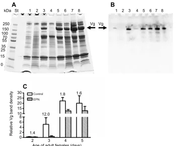
250
150 100 70 55
35 25
**
15
10
1.5
0
Control
Relative Vg band density EPN 0.1
0.2
St 1 2 3
Vg Vg
1 2 3
a
b
c
Fig. 2. Effect ofS. carpocapsaeinfection on vitellogenin protein expression.(A) SDS-PAGE of hemolymph proteins ofP. apterus: St, MW standards; 1, control 4 day old females (for Vg identification); 2, 8 day old males
1 day after control treatment; 3, 8 day old males 1 day afterS. carpocapsae
treatment. Vg bands are indicated by the arrow. (B) Western blotting of
hemolymph proteins ofP. apterus: lanes 1–3 as in A. Arrows indicate bands
showing a positive reaction to anti-Vg antibody; estimated MW: a, 180 kDa; b, 65 kDa; and c, 24 kDa. (C) Relative density (mean±s.d.) of Vg bands (optical density×area, quantified from corresponding gels that are not shown) in the
hemolymph of 8 day oldP. apterusmales 1 day afterS. carpocapsae(EPN) or
control treatment. **Statistically significant difference between infected and
control groups at the 1% level evaluated by Student’st-test (n=6–7). The
number above the bar represents the fold-difference of the relative Vg quantity
between the EPN and control group.
Journal
of
Experimental
[image:3.612.323.555.290.599.2](each consisting of 20 firebugs) for each experimental treatment, as well as for controls, were inspected 24 h post-treatment.
Similarly, the effect ofI. fumosoroseaon mortality of the firebug males and females was examined. The firebugs were injected with a dose of 30,000 blastospores per bug; controls were injected with Ringer saline only. Mortality was monitored 1–3 days post-infection.
Data presentation and statistical analysis
The results were plotted using the graphic software Prism (Graph Pad Software, v6.0, San Diego, CA, USA). The bar graphs represent means±s.d.; the number of replicates (n) is depicted in the figure legends. Statistical differences were evaluated by Student’st-test, two-way ANOVA and one-way ANOVA using Prism software as indicated in the figure legends.
RESULTS
Steinernema carpocapsaeinfection
The first series of experiments focused on measuringVgtranscript levels during nematobacterial infection of the fat body of male
P. apterus. Infection of males with the EPNS. carpocapsaeresulted
in a 7.7-fold increase of Vgtranscript level, 1 day post-infection (Fig. 1). Vg protein level in hemolymph was also increased 1.5-fold, as visualized by SDS-PAGE (Fig. 2A,C). These observations were verified using western blotting with anti-Vg antibody (Fig. 2B). Interestingly, the anti-Vg antibody used for western blots positively recognized not only the main Vg band (about 180 kDa) but also two smaller bands of 65 and 24 kDa, which might be degradation products of EPN toxic actions in the body ofP. apterusmales.
As expected,Vggene expression level was substantially higher in the fat bodies of females than in those of males (Fig. 3).Vg
transcript levels in females continuously increased during the first 5 days of development (Fig. 3), i.e. the critical period for egg formation. Application of EPN radically reduced the level of Vg transcripts in 2–5 day old females 1 day after infection (Fig. 3); the extent of the inhibition ranged from about 9-fold (in 4 day old females) to about 150-fold (in 3 day old females). A similar trend was observed with Vg protein levels in hemolymph, although the differences were not so profound (Fig. 4): a maximal 12-fold change (in 3 day old females) was recorded (Fig. 4C). These results were confirmed by immunoanalysis using western blotting (Fig. 4B). In contrast to the reaction observed in males, the antibody recognized only the 180 kDa Vg band in female hemolymph. This suggests that male and female bodies reacted differently to EPN infection.
Steinernema carpocapsae elicited mortality in the treated
firebugs (Fig. 5A). The mortality rate was about 2.9 times lower in females than in males, 1 day after treatment. In corresponding controls, no mortality was recorded (data not shown), perhaps as a
0 16.9
2 3
Age of adult females (days) 4 5 5
10 40 60 80 700
800 Control
EPN
94.3
151.2 8.9
Relative
Vg
[image:4.612.90.261.60.181.2]transcript level
Fig. 3.Vgtranscript expression in females followingS. carpocapsae infection.RelativeVgtranscript level (mean±s.d.) in the fat body of 2, 3, 4 and
5 day oldP. apterusfemales 1 day afterS. carpocapsae(EPN) and control
treatment. Two-way ANOVA showed a statistically significant difference at the
0.1% level between the EPN groups and controls (n=3). Numbers above the
bars represent fold-differences ofVgtranscript levels between the EPN and
corresponding control group.
kDa
A
B
C
St 1 2 3 4 5 6 7 8 1 2 3 4 5 6 7 8
Vg Vg 250
150 100 70 55 35 25 15
10
0 2 4 6 8 10 15 20 25
30 Control 1.8 1.6
12.0
1.4
2 3
Age of adult females (days)
Relative Vg band density
4 5
EPN
Fig. 4. Vg protein expression in females followingS. carpocapsaeinfection.(A)
SDS-PAGE of hemolymph proteins ofP. apterus
females: St, MW standards; 1 and 2, control (1) andS. carpocapsae-treated (2) 2 day old
females; 3 and 4, control (3) andS. carpocapsae
-treated (4) 3 day old females; 5 and 6, control (5) andS. carpocapsae-treated (6) 4 day old
females; 7 and 8, control (7) andS. carpocapsae
-treated (8) 5 day old females. In each case, measurements were obtained 1 day after
S. carpocapsaeor control treatment. Vg bands are indicated by the arrow. (B) Western blotting of
hemolymph proteins ofP. apterusfemales: lanes
1–8 as in A. Arrow indicates bands showing a
positive reaction to anti-Vg antibody. (C) Relative density (mean±s.d.) of Vg bands (optical density×area, quantified from corresponding
gels that are not shown) in hemolymph of 2–5 day
oldP. apterusfemales 1 day after
S. carpocapsae(EPN) or control treatment. Two-way ANOVA showed a statistically significant difference at the 0.1% level between the EPN
groups and controls (n=6–8). The numbers
above the bars represent the fold-difference of the relative Vg quantity between the EPN and corresponding control group.
Journal
of
Experimental
[image:4.612.51.404.444.733.2]result of the higher level of Vg in the female body. Further, using the disc diffusion method, we tested the antimicrobial effect of Vg on growth of the bacterium Xenorhabdusspp. isolated from
S. carpocapsae body (Fig. 5B). We found that Vg inhibited
Xenorhabdus growth; the inhibition was 2.5-fold more effective
than in controls.
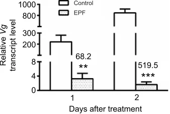
Isaria fumosoroseainfection
Because of the slower development of the EPFI. fumosoroseain the fire bug body, Vg levels were monitored daily for 2–3 days after infection in the tested individuals. In the male fat body,Vgtranscript level nominally increased 1.6-fold the day after infection, although this change did not reach the level of statistical significance (Fig. 6). However, on day 2 after infection, the 2-fold increase in Vg
transcript level was significant. Nevertheless, the infection had no impact on Vg level in hemolymph (Fig. 7), as SDS-PAGE and western blotting results were not significantly different between EPF infection and control groups.
Females exhibited a more pronounced response to EPF infection. Strong reductions of Vg transcript level in the fat body were recorded on both day 1 and day 2 after infection (Fig. 8). Similar significant changes in Vg protein level were detected in the hemolymph during the whole monitored period according to SDS-PAGE analysis (Fig. 9A,C). Identical results were also obtained using immunoblotting (Fig. 9B). Surprisingly, no differences between male and female firebugs were recorded when mortality was monitored for 1–3 days after treatment with I. fumosorosea
(Fig. 10).
DISCUSSION
Pathogenic organisms elicit severe stress in the host body, which results in the disruption of functional homeostasis (Ivanović and Jankovic-Hladni, 1991) and activation of characteristic defense response to eliminate or at least reduce the impact of the stress on the organism. This response occurs at both organismal and cellular levels (Hightower, 1991), and may include both humoral and cellular defenses. The humoral response includes production of various compounds, such as eicosanoids, phenoloxidases, proteinases, proteinase inhibitors and a wide selection of antimicrobial peptides and proteins (Jiang, 2008; Beckage, 2008). The list of the protective compounds also includes Vgs, whose involvement in insect defense systems has recently been described in several insect species (Havukainen et al., 2013; Singh et al., 2013; Salmela et al., 2015; Salmela and Sundstrom, 2017; Park et al., 2018). We have found in this study that in the firebugP. apterus, Vg is probably also involved in the defense reaction against the entomopathogenic nematode
S. carpocapsaeand, partially, against the fungusI. fumosorosea.
To invade their hosts, EPNs usually use oral and anal openings or spiracles. To speed the infection up, we injectedS. carpocapsaeinto the hemocoel. Once the nematodes are inside the insect body, the infection develops quite quickly; therefore, we monitored the effect of the EPN at just 1 day post-infection: 1 day later, mortality reached almost 100% (data not shown). During development in the host body, EPNs produce various venoms and toxins, which are generated by the nematodes themselves and also by symbiotic bacteria (Simões et al., 2000; Duchaud et al., 2003). In the first step of the nematobacterial infection, the toxins protect the EPNs against the defense system of their insect host, and afterwards, they kill the host and use its organs as a source of nutrients for growth and development. The insects protect themselves by clotting cascades, and production of reactive oxygen species and other fast-reacting immune factors (Wang et al., 2010; Hyršl et al., 2011; Arefin et al., 2014; Kodrík et al., 2015).
We found in this study that the nematobacterial complex of
S. carpocapsaeandXenorhabdusspp. affected Vg characteristics in
both male and femaleP. apterus. In males, a significant stimulatory effect of the infection was noted on bothVgtranscript level in the fat body and Vg protein level in the hemolymph. To the best of our knowledge, this is the first report of a stimulatory effect of the
0
Males
Disc area
Area of inhibitory zone (mm
2)
% Mortality
Females 2.9
**
*
2.5
0
Buf fer
Gel eluate Vg eluate
20 40 60 80 100 10 20 30
A
[image:5.612.95.257.58.337.2]B
Fig. 5.Steinernema carpocapsae-induced mortality and Vg antimicrobial effect.(A) Effect ofS. carpocapsaeon mortality ofP. apterusmales (7 days old) and females (4 days old), 1 day after treatment (means±s.d.); there was no
mortality in controls. (B) Inhibitory effect of Vg on the growth ofXenorhabdus
spp. bacteria tested by the disc diffusion method (means±s.d.). Results evaluated 1 day after Vg application are expressed as the area of the inhibitory zone (for details, see Materials and Methods). Asterisks indicate statistically
significant differences between the relevant groups at the 5% level (*A,n=5
groups with 20 adults per each) and 1% level (**B,n=5) evaluated by Student’s
t-test. The numbers above the bars represent the fold-difference between the
relevant groups.
0
Days after treatment
Relative
Vg
transcript level
2 1
2.0
Control EPF
**
2 4 6 8 10
Fig. 6. Effect ofIsaria fumosoroseainfection onVgtranscript expression in males.RelativeVgtranscript level (mean±s.d.) in the fat body of 8 and 9 day
old maleP. apterus, 1 and 2 days, respectively, afterI. fumosorosea(EPF) or
control treatment. **Statistically significant difference between infected and
control males at the 1% level evaluated by Student’st-test (n=3). The number
above the bar represents the fold-difference of theVgtranscript levels between
the EPF and control group.
Journal
of
Experimental
[image:5.612.95.254.554.663.2]nematobacterial complex on Vg production in infected insects. However, at this point, we cannot state, based on our results, whether the effect was primarily elicited by the nematode, its symbiotic bacteriaXenorhabdusspp., or the combined effect of the exposure to both. However, it is known that both organisms are insect pathogens (Herbert and Goodrich-Blair, 2007; Waterfield et al., 2009). Further, it seems that interaction of male Vg with EPN results not only in the stimulation of Vg synthesis but also in the degradation of Vg molecules. Immunoblotting results clearly showed at least two products that positively reacted with the anti-Vg antibody, with molecular weights (24 and 65 kDa) well below the molecular mass of 180 kDa of the intact protein. All these results suggest an active role of Vg against EPN infection in the male body.
Vg plays a key role as an irreplaceable component of yolk in eggs developing in the female body; however, the ( perhaps secondary) role of Vg in immunity seems to be important as well. Evidence for the presence of Vg in the male body has been rather scarce, but Vgs have been identified in P. apterus (Němec et al., 1993), Apis
mellifera (Villar and Grozinger, 2017) and Bombus terrestris
(Jedlička et al., 2016) males. Nevertheless, a comprehensive understanding of the role of Vg in insect males is missing.
Furthermore, the multi-faceted role of Vg in the female body is apparently more complicated: EPN infection significantly decreased both VgmRNA and Vg protein levels. Although it is surprising that EPN infection caused opposite effects in male and femaleP. apterus, this finding is easily explained: one can speculate that a Vg level sufficient for effective defense against pathogens might be much lower than that required for nutritional supply of developing eggs. Thus, during the infection, the female body simply shuts down less important processes to save energy for more significant activities. This trade-off strategy is not so exceptional in insects facing various stressful situations. For example, the resistance of females of the corn earwormHelicoverpa armigera
againstBacillus thuringiensistoxin Cry1Ac was accompanied by the inhibition of reproduction caused by a decrease in Vg gene expression (Zhang et al., 2014, 2015). Similarly, in females of the rice stem borerChilo suppessalis, application of sublethal doses of the insecticide chlorantraniliprole reducedVg mRNA expression (Huang et al., 2016). Additionally, adipokinetic hormone, responsible for energy mobilization during increased energy consumption, suppresses less important processes when the organism is under stress and, in certain conditions, even draws on the mobilized energy (Kodrík, 2008). Moreover, in Locusta
migratoria, adipokinetic hormone inhibits Vg production at the
end of the female reproductive cycle (Moshitzky and Applebaum,
kDa
A
B
C
St 1
10
Control
1 0.15
0.10
0.05
0
3 2
Days after treatment
Relative Vg band density
EPF
15 25 35 55 70 100 150 250
2 3 4 5 6 7
Vg Vg
[image:6.612.47.428.56.363.2]1 2 3 4 5 6 7
Fig. 7. Effect ofI. fumosorosea infection on Vg protein expression in males.(A) SDS-PAGE of hemolymph
proteins ofP. apterus: St, MW
standards; 1, control 4 day old females (for Vg identification); 2 and 3, control (2) andI. fumosorosea-infected (3) 8 day old males 1 day after treatment; 4 and 5,
control (4) andI. fumosorosea-treated
(5) 9 day old males 2 days after treatment; 6 and 7, control (6) and
I. fumosorosea-treated (7) 10 day old males 3 days after treatment. Vg bands are indicated by the arrow. (B) Western blotting of hemolymph proteins of
P. apterus: lanes 1–7 as in A. Arrow indicates bands showing a positive reaction to anti-Vg-antibody. (C) Relative density (mean±s.d.) of Vg bands (optical density×area, quantified from corresponding gels that are not shown) in hemolymph of 8, 9 and 10 day oldP. apterusmales, 1, 2 and 3 days,
respectively, afterI. fumosorosea(EPF)
or control treatment. No statistically significant difference between infected and control groups evaluated by two-way ANOVA at 5% was recorded
(n=6–8).
1 2
Days after treatment
Relative
Vg
transcript level
0 4 8 200 300 800
1000 Control
EPF
519.5 68.2
***
**
Fig. 8. Effect ofI. fumosoroseainfection onVgtranscript expression in females.RelativeVgtranscript level (mean±s.d.) in the fat body of 2 and 3 day oldP. apterusfemales, 1 and 2 days, respectively, afterI. fumosorosea(EPF) or control treatment. Asterisks indicate statistically significant differences between infected and control females at the 1% level (**) and 0.1% level (***)
evaluated by Student’st-test (n=3). The numbers above the bars represent the
fold-difference ofVgtranscript levels between the EPF group and control.
Journal
of
Experimental
[image:6.612.91.260.552.665.2]1990). This process is independent of nutrient mobilization because Vg inhibition occurs at hormone titers about one-tenth those necessary for nutrient mobilization from the fat body: thus, the two activities stimulated by adipokinetic hormone are not overlapping (Carlisle and Loughton, 1986). The mechanism of Vg function during infection is unclear, and perhaps different in males and females–for example, no Vg degradation products were observed by immunoblotting in female hemolymph during infection.
We have demonstrated a bactericidal effect of Vg on the bacterium Xenorhabdusspp. isolated from S. carpocapsae. This clearly suggests that Vg has a certain protective role against the nematobacterial complex, because Vg probably kills entomotoxic bacteria. It has not been established whether Vg affects EPNs; therefore, we cannot definitely exclude this. An antibacterial effect of Vg has already been described in several studies. Singh et al. (2013) showed that Vg of the silkworm Bombyx mori has
antibacterial activity against the gram-positive bacteriumBacillus
subtilis and the gram-negative bacterium Escherichia coli.
Furthermore, Vg ofApis cerana was active against E. coli, and also against the gram-positive bacterium B. thuringiensis (Park et al., 2018). In the latter example, Vg bound to the bacterial surface, inducing structural damage in the cell wall, which resulted in membrane disruption and permeabilization. All these data suggest that Vg is an antibacterial agent with a wide spectrum of action.
EPFs, such asI. fumosoroseaused in this study, usually start their infection by breaking the host cuticle and physically penetrating the host body, using various enzymes, such lipases, proteases, chitosanases and chitinases (Hajek and Leger, 1994; Ali et al., 2010), to dissolve tissues and organs, and the resulting matter is then utilized as a source of nutrition for EPF growth. InI. fumosorosea, the process is facilitated by the production of beauvericin, a toxic depsipeptide that kills the infected cell (Luangsa-Ard et al., 2009). Despite these effective mechanisms, the whole process of EPF infection is rather slow – to speed it up, we used injection of blastospores, in a similar approach to the injection of EPNs (see above). Nevertheless, the EPF infection developed more slowly than the EPN one. However, this circumstance enabled monitoring the EPF effect for 2–3 days after injection. The response of theP. apterus
male body to EPF infection differed from that to EPN infection. The first significant upregulation ofVgtranscription in the fat body was observed 2 days post-infection; however, surprisingly, VgmRNA levels in the hemolymph did not show the same trend, as a similarVg
mRNA expression was recorded in infected and control males. Additionally, the pattern of Vg protein level changes in male hemolymph, as determined by immunoblotting, was apparently different from the results obtained after EPN infection (compare Figs 2B and 7B). This suggests different responses of the male body to these infections: the involvement of Vg in the defense reaction is apparently less intensive in the case of EPF infection. It remains to be
10
0 0.2 0.4 0.6 5 10 15 20 25
Control EPF
1 2
2.5
16.8
3.2
Days after treatment
Vg band density
3 15
25 35 55 70 100 150 250 kDa
A
B
C
St 1 2 3 4 5 6
Vg Vg
[image:7.612.51.391.58.362.2]1 2 3 4 5 6
Fig. 9. Effect ofI. fumosoroseainfection on Vg protein expression in females.(A) SDS-PAGE
of hemolymph proteins ofP. apterusfemales: St,
MW standards; 1 and 2, control (1) and
I. fumosorosea-infected (2) 2 day old females 1 day after treatment; 3 and 4, control (3) and
I. fumosorosea-infected (4) 3 day old females 2 days after treatment; 5 and 6, control (5) and
I. fumosorosea-infected (6) 4 day old females 3 days after treatment. Vg bands are indicated by the arrow. (B) Western blotting of hemolymph
proteins ofP. apterusfemales: lanes 1–6 as in A.
Arrow indicates bands showing a positive reaction to anti-Vg-antibody. (C) Relative density (mean± s.d.) of Vg bands (optical density×area, quantified from corresponding gels that are not shown) in
hemolymph of 2, 3 and 4 day oldP. apterus
females, 1, 2 and 3 days, respectively, after
I. fumosorosea(EPF) or control treatment. Two-way ANOVA test showed a statistically significant difference at the 0.1% level between the EPN
groups and controls (n=6–10). The numbers
above the bars represent the fold-difference of the relative Vg quantity between the EPN and corresponding control group.
1 0 20 40 60 80
Males
Females
Days after treatment
% Mortality
2 3
Fig. 10.Isaria fumosorosea-induced mortality.Effect ofI. fumosoroseaon
mortality ofP. apterusmales (7 days old) and females (4 days old), 1–3 days
after infection (means±s.d.); there was no mortality in controls. No statistically significant difference between males and females evaluated by two-way
ANOVA at the 5% level was recorded (n=11 groups with 20 adults per each).
Journal
of
Experimental
determined whether any other defense systems are involved in responses to infections with EPN and EPF. In contrast, the response of firebug females to EPF infection was quite similar to that elicited by EPN: both mRNA and protein Vg levels were significantly down-regulated. A similar reaction was recorded in the whiteflyBemisia
tabaci, in which bothI. fumosoroseamycelium (in vivo) and fungal
extracts (in vitro) showed a decrease in Vg level, and corresponding damage of the ovaries (Gao et al., 2017). Possible reasons for this phenomenon were discussed above. In addition, an active involvement of Vg in the defense reaction ofA. melliferaagainst the entomopathogenic fungus B. bassiana was recently described by Park et al. (2018), who demonstrated that Vg behaves as a typical anti-microbial peptide.Apis melliferaVg bound toB. bassianacells and induced structural damage of the cell wall leading to anti-microbial activity against the fungus. Interestingly, Vg is also present in the venom of the honey bee and some other hymenopterans (Blank et al., 2013; Park et al., 2018), where it probably serves as an allergen that intensifies venom efficacy by causing an allergic reaction in the stung tissue. Vg is also expressed in the brain of several hymenopteran species, where it controls various processes, including aging (Munch et al., 2015; Lockett et al., 2016; Gospocic et al., 2017).
In conclusion, our findings expand the growing list of Vg functions in insects, which are more complex than previously thought. Clearly, Vg proteins play an important role in the defense against various types of infection, including those caused by EPN and EPF. Furthermore, Vg transcription and protein synthesis are modulated depending on the type of infection and the sex of the infected insect. Vg acts directly againstXenorhabdusspp. bacteria isolated from the entomopathogenic complex of S. carpocapsae. Other mechanisms of Vg activities are not known at present, but they might differ depending on the type of infection, as documented by different Vg characteristics (in males) and a different mortality response (in both sexes) to the two studied pathogens.
Acknowledgements
The authors thank Mrs H. Štěrbováand Miss D. Hlávkováfor their technical assistance. Editage Author Services checked the English grammar in the manuscript prior to submission.
Competing interests
The authors declare no competing or financial interests.
Author contributions
Conceptualization: D.K.; Methodology: E.I., U.K.G., V.K., P.J.; Validation: D.K., R.Č.F.; Formal analysis: D.K., R.Č.F., A.B.; Investigation: D.K., R.Č.F., A.B.; Writing -original draft: D.K.; Writing - review & editing: D.K., R.Č.F.; Supervision: D.K.; Project administration: D.K.; Funding acquisition: D.K.
Funding
This study was supported by grant no. 17-03253S (D.K.) from the Czech Science Foundation (GrantováAgentura ČeskéRepubliky), and by project RVO 60077344 of the Institute of Entomology. A.B. acknowledges Fellowship no. L200961701 from the Program of Support of Promising Human Resources, awarded by The Czech Academy of Sciences (Akademie Věd ČeskéRepubliky) and the European Union, European Structural and Investing Funds Operational Programme Research, Development and Education (CZ.02.2.69/0.0/0.0/18_070/0008772).
Supplementary information
Supplementary information available online at
http://jeb.biologists.org/lookup/doi/10.1242/jeb.202853.supplemental
References
Ali, S., Huang, Z. and Ren, S.(2010). Production of cuticle degrading enzymes by Isaria fumosorosea and their evaluation as a biocontrol agent against diamondback moth.J. Pest. Sci.83, 361-370. doi:10.1007/s10340-010-0305-6 Arefin, B., Kučerová, L., Dobeš, P., Markus, R., Strnad, H., Wang, Z., Hyršl, P.,
Zurovec, M. and Theopold, U.(2014). Genome-wide transcriptional analysis of
Drosophilalarvae infected by entomopathogenic nematodes shows involvement of complement, recognition and extracellular matrix proteins.J. Inn. Immun.6, 192-204. doi:10.1159/000353734
Beckage, N. E.(2008).Insect Immunology. San Diego, USA: Academic Press Elsevier.
Beckman, K. B. and Ames, B. N.(1998). The free radical theory of aging matures. Physiol. Rev.78, 547-581. doi:10.1152/physrev.1998.78.2.547
Bednářová, A., Kodrık, D. and Krishnan, N.́ (2013). Adipokinetic hormone exerts its anti-oxidative effects using a conserved signal- transduction mechanism involving both PKC and cAMP by mobilizing extra- and intracellular Ca2+stores.
Comp. Biochem. Physiol. C158, 142-149. doi:10.1016/j.cbpc.2013.07.002 BenVau, L. R. and Nieh, J. C.(2017). Larval honey bees infected withNosema
ceranaehave increased vitellogenin titers as young adults.Sci. Rep.7, 14144. doi:10.1038/s41598-017-14702-4
Blank, S., Seismann, H., McIntyre, M., Ollert, M., Wolf, S., Bantleon, F. I. and Spillner, E.(2013). Vitellogenins are new high molecular weight components and allergens (Api m 12 and Ves v. 6) ofApis melliferaandVespula vulgarisvenom. PLoS ONE8, e62009. doi:10.1371/journal.pone.0062009
Carlisle, J. and Loughton, B. G.(1986). The inhibition of protein synthesis in Locusta migratoriaby adipokinetic hormone. J. Insect Physiol.32, 573-578. doi:10.1016/0022-1910(86)90074-0
Chapman, R. F.(1998).The Insects, Structure and Function. Cambridge, UK: Cambridge University Press.
Duchaud, E., Rusniok, C., Frangeul, L., Buchrieser, C., Givaudan, A., Taourit, S., Bocs, S., Boursaux-Eude, C., Chandler, M., Charles, J. F. et al.(2003). The genome sequence of the entomopathogenic bacterium Photorhabdus luminescens.Nat. Biotechnol.21, 1307-1313. doi:10.1038/nbt886
Ehlers, R. U.(2003). Entomopathogenic nematodes in the European biocontrol market.Commun. Agric. Appl. Biol. Sci.68, 3-16.
Fridovich, I.(1978). The biology of oxygen radicals.Science201, 875-880. doi:10. 1126/science.210504
Gao, T. N., Wang, Z. L., Huang, Y., Keyhani, N. O. Huang, Z.(2017). Lack of resistance development inBemisia tabacitoIsaria fumosoroseaafter multiple generations of selection.Sci. Rep.7, 42727. doi:10.1038/srep42727
Gospocic, J., Shields, E. J., Glastad, K. M., Lin, Y., Penick, C. C., Hua Yan, H., Mikheyev, A. S., Linksvayer, T. A., Garcia, B. A., Berger, S. L. et al.(2017). The neuropeptide corazonin controls social behavior and caste identity in ants.Cell 170, 748-759. doi:10.1016/j.cell.2017.07.014
Hajek, A. E. and Leger, R. J. S.(1994). Interactions between fungal pathogens and insect hosts. InAnnual Review of Entomology(ed. T.E. Mittler, F.J. Radovsky and V.H. Resh), pp. 293-322. Palo Alto, USA: Annual Reviews, Inc.
Harlow, E. and Lane, D.(1988).Antibodies. A Laboratory Manual. Cold Spring Harbor, USA: Cold Spring Harbor Laboratory.
Havukainen, H., Münch, D., Baumann, A., Zhong, S., Halskau, Ø., Krogsgaard, M. and Amdam, G. V.(2013). Vitellogenin recognizes cell damage through membrane binding and shields living cells from reactive oxygen species.J. Biol. Chem.288, 28369-28381. doi:10.1074/jbc.M113.465021
Herbert, E. E. and Goodrich-Blair, H.(2007). Friend and foe: the two faces of Xenorhabdus nematophila. Nat. Rev. Microbiol. 5, 634-646. doi:10.1038/ nrmicro1706
Hightower, L. E. (1991). Heat-shock, stress proteins, chaperones, and proteotoxicity.Cell66, 191-197. doi:10.1016/0092-8674(91)90611-2
Huang, L., Lu, M., Han, G., Dua, Z. and Wang, K.(2016). Sublethal effects of chlorantraniliprole on development, reproduction and vitellogenin gene (CsVg) expression in the rice stem borer,Chilo suppressalis.Pest Manag. Sci. 72, 2280-2286. doi:10.1002/ps.4271
Hyršl, P., Dobeš, P., Wang, Z., Hauling, T., Wilhelmsson, C. and Theopold, U. (2011). Clotting factors and eicosanoids protect against nematode infections. J. Innate Immun.2, 65-70. doi:10.1159/000320634
Ibrahim, E., Hejnı́ková, M., Shaik, H. A., Doležel, D. and Kodrık, D.́ (2017). Adipokinetic hormone activities in insect body infected by entomopathogenic nematode.J. Insect Physiol.98, 347-355. doi:10.1016/j.jinsphys.2017.02.009 Inman, F. L., Singh, S. and Holmes, L. D.(2012). Mass production of the beneficial
nematodeHeterorhabditis bacteriophoraand its bacterial symbiontPhotorhabdus luminescens.Indian J. Microbiol.52, 316-324. doi:10.1007/s12088-012-0270-2 Ivanović, J. and Jankovic-Hladni, M.(1991).Hormones and Metabolism in Insect
Stress. Boca Raton, USA: CRC Press.
Jedlička, P., Ernst, U. R., Votavová, A., Hanus, R. and Valterová, I.(2016). Gene expression dynamics in major endocrine regulatory pathways along the transition from solitary to social life in a bumblebee,Bombus terrestris.Front. Physiol.7, 574. doi:10.3389/fphys.2016.00574
Jiang, H.(2008). The biochemical basis of antimicrobial responses inManduca sexta.Insect Sci.15, 53-66. doi:10.1111/j.1744-7917.2008.00187.x
Kodrık, D.́ (2008). Adipokinetic hormone functions that are not associated with insect flight. Physiol. Entomol. 33, 171-180. doi:10.1111/j.1365-3032.2008. 00625.x
Kodrık, D., Bedná ́řová, A., Zemanová, M. and Krishnan, N.(2015). Hormonal regulation of response to oxidative stress in insects - an update.Int. J. Mol. Sci.16,
25788-25816. doi:10.3390/ijms161025788
Journal
of
Experimental
Krishnan, N., Večeřa, J., Kodrı́k, D. and Sehnal, F.(2007). 20-hydroxyecdysone prevents oxidative stress damage in adult Pyrrhocoris apterus. Arch. Insect Biochem. Physiol.65, 114-124. doi:10.1002/arch.20182
Laemmli, U. K.(1970). Cleavage of structural proteins during the assembly of the head of bacteriophage T4.Nature227, 680-685. doi:10.1038/227680a0 Lockett, G. A., Almond, E. J., Huggins, T. J., Parker, J. D. and Bourke, A. F. G.
(2016). Gene expression differences in relation to age and social environment in queen and worker bumble bees.Exp. Gerontol.77, 52-61. doi:10.1016/j.exger. 2016.02.007
Luangsa-Ard, J. J., Berkaew, P., Ridkaew, R., Hywel-Jones, N. L. and Isaka, M. (2009). A beauvericin hot spot in the genusIsaria.Mycol. Res.113, 1389-1395. doi:10.1016/j.mycres.2009.08.017
Mahar, A. N., Munir, M., Elawad, S., Gowen, S. R. and Hague, N. G. M.(2005). Pathogenicity of bacterium, Xenorhabdus nematophila isolated from entomopathogenic nematode (Steinernema carpocapsae) and its secretion againstGalleria mellonellalarvae.J. Zhejiang Univ. Sci. B6, 457-463. doi:10. 1631/jzus.2005.B0457
Moshitzky, P. and Applebaum, S. W.(1990). The role of adipokinetic hormone in the control of vitellogenin in locusts.Insect Biochem.20, 319-323. doi:10.1016/ 0020-1790(90)90050-5
Munch, D., Ihle, K. E., Salmela, H. and Amdam, G. V.(2015). Vitellogenin in the honey bee brain: Atypical localization of a reproductive protein that promotes longevity.Exp. Gerontol.71, 103-108. doi:10.1016/j.exger.2015.08.001 Němec, V., Kodrı́k, D., Matolın, S. and Laufer, H.́ (1993). Juvenile hormone effects
of retinoic acid in insect metamorphosis, embryogenesis and reproduction. J. Insect Physiol.39, 1083-1093. doi:10.1016/0022-1910(93)90132-B Park, H. G., Lee, S. K., Kim, B. Y., Yoon, H. J., Choi, Y. S., Lee, K. Y., Wan, H., Li,
J. and Jin, B. R. (2018). Honeybee (Apis cerana) vitellogenin acts as antimicrobial and antioxidant agent in the body and venom. Dev. Comp. Immunol.85, 51-60. doi:10.1016/j.dci.2018.04.001
Sadd, B. M., Kleinlogel, Y., Schmid-Hempel, R. and Schmid-Hempel, P.(2005). Trans-generational immune priming in a social insect.Biol. Lett.1, 386-388. doi:10.1098/rsbl.2005.0369
Salmela, H. and Sundstrom, L.(2017). Vitellogenin in inflammation and immunity in social insects.Inflamm. Cell Signal.4, e1506. doi:10.14800/ics.1506 Salmela, H., Amdam, G. V. and Freitak, D.(2015). Transfer of immunity from
mother to offspring is mediated via egg-yolk protein vitellogenin.PLoS Pathog.11, e1005015. doi:10.1371/journal.ppat.1005015
Seehuus, S.-C., Norberg, K., Gimsa, U., Krekling, T. and Amdam, G. V.(2006). Reproductive protein protects functionally sterile honey bee workers from oxidative stress.Proc. Natl. Acad. Sci. USA103, 962-967. doi:10.1073/pnas. 0502681103
Simões, N., Caldas, C., Rosa, J. S., Bonifassi, E. and Laumond, C.(2000). Pathogenicity caused by high virulent and low virulent strains ofSteinernema
carpocapsaetoGalleria mellonella.J. Invertebr. Pathol.75, 47-54. doi:10.1006/ jipa.1999.4899
Singh, N. K., Pakkianathan, B. C., Kumar, M., Prasad, T., Kannan, M., König, S. and Krishnan, M.(2013). Vitellogenin from the silkworm,Bombyx mori: an effective anti-bacterial agent.PLoS ONE8, e73005. doi:10.1371/journal.pone. 0073005
Sinpoo, C., Paxton, R. J., Disayathanoowat, T., Krongdang, S. and Chantawannakul, P.(2018). Impact ofNosema ceranaeandNosema apison individual worker bees of the two host species (Apis ceranaandApis mellifera) and regulation of host immune response.J. Insect Physiol.105, 1-8. doi:10.1016/j. jinsphys.2017.12.010
Socha, R. and Kodrı́k, D. (1999). Differences in adipokinetic response of Pyrrhocoris apterus(Heteroptera) in relation to wing dimorphism and diapause. Physiol. Entomol.24, 278-284. doi:10.1046/j.1365-3032.1999.00143.x Socha, R., Šula, J., Kodrı́k, D. and Gelbič, I. (1991). Hormonal control of
vitellogenin synthesis inPyrrhocoris apterus.J. Insect Physiol.37, 805-816. doi:10.1016/0022-1910(91)90077-D
Stoscheck, C. M.(1990). Quantitation of proteins. InMethods in Enzymology(ed. M. P. Deutscher), pp. 50-68. London, UK: Academic Press.
Towbin, H., Staehelin, T. and Gordon, J.(1979). Electrophoretic transfer of protein from polyacrylamide gel to nitrocellulose. Proc. Natl. Acad. Sci. USA 76, 4350-4354. doi:10.1073/pnas.76.9.4350
Villar, G. and Grozinger, C.M. (2017). Primer effects of the honeybee, Apis mellifera, queen pheromone 9-ODA on drones.Anim. Behav. 127, 271-279. doi:10.1016/j.anbehav.2017.03.023
Wang, Z., Wilhelmsson, C., Hyršl, P., Loof, T. G., Dobeš, P., Klupp, M., Loseva, O., Mörgelin, M., Iklé, J., Cripps, R. M. et al.(2010). Pathogen entrapment by transglutaminase-A conserved early innate immune mechanism.PLoS. Pathog. 6, 1-9. doi:10.1371/journal.ppat.1000763
Waterfield, N. R., Ciche, T. and Clarke, D.(2009).Photorhabdusand a host of hosts.Annu. Rev. Microbiol.63, 557-574. doi:10.1146/annurev.micro.091208. 073507
Zhang, W. N., Xiao, H. J., Liang, G. M., Guo, Y. Y. and Wu, K. M.(2014). Tradeoff between reproduction and resistance evolution to Bt-toxin in Helicoverpa armigera: regulated by vitellogenin gene expression.Bull. Entomol. Res.104, 444-452. doi:10.1017/S0007485314000066
Zhang, W. N., Ma, L., Zhong, F., Wang, Y. N., Guo, Y. Y., Lu, Y. H. and Liang, G. M. (2015). Fitness costs of reproductive capacity and ovarian development in a Bt resistant strain of the cotton bollworm, Helicoverpa armigera (Hubner) (Lepidoptera: Noctuidae).Pest Manage. Sci.71, 870-877. doi:10.1002/ps.3900 Zimmermann, G.(2008). The entomopathogenic fungiIsaria farinosa(formerly
Paecilomyces farinosus) and theIsaria fumosoroseaspecies complex (formerly Paecilomyces fumosoroseus): biology, ecology and use in biological control. Biocontrol Sci. Technol.18, 865-901. doi:10.1080/09583150802471812