RIBONUCLEIC ACIDS: CAPILLARY
ELECTROPHORESIS
J. Skeidsvoll, University of Bergen, Bergen, Norway
Copyright^ 2000 Academic Press
Introduction
With the introduction of capillary electrophoresis (CE), a new generation of electrophoretic tech-niques has seen the light of day. The scientiRc literature today describes a large number of applica-tions of this powerful analytical technique in the analysis of nucleic acids. For nucleic acids, as for most other analytes, CE offers signiRcant advantages over many of the conventional elec-trophoretic techniques. In general, CE is character-ized by short analysis time, high resolution, accuracy and reproducibility, quantitative online detection and automation. The small sample volumes required and the extreme sensitivity CE offers, represent a large analytical potential for samples of biological origin. The fundamental analytical and operational parameters for the separation of nucleic acids by CE were identiRed around 1990. A decade later, CE is considered a fully developed technology for the anal-ysis of DNA. The rapid development of this applica-tion of CE seems to have been driven by the many practical applications of electrophoretic separation and detection of DNA in both basic and applied science.
The Rrst reported analysis of RNA by CE was published in 1993 by Reyes-Engelet al. and describes the separation and quantiRcation of a speciRc messenger RNA by capillary zone electrophoresis. To date, only a limited number of articles have been published which focus on the application of CE in the analysis of RNA. The reason for this is not obvious, considering the widespread use of conven-tional gel electrophoresis of RNA throughout the biomedical scientiRc Reld. The fact that RNA, in many respects, displays similar characteristics as DNA, should constitute the basis for signiRcant efforts in the development of RNA analyses based on CE. However, the scientiRc literature holds promise for a substantial increase in the use of CE in RNA analyses. The following sections intend to give a basic introduction to CE of RNA, with emphasis on important analytical and operational parameters in the analyses. Finally, examples from a diverse group of applications are presented.
Capillary Electrophoresis of RNA
In general, electrophoretic separation of RNA is based on the differences in electrophoretic mo-bilities of the analytes. As in conventional elec-trophoresis, the rate of migration of a RNA molecule in CE depends on the mass and the dimensions of the molecule, the charge carried, the applied current and the resistance of the medium. In an electric Reld, at moderate pH, negatively charged RNA migrates to-ward the anode. A number of parameters affect the separation of RNA in CE (see below). CE of RNA can be divided into two separate categories based on the principle by which the molecules are separated: capillary zone electrophoresis (CZE) and capillary gel electrophoresis (CGE). In CZE, the RNA molecules are mainly separated by their charge to mass ratio. From the fact that nucleic acids larger than a few nucleotide units have approximately iden-tical charge to mass ratio, CZE provides little or no separation power. Consequently, only single RNA species can be identiRed by this technique, unless multiple labelling is being used. In CGE, the RNA molecules are separated mainly by their molecular dimensions, i.e., the ability of the different analytes to migrate through a gel matrix. CGE is by far the most common technique for RNA ana-lyses. A description of CGE of RNA is given in the following section.
Capillary Gel Electrophoresis of RNA
Analytical parameters of signiRcance for the separation of DNA by CGE, including gel polymer concentration, electrical Reld strength and temperature, have been investigated and optimized for the analysis of RNA molecules ranging from oligomers (10 to 40 bases) to several kilobases (Figure 1).
RNA Migration in Capillary Gel Electrophoresis
Figure 1 Electropherogram of RNA molecular-mass marker. The sample was denatured, injected at 300 V cm\1for 10 s and
subjected to CGE at 200 V cm\1in 1;TBE/8 mol L\1urea
con-taining 0.3%HPMC. AU, arbitrary units. (Reprinted from Skeid-svoll J and Ueland PM (1996) Analysis of RNA by capillary electrophoresis.Electrophoresis 17: 1512}1517. Copyright 1996, with permission from Wiley-VCH Verlag GmbH.)
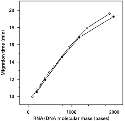
Figure 2 Comparison of migration of RNA and single-stranded DNA. A molecular-mass marker containing RNA and DNA com-ponents was denaturated by pre-incubation at 953C for 3 min in the presence of 80%formamide and subjected to electrophoresis in a separation buffer containing 8 mol L\1urea and 0.3%HPMC.
Migration time is plotted versus molecular mass for RNA (*) and DNA (䢇). (Reprinted from Skeidsvoll J and Ueland PM (1996) Analysis of RNA by capillary electrophoresis.Electrophoresis 17: 1512}1517. Copyright 1996, with permission form Wiley-VCH Verlag GmbH.)
appropriate pore size. Experiments have dem-onstrated that CGE of higher molecular mass RNA (in the range from 100 bases to more than 6 kb) to a large extent resembles CGE of single-stranded DNA. An interesting Rnding is that RNA
and single-stranded DNA of identical length display different migration when co-analysed under com-pletely denaturing conditions, DNA having a slightly higher migration rate than RNA (Figure 2). The shift in migration for DNA vs. RNA is found constant for molecules ranging from 100 to approximately 1000 bases. The phenomenon is explained by the higher charge to mass ratio of single-stranded DNA.
An inherent property of the (single-stranded) RNA molecule is the potential to form secondary structures or intramolecular and intermolecular hydrogen bonds. To what extent the reaction takes place is primarily a function of the RNA sequence. The pre-dictable determination of RNA molecular mass is essential in most RNA techniques based on elec-trophoretic separation. Consequently, in order to pre-vent unpredictable migration of RNA due to the formation of secondary structures, CE should be car-ried out under completely denaturing conditions. Such conditions can be accomplished through optim-ization of physical and/or chemical parameters. For example, heating the sample in the presence of a denaturant prior to electrophoresis and addition of a denaturant in the electrophoresis and separation buffers combined with high temperature during electrophoresis should have a strong denaturing effect. Denaturants are chemical compounds that disrupt hydrogen bonds. The most commonly used denaturant, urea, is often added to the separ-ation buffer in very high concentrsepar-ations (up to 8 mol L\1). Despite an extensive use of buffer additives, data from both conventional RNA gel elec-trophoresis and CE of RNA indicate that even the presence of 8 mol L\1 urea in the separation buf-fer is not sufRcient to completely disrupt intra-molecular or intermolecular hydrogen bonds. Addition of the stronger denaturant formamide in concentrations up to 30% (in addition to 3.5 mol L\1 urea) and performing CE at 603C has been necessary to disrupt extensive secondary structures in a ham-merhead ribozyme (37 nucleotides) and to provide appropriate separation from its substrate (17 nucleo-tides). In addition, a decrease in ionic (cationic) strength and an increase in pH are known to have a denaturing effect on RNA. Common problems related to inefRcient separation, detection and identiRcation of RNA in CE, probably result from incomplete denaturation of RNA.
Important Analytical and Operational
Parameters
[image:2.568.59.274.398.606.2]strength (E, V cm\1) and temperature (t,3C) have to be chosen carefully to optimize the separation of RNA. In most applications of CE in RNA analyses, the electroosmoticSow is eliminated through the use of surface-modiRed (coated) capillaries. This consider-ably simpliRes the experimental design and leaves the scientist with a limited number of variable analytical and operational parameters.
Buffer Composition
In general, all buffer systems that are used for CZE can also be used for CGE. The most common buffers are the Tris-borate buffers (i.e., TBE) with a pH range of 7.5}9.0. Buffer additives like methanol and acetonitrile are used in separation buf-fers optimized for low-molecular-mass RNA. Urea and formamide are mainly added as denaturants. Moreover, the addition of urea to the separation buffer has been found to increase the resolution of RNA (except for oligoribonucleotides less than 5 bases).
Gel-forming Polymers
A number of different gel-forming polymers have successfully been used in both DNA and RNA separations by CE. The separation matrices comprise both cross-linked gel polymers like polyacrylamide and noncross-linked gel polymers like linear polyac-rylamide and cellulose derivatives. Through the op-timization of composition and concentration, noncross-linked polymers have now taken over as the predominant separation matrices for most RNA ana-lyses. These materials have demonstrated signiRcant advantages over cross-linked polymers, including ease of preparation and use, physical stability and uncomplicated washing and reRlling procedures be-tween analyses. The resolving power of these gels mainly depends on the concentration of the dissolved polymer } dilute gels are used for high-molecular-mass RNA molecules and more concentrated gels for low-molecular-mass RNA.
A systematic study of the electrophoretic separ-ation of RNA at different concentrsepar-ations of a noncross-linked polymer gel demonstrated that high concentrations ('0.3%) hydroxypropylmethyl-cellulose (HPMC) were optimal for the separation of RNA less than 1000 bases and low concentrations were optimal for the separation of higher molecular-mass RNA. The results are consistent with data from the separation of DNA by CE.
A number of separation matrices, optimized for different ranges of RNA molecular mass, are commercially available. Additionally, matrices are available which contain denaturants.
Electrical Field Strength
ElectricalReld strength is recognized as an important operational parameter in CE of RNA. An in-crease in electrical Reld strength is found to result in a logarithmic decline in migration times. EfR -ciency,N (number of theoretical plates) and resolu-tion,Rs, are found to have a more complex relation to the electrical Reld strength, although a clear tendency towards a decline in both parameters with increased electrical Reld strength has been demonstrated. In general, low electrical Reld strengths are preferable for the optimal separation of RNA molecules larger than 100 bases. With the increase in electrical Reld strength, an increased current will result in the production of heat (Joule heating), which, if excessive, adversely affects the separation by causing broadening of the migrating zones.
Temperature
Temperature, an important analytical and opera-tional parameter, inSuences both total analysis time and system efRciency. The effect is mainly mediated by a decrease in the separation buf-fer viscosity. A linear decrease in migration time for RNA molecules ranging from 200 to 2000 bases has been observed for temperatures ranging from 20 to 503C. The separation efRciency and resolution were found essentially constant over the temperature range. In addition, temperature is a parameter of signiRcant importance in the CE of RNA due to its denaturing effect on intra- and intermolecular hydrogen bonds.
Quantitative Aspects
For a general description of the quantitative aspects of injection in CE, see ‘DNA: Capillary Electrophor-esis’. Electrokinetic injection is the most common injection mode for RNA in CE. In order to obtain quantitative data, an external reference should be added to or co-injected with the RNA sample. Ideally, the external reference should resemble the sample of interest, but be readily identiRable. Hydrodynamic injection is often used in experiments for the deter-mination of reaction kinetics or in studies of enzy-matic activity. Hydrodynamic injection provides representative samples for analysis.
Figure 3 CGE analysis of a mixture of 12 oligoribonucleotides from 4 to 18 units under nondenaturing conditions. (Reprinted from Kolesar JM, Allen PG and Doran CM (1997) Direct quantification of HIV-1 RNA by capillary electrophoresis with laser-induced fluorescence.Electrophoresis 697: 189}194. Copyright 1997, with permission from Elsevier Science.)
and sensitivity required for a number of analyses where the concentration of analytes is low. However, this detection principle normally requires the co-valent attachment of Suorophores to target mole-cule(s) orSuorogenic buffer additives.
Applications
The application of CE to RNA includes a diverse group of analyses, which often includes one or a com-bination of the following elements:
E Characterization of RNA molecular dimensions (mass or spatial structure).
E Characterization of RNA sequence. E Characterization of RNA reaction kinetics. E Characterization of RNA-binding constants.
An example of a group of CE-based RNA analyses that combines more than one of these elements is the hybridization techniques, which both rely on molecular mass determination and sequence-speciRc detection of the RNA of interest. In the applica-tions described, RNA samples originate either from chemical synthesis (oligoribonucleotides) or are ex-tracted from biological material. The last group comprise RNA of eukaryotic, prokaryotic and viral origin.
Characterization of RNA Molecular Dimensions (Mass or Spatial Structure)
Capillary electrophoresis analysis of synthetic short-chain oligoribonucletides(Figure 3) Thirty synthetic oligoribonucleotides, ranging from 3 to 18 nucleotides were analysed by CE in a nondenaturing noncross-linked gel polymer. An equation was developed, based on the experimental data which, under Rxed condi-tions, accounts for the inSuence of charge to mass ratio (i.e., net charge and base composition) on migration time. High resolution (1 nucleotide unit) was
obtained for homologous series of oligoribonucleo-tides, and, to some extent, for groups of oligoribonuc-leotides of identical length, but different sequence.
CGE is often used to determine the quality of chemically synthesized oligoribonucleotides and can be used in conjunction with HPLC to develop an effective method for the puriRcation of crude oligonucleotide solutions.
Low-molecular-mass RNAVngerprinting of bacteria by capillary electrophoresis RNA proRling provides a direct genotypicRngerprint technique for the identi-Rcation and classiRcation of bacteria by generating an electropherogram including three groups of mole-cules of taxonomic signiRcance, small tRNAs, large tRNAs, and 5S rRNA (ranging from 70 to 135 nu-cleotides). The technique is of particular importance for molecular ecology and taxonomic studies, and can also be applied directly to analyses of environ-mental samples. CGE using both noncross-linked polymer gels (HPMC) and cross-linked polyacrylam-ide gels have been investigated and optimized for their applicability in the separation of this class of RNA molecules. Good resolution was obtained only for small tRNAs up to approximately 80 nucleotides using cross-linked gels, larger tRNAs and 5S rRNA could not be resolved with this experimental set-up. The use of noncross-linked polymer gels resolved tRNAs and 5S rRNA under nondenaturing condi-tions, even when they possessed only different secondary structure or small differences in length (1}5 nucleotides). CE using HPMC in the separation buffer resulted in both good peak resolution and reproducibility and was suitable for routine R nger-printing of bacterial low-molecular-mass RNA.
cellular uptake and degradation of aSuorescein label-led chemically stabilized ribozyme (37-mer). After internalization by transfection or uptake of free ribozyme, electrophoretic peaks of intact ribozyme and different degradation products were easily resolved and the amount of intracellular intact ribozyme quantiRed. Using laser-induced S uores-cence for detection, the method offered extreme sensitivity with estimated limit of detection: 10 and 200 pmol L\1 ribozyme from cell extracts and cell medium, respectively.
A third example include the direct quantiRcation (by UV absorbance measurements) of HIV-1 RNA in human plasma by CZE.
Characterization of RNA Sequence
An important and diverse group of analytical RNA techniques is based on sequence-speciRc hybridiza-tion between two single-stranded nucleic acids and the electrophoretic separation, detection and quantiRcation of the intermolecular reaction product (hybrid). Consequently, the analyses involves charac-terization of RNA in two dimensions, size and se-quence. The Northern (RNA) blotting technique, the nuclease- (S1 or RNase) protection assays and other RNA-hybridization techniques play an important role in the qualitative and quantitative analysis of all classes of RNA in biological systems. The techniques often involve use of radioisotope labels in detection. CE-based hybridization analyses of RNA has been successfully demonstrated for a number of applica-tions. In general, the hybridization reactions are car-ried out pre-column (in solution) and the separation and detection of the hybrids on-column. It is demand-ing to transfer the conventional hybridization tech-niques to the capillary format and important challenges are related to the development of selective and compatible conditions for both the pre-column and on-column elements of the analyses. Addition-ally, the low sample volumes injected in CE represent signiRcant analytical and instrumental challenges.
Direct quantiVcation of a speciVc messenger RNA by capillary zone electrophoresis Total RNA was isolated from whole blood and hybridized with a biotinylated oligonucleotide speciRc for a receptor mRNA (angiotensin II). The hybrid wasRrst captured on streptavidin-conjugated magnetic beads, then eluted and Rnally quantiRed by UV absorbance in CZE. Using this procedure, quantiRcation of the ex-pression of low expressed genes is easy and fast and subject to two limiting factors: the speciRcity of the capturing oligonucleotide or probe selected and the amount of total RNA. The procedure represents an nonradioactive alternative to conventional RNA
ana-lyses like Northern blotting, RT-PCR or the nuclease-(S1 or RNase) protection assays.
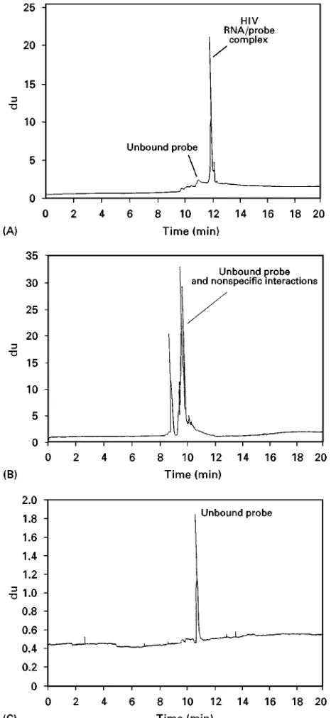
Direct quantiVcation of HIV-1 RNA by capillary elec-trophoresis with laser-inducedWuorescence (LIF) de-tection (Figure 4) A hybridization method using a HIV-speciRc probe with analysis by CE}LIF was de-veloped. Plasma samples from HIV-seropositive pa-tients were lysed to obtain RNA, hybridized with aSuorescein-labelled HIV-speciRc DNA probe, diges-ted by a speciRc RNase to remove nonhybridized RNA and analysed by CE-LIF in presence of the Suorescent intercalator thiazole orange (TO). 19 fg (corresponding to 1710 copies per mL of starting plasma) of HIV RNA was quantitatively detected. The technique, analogous to the conventional RNase protection assay, takes advantage of signal ampliR ca-tion by using the RNA-bindingSuorescent intercala-tor TO. Calibration is done through the analysis of a Suorescein-labelled RNA marker. The actual ap-proach appears to be an efRcient, sensitive and reliable method to speciRcally and quantitatively ana-lyse RNA from a variety of sources.
Detection of oligonucleotide N3}P5 phosphor-amidate/RNA duplexes with capillary gel electro-phoresis The DNA analogues N3}P5 phosphoramidates (3-phosphoramidates) has dem-onstrated favourable properties as hybridization probes, including high melting temperature of du-plexes with RNA and high reaction rate at low ionic strengths. The RNA hybridization technique takes advantage of the 3-phosphoramidate oligomer prop-erties as hybridization probes through duplex forma-tion with short complementary strands of RNA of identical length (9 nucleotides). Hybrids were found to have unique relative mobilities in CGE, compared to the reactants. The ability of CGE to detect the presence of, and to discriminate between, perfect du-plexes and dudu-plexes that contained a base mismatch were demonstrated under routine electrophoretic running conditions. In conclusion, the study indicates that 3-phosphoramidate oligonucleotides may have application in nucleic acid based diagnostics.
Characterization of RNA Reaction Kinetics
Figure 4 Electropherogram from a hybridization experiment. RNA samples obtained from a HIV-seropositive patient and a seronegative volunteer were hybridized with a HIV-specific probe and analysed by CGE: (A) HIV RNA/probe complex (HIV-positive patient); (B) seronegative volunteer; (C) negative control containing all reaction components except RNA. (Reprinted from Kolesar JM, Allen PG and Doran CM (1997) Direct quantification of HIV-1 RNA by capillary electrophoresis with laser-induced fluorescence,Journal of Chromatography B 697: 189}194. Copy-right 1997, with permission form Elsevier Science.)
Figure 5 Typical electropherograms demonstrating different stages in a ribozyme-mediated catalytic breakdown of a RNA oligonucleotides substrate. (Reprinted from Saevels J, Schepdael AV and Hoogmartens J (1999) Capillary electrophoresis of RNA oligonucleotides: catalytic activity of a hammerhead ribozyme. Analytical Biochemistry 266: 93}101. Copyright 1999, with per-mission from Academic Press.)
A thermostated and closed sample vial and a computer-controlled injection system is equivalent to a chemical reaction chamber and an automatic
sampling operation, respectively. CE has developed into an effective technique, for example, deter-mination of apparent equilibrium constants for mo-lecular association in solution. Examples of CE being used in the characterization of RNA reaction kinetics are described below.
the substrate exhibited linear detector response. RNA detection by UV absorbance was found to be a limit-ing factor in the Michaelis}Menten analysis.
Characterization of pre-tRNA maturation by RNase using capillary gel electrophoresis A CGE-based technique has been developed in order to characterize the reaction kinetics and mechanism for maturation of a set of pre-tRNAfMetmutants. At all steps of the study of RNase P, including the preparation of the pre-tRNA (quality), the kinetic analysis and the con-trol and yield of the puriRcation steps, CGE was found appropriate and reliable.
Analysis of a ribonuclease H digestion of N3}P5 phos-phoramidate}RNA duplexes by capillary gel elec-trophoresis The activity of a ribonuclease H (RNase H)-mediated RNA hydrolysis of duplexes formed by oligodeoxyribonucleotides or their analogue, N3}P5 phosphoramidates and complementary RNA strands, have been investigated. The enzymatic assay conditions were carefully optimized enabling sampling directly from the reaction mixture. CGE electropherograms revealed that RNA}N3}P5 phosphoramidates du-plexes remained intact and therefore did not appear to be a substrate for RNase H.
Conclusion
Today, CE of nucleic acids has become an important analytical technique for biochemists and molecular
biologists and the scientiRc studies described here clearly illustrate the applicability of CE in the analysis of RNA. Through efRcient separations of RNA molecules ranging from a few bases to several kilobases, the speciRc and sensitive detection of RNA sequences and the study of RNA reaction kinet-ics, scientists have taken advantage of the prominent characteristics of CE. Compared to the analysis of DNA, additional challenges exist in the analysis of RNA, challenges mainly related to RNA stability and conformation. However, efforts should be made to overcome problems related to inefRcient separ-ation, identiRcation and detection of RNA in CE. Extended insight into these phenomena will realize the inherent potential of CE for a diversity of RNA analyses.
Further Reading
Cellai L, Onori AM, Desiderio C and Fanali S (1998)
Electrophoresis19: 3160}3165.
Dedonisio L, Raible AM and Gryaznov SM (1998) Elec-trophoresis19: 1265}1269.
Katsivela E and HoKSe MG (1995)Journal of Chromatogra-phy A717: 91}103.
Kolesar JM, Allen PG and Doran CM (1997)Journal of
Chromatography B697: 189}194.
Saevels J, Schepdael AV and Hoogmartens J (1999)Journal of Analytical Biochemistry266: 93}101.
Skeidsvoll J and Ueland PM (1996) Electrophoresis 17: 1512}1517.
RNA
See III / DEOXYRIBONUCLEIC ACID PROFILING: Capillary Electrophoresis
SELECTIVITY OF IMPRINTED POLYMERS:
AFFINITY SEPARATION
O.RamstroKm, ISIS}Universite& Louis Pasteur, Strasbourg, France
Copyright^ 2000 Academic Press
Ever since the discovery of antibodies and receptors, and their remarkable selectivities for almost any given chemical structure, scientists have been intrigued by the quest of mimicking their properties in synthetic or