Original Research Article
Evaluation of laboratory markers of sepsis screen in the diagnosis of
early onset neonatal septicemia
Zeeba Zaka-ur-Rab
1*, Monika Kar
2, Veena Gupta
2, Brahm P. Kalra
2, Kafil Akhtar
3INTRODUCTION
The inability of neonates to effectively muster the requisite minimum inflammatory response makes them more susceptible to bacterial invasion of the blood stream in comparison to older children and adults.1 However, since, early features of neonatal septicemia are often
non-specific, minimal and subtle, distinguishing between infected and uninfected babies may not always be easy. Whereas on the one hand, indiscriminate use of antibiotics (for presumed bacterial infection) may lead to unnecessary and prolonged treatment of many uninfected babies, besides heightening the risk of emergence of multidrug resistant strains, delaying or withholding
ABSTRACT
Background: In the absence of accessibility to more recent, sophisticated techniques for early detection of neonatal sepsis, most neonatal health care facilities are forced to still rely on various routinely available hematological markers. However, reports on the diagnostic utility of these markers in the septic screen are rather conflicting. Aim of the study was to evaluate the diagnostic accuracy of routinely available laboratory markers of sepsis screen, and their combinations in diagnosis of early onset neonatal septicemia (EOS).
Methods: This prospective, hospital based, cross-sectional study was performed in the Department of Pediatrics of two Tertiary Care Teaching Hospitals, North India. The study involved term neonates admitted with either suspected EOS/ risk factors for sepsis in neonatal ICU, post-natal ward, or the pediatric ward. Control group comprised apparently healthy neonates without risk factors for sepsis. Blood cultures and sepsis screen were performed in all babies at enrollment.
Results: The study group included 161 babies. Of these, 56 (34.78%) were blood culture positive. Control group comprised 102 neonates. Sepsis screen was positive in 69.57% subjects with clinically suspected sepsis, 83.93% cases of true sepsis, and 54.92% controls. Sensitivity, specificity, positive predictive value and negative predictive value of sepsis screen were 83.93%, 35.29%, 41.59% and 80.00%, respectively. Amongst individual markers of sepsis screen, CRP had best diagnostic utility with highest sensitivity (82.14%), specificity (89.22%), PPV (80.70%), NPV (90.10%), and LR+ (7.62), and the lowest LR- (0.20). The combination of CRP plus I/T had the highest sensitivity (78.57%), specificity (100%), PPV (100%) and NPV (89.47%), and the lowest LR- (0.21) amongst all other 2 marker combinations.
Conclusions: Combination of CRP plus I/T can serve as a reliable and useful tool for rapid diagnosis of EOS in term neonates. However, all other two test combinations do not have requisite high sensitivity, specificity and negative predictive value.
Keywords: Neonate, Sepsis screen, Septicemia
1Department of Pediatrics, Jawaharlal Nehru Medical College, A.M.U., Aligarh, Uttar Pradesh, India 2Department of Pediatrics, Himalayan Institute of Medical Sciences, S.R.H.U., Dehradun, Uttarakhand, India 3
Department of Pathology, Jawaharlal Nehru Medical College, A.M.U., Aligarh, Uttar Pradesh, India
Received: 23 August 2016
Accepted: 29 August 2016
*Correspondence:
Dr. Zeeba Zaka-ur-Rab, E-mail: zzrab@yahoo.co.in
Copyright: © the author(s), publisher and licensee Medip Academy. This is an open-access article distributed under the terms of the Creative Commons Attribution Non-Commercial License, which permits unrestricted non-commercial use, distribution, and reproduction in any medium, provided the original work is properly cited.
antibiotics from truly septicemic babies, on the other hand, could also prove catastrophic, given the rapidity of progression of the illness.2
Although a positive blood culture still remains „the gold standard‟ for diagnosing sepsis, it is very often negative. Moreover, microbiological culture facilities in many developing countries are still far from optimal. Most pediatricians, therefore, are forced to rely, even today, on a sepsis screen (which includes various hematological and biochemical markers) for a quick and reliable diagnosis. However, the reported sensitivity and specificity of these individual markers, is rather low. An
elaborate haematological scoring system (HSS)
formulated earlier, could also not find wide acceptance amongst clinicians because of its unfavorable diagnostic values, complexity of the scoring method, and the fact that some of these tests were labour intensive and required a highly trained technician to produce an accurate result.2,3 Recent investigations have, largely, focused on various groups of chemokines, cytokines, adhesion molecules, components of the immune pathway, and molecular genetics techniques which could be used
as early markers to diagnose neonatal infection.4
Howsoever promising though these advanced techniques might seem, many of these potential markers of sepsis besides being expensive and complex to perform, are still not routinely available to the laboratory especially in
developing countries. Furthermore, the greatest
predictability usually results from not a single assay, but a combination of assays.4
Conflicting observations from different studies, coupled with a shift in focus of researchers towards more advanced techniques (that are of little help to most clinicians from developing countries in routine decision making) have made it rather imperative to have a fresh look at various routinely available hematological and biochemical markers of sepsis screen. It was with this in view that the present study was undertaken to evaluate the diagnostic accuracy of the routinely available markers of sepsis screen, and their combinations in the diagnosis of early onset neonatal septicemia (EOS).
METHODS
This prospective, hospital based, cross-sectional study was performed in the Department of Pediatrics of two Tertiary Care Teaching Hospitals in North India. The study involved term neonates admitted in the Neonatal Intensive Care Unit, post-natal ward, or the Pediatric ward with clinical suspicion of early onset (onset <72 hours) septicemia or more than 3 risk factors for sepsis. Risk factors for sepsis included birth weight <2000 g, febrile illness in the mother within 2 weeks of delivery, foul smelling and/ or meconeum stained liquor amnii, prolonged rupture of membranes >12 hours, more than three vaginal examinations during labour, prolonged and difficult delivery with instrumentation, birth asphyxia, and difficult resuscitation.5 The following signs and
symptoms either alone or in combination were considered suggestive of clinical sepsis: hypothermia, fever or temperature instability, lethargy, refusal to feed, irritability; gastrointestinal dysfunction with milk intolerance, vomiting, abdominal distension or bloody stool; respiratory dysfunction as evidenced by a progressive increase in ventilator settings or oxygen requirement in a previously stable infant, apnoeic spells, sudden increase in respiratory rate or persistent tachypnoea; cardiovascular dysfunction including sudden increase or decrease in heart rate or persistent tachycardia or bradycardia, poor peripheral circulation, prolonged capillary filling time, hypotension or sudden increase in requirement of inotropic support; and unexplained abnormal biochemical and haematological parameters such as persistent metabolic acidosis, hyperglycaemia,
thrombocytopenia, leukopenia.6 Neonates already on
antibiotic therapy, gestational age <37 weeks or with history of maternal antibiotics use within 2 weeks prior to delivery were excluded from the study. The control group was constituted by apparently healthy blood culture negative neonates without any clinical features or risk factors for sepsis.
The study was approved by the Institutional Ethics Committee, and all efforts were made to remain true to guidelines in „The Declaration of Helsinki.‟ Informed written consent was obtained from the parents of all subjects for participation in the study. A detailed history was taken from the parents/care givers and meticulous physical examination of the neonates performed. Two ml venous blood samples were drawn, aseptically for blood culture and sepsis screen at the time of enrollment. A diagnosis of true sepsis was made in the presence of a positive blood culture. Sepsis screen included various laboratory markers, viz. total leukocyte count (TLC), platelet count, immature to total neutrophil (I/T) ratio, micro-erythrocyte sedimentation rate (ESR), gastric aspirate cytology (GAC), and C-reactive protein (CRP). Values of TLC <5000/mm3, (I/T) ratio >0.2, platelet count <150,000/mm3, micro-ESR >15 mm, CRP >8 mcg/ml, GAC >5 polymorphs/Hpf were taken as abnormal.5 Sepsis screen was taken as positive when 2 or more of these markers were abnormal.7
Statistical analysis
Continuous variables were expressed as means and standard deviation. Sensitivity, specificity, positive predictive value (PPV), negative predictive value (NPV), likelihood of a positive test (LR+), and likelihood of a negative test (LR-) along with their 95% Confidence Intervals were calculated for each individual laboratory marker as well as combination of two markers.
RESULTS
(true sepsis). Control group comprised 102 neonates. There was an obvious male preponderance with males accounting for 54.67% % and 60.78% of patients in the study and control group, respectively (Table 1).
Sepsis screen was positive in 112 (69.57%) of subjects with clinically suspected sepsis/risk factors for sepsis, 47 (83.93%) neonates with true sepsis, and 56 (54.92%) controls. The sensitivity, specificity, positive predictive value and negative predictive value of sepsis screen was 83.93%, 35.29%, 41.59% and 80.00%, respectively.
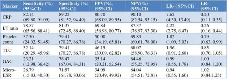
“Amongst all individual markers, CRP was observed to be the best diagnostic marker having the highest sensitivity (82.14%, 95% CI: 69.60 to 91.09 ), specificity (89.22%, 95% CI: 81.52 to 94.49), PPV (80.70%, 95% CI: 68.09 to 89.95), NPV ( 90.10 %, 95% CI: 82.54 to 95.15), and LR+ ( 7.62, 95% CI: 4.30 to 13.49), and the lowest LR- (0.20, 95CI: 0.11 to 0.35) followed by I/T ratio (sensitivity: 78.57, 95% CI: 65.56 to 88.41; specificity: 81.37%, 95% CI: 72.45 to 88.40; PPV: 69.84%, 95% CI: 56.98 to 80.77; NPV: 87.3%, 95% CI: 78.97 to 93.30; LR+ : 4.22, 95% CI: 2.75 to 6.47; and LR-: 0.26, 95% CI: 0.16 to 0.44). On the other hand,
Micro ESR was observed to be the least diagnostic single marker having the lowest specificity (71.57%, 95% CI: 61.78 to 80.06); PPV (34.09%, 95% CI: 20.49 to 49.92), LPV (64.04%, 95% CI: 54.51 to 72.81) and, LR+ (0.94, 95% CI: 0.55 to 1.60), and highest LR- (1.02, 95% CI: 0.84 to 1.25). GAC was observed to have the least sensitivity (23.21%, 95% CI: 12.98 to 36.42) (Table 2).”
Table 1: Clinical characteristics of study population.
Characteristics Study group (n=161)
Control group (n=102)
Mean birth
weight (g) 2580.63±372.55 2590.71±434.65
Mean gestational
age (weeks) 38.32±1.15 38.33±1.13
In born:out born 2.74:1 2.92:1
Male:female 1.21:1 1.55:1
Clinical features
of sepsis 62 (38.51%) 0
Mean age at
sampling (days) 2.09±0.80 2.0±0.82
Blood culture
positive 56 (34.78%) 0
Table 2: Diagnostic accuracy of various laboratory markers of sepsis screen.
Marker Sensitivity (%) (95%CI)
Specificity (%) (95%CI)
PPV(%), (95%CI)
NPV(%)
(95%CI) LR- ( 95%CI)
LR- (95%CI)
CRP 82.14
(69.60, 91.09)
89.22 (81.52, 94.49)
80.70 (68.09, 89.95)
90.10 (82.54, 95.15)
7.62 (4.30, 13.49)
0.20 (0.11, 0.35)
I/T ratio 78.57 (65.56, 88.41)
81.37 (72.45, 88.40)
69.84 (56.98, 80.77)
87.37 (78.97, 93.30)
4.22 (2.75, 6.47)
0.26 (0.16, 0.44) Platelet
count
37.50 (24.92, 51.45)
79.41 (70.27, 86.78)
50.00 (34.19, 65.81)
69.83 (60.61, 78.00)
1.82 (1.09, 3.03)
0.79 (0.63, 0.99)
TLC 32.14
(20.29, 45.96)
79.41 (70.27, 86.78)
46.15 (30.09, 62.82)
68.07 (58.90, 76.31)
1.56 (0.91, 2.68)
0.85 (0.70, 1.05)
GAC 23.21
(12.98, 36.42)
76.47 (67.04, 84.31)
35.14 (20.21, 52.54)
64.46 (55.25, 72.95)
0.99 (0.55, 1.78)
1.00 (0.84, 1.20)
Micro-ESR
26.79 (15.83, 40.30)
71.57 (61.78, 80.06)
34.09
(20.49, 49.92)
64.04 (54.51, 72.81)
0.94 (0.55, 1.60)
1.02 (0.84,1.25)
Amongst combinations of 2 markers, CRP plus I/T was to observed have the best diagnostic accuracy with the highest sensitivity (78.57%, 95% CI: 65.56 to 88.41), specificity (100%, 95% CI: 96.45, 100), PPV (100%, 95% CI: 91.96 to 100) and NPV (89.47%, 95% CI: 82.33 to 94.44), and the lowest LR- (0.21, 95% CI: 0.13 to 0.35) (Table 3). On the other hand, combination of I/T Ratio plus GAC was observed to have the least diagnostic utility with the lowest specificity (77.45%, 95% CI: 68.11 to 85.14), PPV (25.81%, 95% CI: 11.86 to 44.61), NPV (62.20%, 95% CI: 53.17 to 70.65), and LR+ (0.63, 95% CI: 0.30 to 1.32), and the highest LR- (1.11, 95% CI: 0.95 to 1.29). Combination of I/T Ratio plus Micro-ESR had the poorest sensitivity (8.93, 95% CI: 2.96 to 19.62).
DISCUSSION
to minimize unnecessary use of antibiotics in false positive cases, a competent diagnostic marker also needs to have a reasonably high specificity (the test is negative if infection is absent) and a good positive predictive value (infection is present when the test is positive), preferably better than 85%.2,8 The ideal early diagnostic test for
infection would have 100% sensitivity and specificity. Such an ideal test, however, is rather unlikely to be discovered, since most tests are measured on a continuous scale with an overlap between infected and non- infected infants.4
Table 3: Diagnostic accuracy of combinations of two laboratory markers of sepsis screen.
Markers Sensitivity (%), (95%CI) Specificity (%), (95%CI) PPV(%), (95%CI) NPV(%),
(95%CI) LR- (95%CI)
LR- (95%CI)
CRP+I/T Ratio 78.57
(65.56, 88.41) 100.00 (96.45, 100) 100.00 (91.96, 100) 89.47
(82.33, 94.44) -
0.21 (0.13, 0.35)
CRP+TLC 26.79
(15.83, 40.30) 87.25 (79.19, 93.04) 53.57 (33.87, 72.49) 68.46 (58.73, 76.33) 2.10 (1.08, 4.10) 0.84 (0.70, 1.00) CRP+Micro- ESR 10.71 (4.03, 21.88) 92.16 (85.13, 96.55) 42.86 (17.66, 71.14) 65.28 (56.90, 73.01) 1.37
(0.50 to 3.74)
0.97 (0.87, 1.08)
CRP+GAC 14.29
(6.38, 26.22 )
86.27 (78.04, 92.29) 36.36 (17.20, 59.34) 64.71 (56.05, 72.70) 1.04 (0.47, 2.33) 0.99 (0.87, 1.13)
CRP+Platelets 28.57
(17.30, 42.21) 80.39 (71.35, 87.59) 44.44 (27.94, 61.90) 67.21 (58.13, 75.44) 1.46 (0.82, 2.58) 0.89 (0.73, 1.08)
TLC+I/T Ratio 25.00
(14.39, 38.37) 84.31 (75.78, 90.76) 46.67 (28.34, 65.67) 67.19 (58.33,75.22) 1.59
(0.84 to 3.02)
0.89 (0.75, 1.06) TLC+Micro- ESR 10.71 (4.03, 21.88) 88.24 (80.35, 93.77) 33.33 (13.34, 59.01) 64.29 (55.75, 72.20) 0.91 (0.36, 2.29) 1.01 (0.90, 1.14)
TLC+GAC 23.21
(12.98, 36.42) 80.39 (71.35, 87.59) 39.39 (22.91, 57.86) 65.60 (56.58, 73.86) 1.18 (0.64, 2.20) 0.96 (0.80, 1.14)
TLC+Platelets 17.88
(8.91, 30.40) 80.39 (71.35, 87.59) 33.33 (17.29, 52.81) 64.06 (55.11, 72.35) 0.91 (0.46, 1.81) 1.02 (0.87, 1.19) I/T Ratio+Micro-ESR 08.93 (2.96, 19.62) 89.22 (81.52, 94.49) 31.25 (11.02, 58.66) 64.08 (55.61,71.96) 0.83 (0.30, 2.26) 1.02 (0.92, 1.14)
I/T Ratio+GAC 14.29
(6.38, 26.22) 77.45 (68.11, 85.14) 25.81 (11.86, 44.61) 62.20 (53.17, 70.65) 0.63 (0.30, 1.32) 1.11 (0.95, 1.29) I/T Ratio+Platelets 28.57 (17.30, 42.21) 79.41 (70.27, 86.78) 43.24 (27.10, 60.51) 66.94 (57.81, 75.22) 1.39 (0.79, 2.44) 0.90 (0.74, 1.09)
Platelets+GAC 17.86
(8.91,30.40) 78.43 (69.19, 85.96) 31.25 (16.12, 50.01) 63.49 (54.45, 71.88) 0.83 (0.42, 1.62) 1.05 (0.89, 1.23) Platelets+Micro-ESR 10.71 (4.03, 21.88) 89.22 (81.52, 94.49) 35.29 (14.21, 61.67) 64.54 (56.05, 72.41) 0.99 (0.39, 2.54) 1.00 (0.89, 1.12) GAC+Micro-ESR 14.29 (6.38, 26.22) 90.20 (82.71, 95.20) 44.44 (21.53, 69.24) 65.71 (57.23, 73.52) 1.46 (0.61, 3.48) 0.95 (0.84, 1.08)
Current evidence suggests that promising markers may be useful for early termination of antimicrobial treatment, but none of the current diagnostic tests are sensitive and specific enough to influence the clinical decision for withholding antibiotic treatment at the onset of suspected infection.2 Some researchers have even stated that the decision to start antibiotics need not necessarily be conditional to the results of sepsis screen in the presence of a strong clinical suspicion of sepsis.7 According to some authors, a negative screen must be repeated within 12 hours in case of persisting clinical suspicion of septicemia. Two sepsis screens performed 12-24 hours apart had a negative predictive value of 100%.9
Conflicting results have been obtained in different studies on diagnostic accuracy of individual markers of sepsis screen. TLC, total neutrophil count, immature neutrophil count, I/T ratio, immature to mature neutrophil (I/M) ratio, morphological or degenerative changes in neutrophil such as vacuolisation, Döhle bodies, intracellular bacteria, toxic granulation, and platelet count have been studied either singly or in combination.2 Since no currently available test or groups of test provided the ideal of high sensitivity, specificity, and NPV, an elaborate haematological scoring system (HSS) involving seven of the above variables (one point allocated to each abnormal variable) was formulated.3 The higher the score, the greater the certainty that the suspected septic episode was genuine. Using a cut off of >3, the score had a high sensitivity of 96%, but a disappointingly low positive predictive value of 31%.3 This scoring system, however, has not been adopted widely.2
White cell counts and ratios of haematological parameters are reported to vary widely across studies, with sensitivities and specificities ranging from 17% to 90% and 31% to 100% respectively.10 In general, the abnormal leukocyte ratios, including the I/T ratio >0.2, tend to have high sensitivity, whereas abnormal leukocyte counts, such as leukopenia and neutropenia, tend to have high specificity.2 TLC <10 x 109/L, TNC <8 x 10⁹/L, I/M >0.25, I/T >0.14, band count >15% and platelet count <150 x 109 were found to have optimal sensitivities and negative predictive values by some authors.11 Similarly, an abnormal I/T ratio followed by an abnormal I/M ratio were reported to be the most sensitive indicators in identifying infants with sepsis.1,12,13 These two criteria along with thrombocytopenia were reported to have a high negative predictive value over 94%.1 On the other hand, I/M ratio followed by I/T ratio was reported to have the highest high sensitivity for identifying neonates with sepsis as per some authors.14 In another study, immature PMN count was reported to have the highest sensitivity followed by Immature:Total (I/T) ratio and total PMN count; while I/M ratio followed by I/T ratio, degenerative changes and platelet count were reported to be highly specific tests for diagnosing sepsis in neonates. I/M was also reported to have the highest PPV.15 The authors further stated that HSS had much higher sensitivity, specificity, PPV and NPV in preterm babies in comparison to term neonates. Some workers were of the view that during the first three days of life leukopenia, neutropenia, elevated I/M ratio and CRP were good diagnostic aids for neonatal sepsis, whereas after 3 days of life, CRP was the best single test.16 However, Abnormalities in these markers soon after a birth complicated by clinical signs and obstetric risk factors of sepsis were highly suggestive of early onset neonatal sepsis.4 These reports, therefore, advocate the use of multiple indicators for detection of sepsis. Although CRP was found to have highest sensitivity (82.14 %) amongst individual markers of sepsis screen in the present study, a substantial proportion of septicemic patients were likely to be missed if it were used alone.
Moreover, it was not found to have optimal specificity, PPV and NPV. Sensitivity and specificity of CRP for diagnosis of early onset sepsis have been reported to range from, 43 to 90% and 70 to 78%, respectively in various other studies.4 In late onset sepsis, the specificity and positive predictive value of CRP reportedly range from 93% to 100%. Thus CRP is a „„specific‟‟ but „„late‟‟ marker of neonatal infection.6 CRP had higher sensitivity and specificity than total neutrophil count and I/T ratio as a diagnostic marker in neonates.10 It is synthesized within six to eight hours of exposure to an infective process or tissue damage, with a half-life of 19 hours, and may increase more than 1000-fold during an acute phase response.17 CRP levels have been shown to rise during the initial 24 hours in many babies irrespective of administration of antibiotics.18,19 These were considered the best tools available for assessment of effectiveness of antibiotic therapy, its duration, and whether or not recurrence of infection had occurred following cessation of treatment.20 Despite its promising characteristics the test has been reported to be falsely negative in cases of life threatening central nervous system candidiasis (probably because of the localised and chronic low grade nature of these infections).21 Conversely, increased CRP concentrations have been observed in some non-infective clinical conditions such as meconium aspiration, tissue necrosis, recent vaccination, and post-surgery.2,6
It has been observed that specificity of two test combinations was higher than that of individual tests, but three test combinations had no overall advantage over two test combinations.22 Some authors, on the other hand, were of the view that a combination of three tests enhances the sensitivity of these tests.23 CRP with any other marker (gastric aspirate, band cell / neutrophil ratio, toxic granules) except micro-ESR has been reported to have a sensitivity of 100% but a lower specificity.22 In an earlier study, CRP with gastric aspirate was found to be the best combination with sensitivity of 80% and specificity of 70%.24 The combination of CRP (0.10 mg/l) with full blood examination (abnormal film and/or
I/T ratio >0.2) and/or gastric aspirate (>5
polymorphs/high power field or potential pathogen on gram stained smear and/or culture of potential pathogen) has been reported to have a sensitivity of 97%, specificity of 61%, NPV of 98%, and likelihood ratio of 49 for early onset neonatal sepsis.25 In the present study, amongst combinations of 2 markers, CRP plus I/T was observed have the best diagnostic value with the highest sensitivity (78.57%), specificity (100%), PPV (100%) and NPV (89.47), and the lowest LR- (0.21). On the basis of its high diagnostic accuracy, this combination appeared to be the most suitable one for identification of cases of EOS. However, these findings are contrary to observations of some others who reported that a combination of CRP with haematological parameters decreased the sensitivity and NPV of HSS.11
CRP in combination with I/T ratio could serve as a simple and reliable tool for rapid detection of neonatal sepsis. However, all other two test combinations did not have the sensitivity, specificity and negative predictive value required for precise diagnosis of neonatal septicemia. Relying only on abnormality of any 2 markers of sepsis screen is, therefore, liable to result in inadvertent over treatment of a large majority of uninfected neonates. However, since preterm babies as well as those on prior antibiotic therapy had been excluded in the present study, further large scale studies need to be conducted on combinations of different tests in these populations groups.
Funding: No funding sources Conflict of interest: None declared
Ethical approval: The study was approved by the Institutional Ethics Committee
REFERENCES
1. Ghosh S, Mittal M, Jaganathan G. Early diagnosis of neonatal sepsis using a hematological scoring system. Indian J Med Sci. 2001;55:495-500.
2. Ng PC. Diagnostic markers of infection in neonates. Arch Dis Child Fetal Neonatal Ed. 2004;89:F229-35.
3. Rodwell RL, Leslie AL, Tudehope Dl. Early diagnosis
of neonatal sepsis using a hematological scoring system. J Pediatr. 1988;112:761-6.
4. Mishra UK, Jacobs SE, Doyle LW, Garland SM.
Newer approaches to the diagnosis of early onset neonatal sepsis. Arch Dis Child Fetal Neonatal Ed. 2006;91(3):F208-12.
5. Singh M. Perinatal infections. In: Singh M, ed. Care of Newborn. 6th ed. New Delhi: Sagar Publications, 2004:196-216.
6. Ng PC, Cheng SH, Chui KM, Fok TF, Wong MY,
Wong W, et al. Diagnosis of late onset neonatal sepsis with cytokines, adhesion molecule and C reactive protein in preterm very low birth weight infants. Arch Dis Child Fetal Neonatal Ed. 1997;77(3):F221-7. 7. Sankar MJ, Agarwal R, Deorari AK, Paul VK. Sepsis
in the newborn. Indian J Pediatr. 2008;75:261-6. 8. Ng PC, Li K, Wong RP, Chui KM, Wong E, Fok TF.
Neutrophil CD64 expression: a sensitive diagnostic marker for late-onset nosocomial infection in very low birth weight infants. Pediatr Res. 2002;51(3):296-303. 9. Kumar A, Singh M, Paul U. Clinical approach to
neonatal sepsis. Pediatr Today. 2001;4:493-515. 10. Da Silva O, Ohlsson A, Kenyon C. Accuracy of
leukocyte indices and C-reactive protein for diagnosis of neonatal sepsis: a critical review. Pediatr Infect Dis J. 1995;14:362-6.
11. Manucha V, Rusia U, Sikka M, Faridi MM, Madan N.
Utility of haematological parameters and C-reactive
protein in the detection of neonatal sepsis. J Paediatr Child Health. 2002;38(5):459-64.
12. Narasimha A, Harendra Kumar MLH. Significance of
Hematological Scoring System (HSS) in Early Diagnosis of Neonatal Sepsis. Indian J Hematol Blood Transfus. 2011;27(1):14-7.
13. Majumdar A, Jana A, Jana A, Biswas S, Bhattacharyya
S. Hematologic scoring system (HSS): A guide to decide judicious use of antibiotics in neonatal septicemia in developing countries. J Appl Hematol. 2013;4:110-3.
14. Supreetha MS, Alva SR, Shivendra VS, Kariappa TM.
Evaluation of neonatal septicaemia using
haematological parameters. Internat J of Recent Sci Res. 2015;6:2775-8.
15. Makkar M, Gupta C, Pathak R, Garg S, Mahajan NC. Performance evaluation of hematologic scoring system in early diagnosis of neonatal sepsis. J Clin Neonatol. 2013;2:25-9.
16. Varsha, Rusia U, Sikka M, Faridi MM, Madan N. Validity of hematologic parameters in identification of early and late onset neonatal infection. Indian J Pathol Microbiol. 2003;46:565-8.
17. Vigushin DM, Pepys MB, Hawkins PN. Metabolic and
scintigraphic studies of radioiodinated human C-reactive protein in health and disease. J Clin Invest. 1993; 91:1351–7.
18. Mathai E, Christopher U, Mathai M, Jana AT, Rose D,
Bergstrom S. Is C reactive protein useful in differentiating infected from uninfected neonates among those at risk of infection. Indian J Pediatr. 2004;41:895-9.
19. Philip AG, Mills PC. Use of C-reactive protein in minimizing antibiotic exposure: experience with infants initially admitted to a well-baby nursery. Pediatrics. 2000;106:E4.
20. Ainbender E, Cabatu EE, Guzman DM, Sweet AY.
Serum C-reactive protein and problems of newborn infants. J Pediatr 1992; 3: 438-42.
21. Ng PC, Lee CH, Fok TF, Chui K, Wong W, Cheung KL, et al. Central nervous system candidiasis in preterm infants: limited value of biochemical markers for diagnosis. J Paediatr Child Health. 2000;36:509-10.
22. Sharma A, Kutty CV, Sabharwal U, Rathee S, Mohan
H: Evaluation of sepsis screen for diagnosis of neonatal septicemia. Indian J Pediatr. 1993;60:559-63.
23. Ahmed Z, Ghafoor T, Waqar T, Ali S, Aziz S,
Mahmood S. Diagnostic value of C- reactive protein and haematological parameters in neonatal sepsis. J Coll Physicians Surg Pak. 2005;5(3):152-6.
24. Chandana A, Rao MN, Srinivas M, Shyamala S. Rapid
diagnostic tests in neonatal septicemia. Indian J Pediatr. 1988;55(6):947-53.
25. Garland SM, Bowman ED. Reappraisal of C-reactive protein as a screening tool for neonatal sepsis. Pathology. 2003;35:240-3.