R E S E A R C H
Open Access
Investigating the role of MRGPRC11 and
capsaicin-sensitive afferent nerves in the
anti-influenza effects exerted by
SLIGRL-amide in murine airways
Amy Y. Chang
1,2, Tracy S. Mann
1, Peter K. McFawn
2, Liang Han
3, Xinzhong Dong
3and Peter J. Henry
1*Abstract
Background:The hexapeptide SLIGRL-amide activates protease-activated receptor-2 (PAR-2) and mas-related G protein-coupled receptor C11 (MRGPRC11), both of which are known to be expressed on populations of sensory nerves. SLIGRL-amide has recently been reported to inhibit influenza A (IAV) infection in mice independently of PAR-2 activation, however the explicit roles of MRGPRC11 and sensory nerves in this process are unknown. Thus, the principal aim of this study was to determine whether SLIGRL-amide-induced inhibition of influenza infection is mediated by MRGPRC11 and/or by capsaicin-sensitive sensory nerves.
Methods:The inhibitory effect of SLIGRL-amide on IAV infection observed in control mice in vivo was compared to effects produced in mice that did not express MRGPRC11 (mrgpr-clusterΔ−/−mice) or had impaired sensory nerve function (induced by chronic pre-treatment with capsaicin). Complementary mechanistic studies using both in vivo and ex vivo approaches investigated whether the anti-IAV activity of SLIGRL-amide was (1) mimicked by either activators of MRGPRC11 (BAM8-22) or by activators (acute capsaicin) or selected mediators (substance P, CGRP) of sensory nerve function, or (2) suppressed by inhibitors of sensory nerve function (e.g. NK1 receptor antagonists). Results:SLIGRL-amide and BAM8-22 dose-dependently inhibited IAV infection inmrgpr-clusterΔ−/−mice that do not express MRGPRC11. In addition, SLIGRL-amide and BAM8-22 each inhibited IAV infection in capsaicin-pre-treated mice that lack functional sensory nerves. Furthermore, the anti-IAV activity of SLIGRL-amide was not mimicked by the sensory neuropeptides substance P or CGRP, nor blocked by either NK1 (L-703,606, RP67580) and CGRP receptor (CGRP8-37) antagonists. Direct stimulation of airway sensory nerves through acute exposure to the TRPV1 activator capsaicin also failed to mimic SLIGRL-amide-induced inhibition of IAV infectivity. The anti-IAV activity of SLIGRL-amide was mimicked by the purinoceptor agonist ATP, a direct activator of mucus secretion from airway epithelial cells. Additionally, both SLIGRL-amide and ATP stimulated mucus secretion and inhibited IAV infectivity in mouse isolated tracheal segments.
Conclusions:SLIGRL-amide inhibits IAV infection independently of MRGPRC11 and independently of
capsaicin-sensitive, neuropeptide-releasing sensory nerves, and its secretory action on epithelial cells warrants further investigation.
Keywords:SLIGRL-amide, Influenza, Airway sensory nerves, Capsaicin, Mas-related G protein-coupled receptor C11, Neuropeptides, Mucin
* Correspondence:peter.henry@uwa.edu.au
Amy Y. Chang and Tracy S. Mann are joint first authors
1School of Medicine and Pharmacology, University of Western Australia,
Crawley, WA 6009, Australia
Full list of author information is available at the end of the article
Background
Worldwide, influenza A virus (IAV) infections are esti-mated to cause up to 5 million cases of severe illness, and between 250,000 and 500,000 deaths each year [1]. The potentially devastating impact of seasonal influenza epidemics is lessened through vaccination, which signifi-cantly reduces the rate of hospitalisation and death from influenza. Unfortunately, annual vaccination does not provide protection against irregular influenza pandemics caused by antigenically novel strains of IAV transmitted from other animal species. Thus, considerable effort has been directed towards preventing and treating IAV in-fection with antiviral agents including the neuraminidase inhibitors oseltamivir (Tamiflu) and zanamivir (Relenza) [2, 3]. Despite these efforts, our capacity to control in-fluenza infection remains threatened due to the rapid emergence of resistance to oseltamivir and other conventional antiviral agents such as the adamantanes [4–6]. Hence, there is an urgent unmet need to identify new anti-viral drugs to provide a frontline defence to in-fection, especially in the event of an unpredictable but inevitable influenza pandemic.
Our laboratory has recently shown that the hexapeptidic sequence SLIGRL-amide (Ser-Leu-Ile-Gly-Arg-Leu-NH2)
inhibits IAV infection in mice [7]. SLIGRL-amide is a well-established activator of protease-activated receptor-2 (PAR-2) [8]. However, SLIGRL-amide inhibits IAV infec-tion independently of PAR-2 as anti-viral effects were pre-served in PAR2 (−/−) mice [7]. SLIGRL-amide has also been reported to activate MRGPRC11, a subtype of MAS-related G protein-coupled receptor (MRGPR) [8–10]. Whether SLIGRL-amide inhibits IAV infection via MRGPRC11 is currently unknown.
The MRGPR family comprises approximately 40 mem-bers that are grouped into nine subfamilies (MRGPRA to–H, and–X), based on sequence similarities. Subfam-ilies A, B, C, and H exist only in rodents, whereas sub-family X is specific to primates including humans [10]. Mouse and rat MRGPRC have been found to exhibit high similarities with human MRGPRX1 in terms of ex-pression pattern, sequence homology and binding profile [11–14]. For example, both rodent MRGPRC11 and human MRGPRX1 are activated by the agonist BAM8-22. The expression of these receptors is primarily restricted to the small-diameter nociceptive sensory neu-rons [11, 12, 15, 16].
Sensory nerves of the respiratory system innervate a range of important structures, including the airway epi-thelium, submucosal glands and airway smooth muscle [17]. The predominant subtype of airway sensory nerve is the vagal bronchopulmonary C-fibre, whose cell bod-ies are located within the jugular and nodose ganglia [18]. Jugular C-fibres are more likely to innervate the upper airways [19] and to express neuropeptides when
activated [20]. Noxious substances such as the TRPV1 activator capsaicin can activate populations of sensory nerves and may promote the release of neuropeptides such as substance P and CGRP via an axonal reflex [21]. Released neuropeptides, via activation of their cognate neurokinin (NK) and CGRP receptors, can in turn in-duce a wide range of biological effects, such as increased cilia beating and mucus secretion [22]. Several compo-nents of mucus, including surfactants, secretory IgA, defensins and MUC5B mucin have been reported to ex-hibit anti-IAV activities [23].
Thus far, the molecular target mediating the anti-IAV effects of SLIGRL-amide is unknown. The current study tests the hypotheses that SLIGRL-amide inhibits IAV in-fection by activating MRGPRC11, and that the antiviral effects of SLIGRL-amide are mediated by neuropeptides, which stimulate the secretion of endogenous substances with anti-IAV activities from mucus-producing airway cells. If the hypotheses are correct, then SLIGRL-amide induced anti-IAV effects should be mimicked by activa-tors of MRGPRC11 (such as BAM8-22) and by exogen-ous neuropeptides (such as substance P and CGRP), as well as by other stimuli that directly activate sensory nerves (TRPV1 activator capsaicin). Furthermore, the anti-IAV effects of SLIGRL-amide should be reduced in mice lacking MRGPRC11 (Mrgpr-clusterΔ−/− mice, [8, 24]) and by processes (capsaicin-induced desensitisation) or agents (neurokinin receptor antagonists) that sup-press sensory nerve function. In the current study, the effect of these interventions on SLIGRL-amide-induced anti-IAV activity was tested using a combination of in vivo (airway inflammation in IAV-exposed mice) and novelex vivoapproaches (immunohistochemical staining for IAV in viable perfused tracheal explants; [7]). Utilisa-tion ofex vivoapproaches also facilitated preliminary in-vestigations of the anti-IAV activity of SLIGRL-amide in human isolated airways.
Methods
Ethics statement
studies were specifically approved by the Sir Charles Gairdner Hospital Human Research Ethics Committee (approval number 2011–128), the Mount Hospital Ethics Committee (approval number EC71.1) and The Univer-sity of Western Australia Human Ethics Office (approval numbers RA/4/1/7256 and RA/4/1/7220).
Influenza virus
Mouse-adapted influenza A/PR/8/34 virus was propa-gated in the allantoic fluid of 9-day old embryonated chicken eggs (Altona Hatchery, Forrestfield, Australia) at 37 °C for 3 days, as described previously [25]. Viral infectivity was assessed using allantois-on-shell titra-tion and quantitated via hemagglutinatitra-tion assay [26]. The TCID50 of the harvested allantoic fluid was 10-5.8/ml.
In vivo infection of mice with IAV
Unless otherwise stated, male BALB/c mice (specified pathogen-free) aged 7 to 8 weeks (Animal Resource Centre, Murdoch, WA) were housed at the University of Western Australia Animal Care Unit under a 12 h light/ dark cycle and received food and waterad libitum. The role of MGRPRC11 in SLIGRL-amide-induced anti-IAV activity was investigated using Mrgpr-clusterΔ−/− and wild-type mice, generated as previously described [24]. Groups of mice were lightly anaesthetised (methoxyflurane) and intranasally (i.n.) inoculated with a 20μl solution con-taining (a) influenza A/PR/8/34 virus (1:800 dilution of stock IAV) alone, (b) IAV plus peptide (SLIGRL-amide, SLIGR-amide, BAM8-22) or (c) 1:800 dilution of allantoic fluid (vehicle). Mice were killed with an overdose of pentobarbitone (160 mg/kg i.p. injection) at day 4 post-inoculation for determination of IAV-induced inflam-mation using differential cell counting of leukocytes recovered from bronchoalveolar lavage (BAL) fluid [7].
Bronchoalveolar lavage
BAL was performed by intratracheal instillation of 2.5 ml of cold phosphate-buffered saline (PBS) pH 7.4, in 0.5 ml volumes via a tracheal cannula. After each in-stillation, BAL fluid was recovered, pooled and centri-fuged at 400 ×g for 5 mins at 4 °C. Supernatant was removed and the cell pellet resuspended in PBS + 1.0 % bovine serum albumin. Total cell counts and viability were determined by use of a haemocytometer and 0.4 % trypan blue exclusion. Cytospin preparations of each cell sample were stained with DIFF-Quik (Thermo Fisher Scientific, Waltham, MA), and differential cell counts of macrophages, neutrophils, eosinophils and lymphocytes were determined by counting 400 cells under a light microscope using standard morphological criteria.
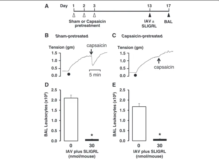
Capsaicin-induced attenuation of sensory nerve function in mice
Administration of multiple subcutaneous injections of capsaicin to anaesthetised mice is an effective means of impairing sensory nerve function [27–30]. On day one, mice were anaesthetized (130 mg/kg ketamine and 13 mg/kg xylazine, i.p.) and administered the bronchodi-lators terbutaline (0.1 mg/kg, i.p.) and theophylline (10 mg/kg, s.c.). Twenty mins later, mice were injected subcutaneously with capsaicin (25 mg/kg, s.c.) in the neck region between the shoulder blades. On days two and three, the procedure was repeated with an increased dose of capsaicin (50 mg/kg, s.c) (capsaicin-pretreated). Mice were left for 10 days before being intranasally inoc-ulated with IAV ± SLIGRL-amide, as described above. Control mice received injections of vehicle (1:1:8 vol/ vol/vol of ethanol/Tween 80/saline) (sham-pretreated). The protocol is shown in Fig. 3a. The effectiveness of the capsaicin desensitization protocol was determined in isometric tension recording studies by examining the capsaicin-induced relaxation responses in mouse isolated tracheal smooth muscle preparations.
Isometric tension recording studies
As previously demonstrated in our laboratories [31, 32] isometric tension recording studies can be used to deter-mine the functionality of the sensory nerves within the mouse trachea. Groups of mice exposed to either capsa-icin or saline (sham) mice were euthanased using pento-barbitone (160 mg/kg, i.p.). Tracheal segments were excised and suspended under a resting tension of 0.2 g in organ baths containing 20 ml of Krebs bicarbonate solu-tion (117 mM NaCl, 5.36 mM KCl, 25 mM NaHCO3,
1.03 mM KH2PO4, 0.57 mM MgSO4.7H2O, 2.5 mM
CaCl2, 11.1 mM D-glucose) maintained at 37 °C and
bub-bled continuously with 5 % CO2and 95 % O2. Changes in
tension were recorded via a 751mT miniTOBs organ bath system (DMT, Aarhus, Denmark) connected to a Powerlab system (ADInstruments Pty Ltd., Castle Hill, Australia). Tracheal preparations were allowed to equili-brate for 30 mins and the viability of the tracheal smooth muscle was determined by cumulative exposure to a sub-maximal (0.2 μM) and maximal (10 μM) dose of carba-chol. Tracheal segments were then repeatedly washed and allowed to rest for 20 mins. Preparations were precon-tracted using 1μM carbachol and upon reaching a plateau level of tension were exposed to a single 20μM bolus dose of capsaicin, and tension recordings continued until the peak response was obtained.
Ex vivo infection of mouse isolated tracheal segments with IAV
effects of selected peptides (SLIGRL-amide, BAM8-22, substance P, CGRP) on the capacity of IAV to infect and replicate in murine isolated airways, as previously de-scribed [7]. Briefly, tracheae harvested from untreated mice (male BALB/c mice at 7 to 8 weeks of age) were mounted onto an 18G blunted needle and perfused for at least half an hour at 37 °C with complete RPMI (cRPMI) medium at the rate of 100 μL/min. Complete RPMI (cRPMI) medium consisted of RPMI 1640 medium (Gibco, Life Technologies-Thermo Fisher Scientific Inc, MA, USA) supplemented with 20 mM HEPES, 2 mM GlutaMAX™, 2.0μg/mL amphotericin B, 20μg/mL gentamycin, and 1 % foetal calf serum (FCS). Following this equilibration period, the perfusion was stopped and the tracheal lumen gently flushed first with 0.5 ml of sterile saline, and then air, before being ex-posed to IAV (1/800 dilution of stock) ± peptides for 15 mins, without perfusion. The lumen of the airway was then washed with 0.5 mL of sterile saline to remove un-attached IAV and peptides, and perfused with cRPMI (100 μL/min at 37 °C) for 48 h. Airway segments were removed from the needle and fixed in 2 % paraformalde-hyde (containing 0.2 % picric acid) in PBS (pH 7.4), for 48 h at 4 °C. Tissues were dehydrated (starting from 50 % ethanol) and processed to paraffin wax on a stand-ard 15 h cycle in a Leica ASP200S automated tissue pro-cessor (Wetzlar, Germany).
Isolated airways from human lung
The anti-IAV activity of SLIGRL-amide was also exam-ined in small airway segments obtaexam-ined from humans. Macroscopically normal samples of lung were obtained from patients undergoing lung surgery, usually for lung cancer. Segments of small airways (1–2 mm internal diameter) were dissected free from surrounding lung tissue and mounted onto the airway explant perfusion system. Perfused airways were exposed to IAV ± SLIGRL-amide, as outlined above for mouse studies.
Immunohistochemistry and imaging
The presence of immunoreactive IAV in isolated trachea of IAV-exposed mice was visualised using standard immu-nohistochemical procedures, as previously reported [7]. Briefly, 5μM wax sections were dewaxed, rehydrated and permeablized with 1 % Triton X-100 for 15 mins. Tissues were then blocked with 20 % normal rabbit serum or 3 % fish skin gelatin for 1 h and further exposed to avidin/biotin blocking as per manufacturer’s instruc-tions (Avidin/Biotin Blocking Kit; Vector Laboratories, Burlingame, CA). Goat anti-influenza A polyclonal antibody, 1/1000 dilution (5 μg/ml) (Millipore Corpor-ation, Billerica, MA); mouse anti-influenza A monoclonal antibody, 1/500 dilution (14.5μg/ml) (Abcam, Cambridge, ENG); normal goat IgG isotype control or normal mouse
serum were applied to sections and incubated overnight at 4 °C. After overnight incubation, sections were thoroughly washed in Tris-buffered saline (TBS) for one hour and en-dogenous peroxidase was quenched with 0.3 % H2O2 in
TBS for 15 mins. The sections were then exposed to bio-tinylated rabbit anti-goat secondary IgG, 1/200 dilution (Vector Elite ABC kit; Vector Laboratories) or biotinylated rabbit anti-mouse IgG F(ab')2, 1/200 dilution (3.65μg/ml) (Dako, Denmark) for 45 mins, followed by avidin-biotin-horseradish-peroxidase complex (Vector Elite ABC kit; Vector Laboratories) for 45 mins. The bound complex was visualised with diaminobenzidine (DAB, 0.4 mg/ml). Sections were counterstained with Mayers haematoxylin and blued with Scotts Tap Water Substitute, washed and dehydrated through graded alcohols to xylene, then cover-slipped with Depex mounting medium. Digital images were acquired on an Aperio ScanScope XT digital slide scanner (Leica (Aperio) Technologies, Vista, CA). Images represent at least three animals per treatment.
Visualisation of immunoreactive IAV in human iso-lated bronchioles was achieved following the same protocol as for mouse tissue (described above) but with the following modifications - the diluent for both the primary and secondary antibodies was 3 % fish skin gel-atin (FSG) (Sigma-Aldrich, St. Louis, MO) in 50 mM Tris + 0.5 M NaCl + 0.01 % Triton-X-100 . The blocking solution was 0.3 M glycine in 3 % FSG, and the wash buffer (50 mM Tris + 0.5 M NaCl + 0.01 % Triton-X-100) contained 0.3 M glycine. Avidin/Biotin blocking was not performed. All antibody concentrations, incuba-tion and washing times, and visualisaincuba-tion with DAB remained the same.
Quantitative analysis of immunoreactive IAV in mouse tracheal epithelium
Attenuation of sensory nerve function in mouse isolated tracheal segments
To investigate whether capsaicin-induced inhibition of sensory nerve function blocks the anti-IAV effects of SLIGRL-amidein vitro, mouse tracheas from naïve mice were connected to the airway explant perfusion system and then exposed to capsaicin (20 μM) for 10 mins. Capsaicin-exposed preparations were washed and per-fused with cRPMI for a further 10 mins prior to a stand-ard 15 min exposure to IAV ± SLIGRL-amide. Tracheal segments were washed and perfused for 48 h with cRPMI, and then processed to determine levels of im-munohistochemical staining for IAV. Complementary isometric tension recording studies were performed to confirm that in vitro capsaicin pre-treatment inhibited TRPV1-mediated activation of airway sensory nerves.
Mucin secretion studies
The capacity of selected compounds to stimulate mucin release from epithelial stores of mucin was determined using the airway explant perfusion system. Following a 24 h period of perfusion with cRPMI, mouse isolated tracheal segments were exposed for 15 mins to SLIGRL-amide (200 μM), BAM8-22 (25 μM), substance P (1 μM), CGRP (1 μM) or to the known mucin secreta-gogue ATP (100 μM), or to saline. Tissues were then fixed in 2 % paraformaldehyde (containing 0.2 % picric acid) in PBS (pH 7.4), for 48 h at 4 °C, processed to paraffin wax and 5 μm sections stained with Alcian Blue-Periodic acid-Schiff’s reagent (AB-PAS) to demon-strate the presence of mucins (the chief glycoprotein constituent of mucus) in the epithelial layer. The chosen concentration and period of exposure to ATP were based on those employed in mucin release studies con-ducted in mice [33, 34].
Quantitative analysis of AB-PAS staining in mouse tracheal epithelium
Digital images of AB-PAS stained sections were gener-ated using a ScanScope XT digital slide scanner (Leica (Aperio) Technologies, Vista, CA) and analysed using ImageScope v11.1.2 and the Positive Pixel Count v9 al-gorithm (Aperio Technologies). Input parameters (color saturation threshold (0.04), hue value (0.1) and intensity ranges for weak positive (188–238), positive (176–188) and strong positive (0–176) pixels) were defined to gen-erate an algorithm able to quantify AB-PAS-positive stores of mucin stained bright pink (as “strong positive pixels”). The number of strong positive pixels (SPP) in the epithelium was counted by the algorithm and expressed as a percentage of the total pixels within the epithelial layer (%SPP). %SPP was calculated for each tracheal section and provided a means of quantifying changes in levels of mucin staining.
Materials
SLIGRL-amide, BAM8-22, SLIGR-amide and CGRP8-37 were supplied by Auspep (Melbourne, Australia). Stock solutions were prepared in sterile high-purity water with subsequent dilutions made in sterile saline. The concen-tration, purity, and composition of the peptides were de-termined by high-performance liquid chromatography, mass spectrometry, and quantitative amino acid analysis. Carbachol (carbamylcholine chloride), ATP, indometh-acin, atropine, propranolol, terbutaline, theophylline and L-703,606 were purchased from Sigma–Aldrich (St. Louis, MO), whilst RP-67580 ((3aR,7aR)-Octahydro-2-[1-imino-2-(2-methoxy-phenyl)ethyl]-7,7-diphenyl-4H-isoindol), sub-stance P, CGRP and L-733,060 were obtained from Tocris Bioscience (Ellisville, MO). Sodium pentobarbitone was supplied by Virbac Australia (Peakhurst, NSW), methoxy-flurane by Medical Developments International Ltd (Springvale, Australia) and Schiff reagent by Australian Biostain (Vic, Australia).
Statistical analysis
Unless otherwise stated, grouped data are expressed as mean ± SEM. Statistical comparisons between groups were made using one-way ANOVA with post-hoc testing (Holm procedure for all pairwise comparisons or Dunnett’s test for comparisons to single control) using InVivoStat software. Differences between groups were considered statistically significant atp<0.05.
Results
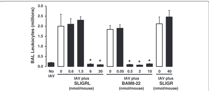
Effect of selected peptides on IAV infection in mice in vivo
SLIGRL-amide and BAM8-22 may not specifically acti-vate MRGPRC11, and other receptors may be involved in mediating their anti-IAV effects.
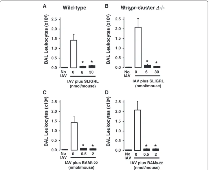
Anti-IAV activity of SLIGRL-amide and BAM8-22 in Mrgpr-clusterΔ−/−and wild-type mice
To definitively evaluate the role of MRGPRC11 in the anti-IAV activities of SLIGRL-amide and BAM8-22, studies were conducted using Mrgpr-clusterΔ−/− mice that do not express MRCPRC11 [8, 24]. Intranasal in-oculation of mice (Mrgpr-clusterΔ−/−and wild-type) with IAV (in the absence of peptides) was associated with a marked increase in total BAL cell number on day 4 post-inoculation (Fig. 2). As expected, both SLIGRL-amide (6 and 30 nmol/mouse) and BAM8-22 (0.5 and 2 nmol/mouse) inhibited the IAV-induced increase in total BAL cell number in wild-type mice [24] (Fig. 2a and 2c). However, contrary to the hypothesis that MRGPRC11 mediates the anti-IAV effects of SLIGRL-amide and BAM8-22, doses of these peptides that inhib-ited IAV-induced increases in total BAL cell number in BALB/c mice (Fig. 1a) and wild-type mice (Fig. 2), also exhibited anti-IAV activity in Mrgpr-clusterΔ−/− mice (Fig. 2b and 2d). These findings provide strong evidence that the anti-IAV activities of either SLIGRL-amide or BAM8-22 are not mediated by MRGPRC11.
Role of sensory nerves in SLIGRL-amide-induced anti-IAV activity
Further studies were completed to evaluate the possibil-ity that the anti-IAV activpossibil-ity of SLIGRL-amide was
mediated by capsaicin-sensitive, neuropeptide-releasing airway sensory nerves. In these studies, the activity of SLIGRL-amide was investigated in mice in which sen-sory nerve function had been attenuated by either (1) chronic exposure to capsaicin or (2) administration of a neurokinin receptor antagonist.
Repeated daily exposure to capsaicin has previously been reported to cause chemical ablation of sensory nerves in mice [27, 29, 36–38]. In our studies, anaesthe-tized mice were exposed to capsaicin on 3 consecutive days (see Fig. 3a for protocol), and confirmation of the effectiveness of this in vivo protocol to induce airway sensory nerve dysfunction was obtained from isometric tension recording experiments (Fig. 3b and 3c, lower panels). Tracheal smooth muscle segments from capsaicin-pre-treated mice did not relax in response to capsaicin, compared to the large relaxation response ob-tained in sham-pre-treated (no capsaicin) mice (Fig. 3b and 3c), consistent with sensory nerve dysfunction.
Inoculation with IAV on day 13 of the protocol pro-duced similar increases in BAL leukocytes in sham and capsaicin-pretreated mice (Fig. 3d and 3e). Moreover, IAV-induced increases in BAL leukocytes were inhibited by SLIGRL-amide in both sham (Fig. 3d) and capsaicin-pretreated mice (Fig. 3e). These findings do not support a role for classic TRPV1-expressing, sensory C-fibres in SLIGRL-amide-induced inhibition of IAV infectivity.
Some populations of activated airway sensory nerves release neuropeptides such as the tachykinin substance P, which transmits responses via activation of neurokinin (NK) receptors. Consistent with this, NK1 receptor
*
*
*
*
*
0.0 0.5 1.0 1.5 2.0 2.5 3.0
No IAV
0 0.6 1.5 6 30 0 0.05 0.5 2 10 0 40
BAL Leukocytes (millions)
IAV plus SLIGRL (nmol/mouse)
IAV plus BAM8-22 (nmol/mouse)
IAV plus SLIGR (nmol/mouse)
antagonists inhibit substance P-induced effects on micro-vascular leakage, mucus secretion and bronchomotor tone in the airways (see [39]). In the current study, administra-tion of the NK1 receptor antagonist L-703,606 (8.5 mg/kg; [40, 41]) did not block the capacity of SLIGRL-amide to inhibit IAV-induced increases in total BAL cell number in mice (Fig. 4), indicating that the actions of SLIGRL-amide were not mediated via activation of the sensory nerve – substance P–NK1 receptor axis.
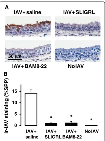
Effect of SLIGRL-amide and BAM8-22 on IAV infectivity in mouse isolated trachea ex vivo
Our laboratories have previously demonstrated that the antiviral activity of SLIGRL-amide observed in intact mice can be replicated in mouse isolated tracheaex vivo [7]. The novelex vivoperfused tracheal system preserves
tissue viability, architecture and microenvironment of the airways, and enables the study of the cellular mecha-nisms through which SLIGRL-amide inhibits IAV virus infection. Furthermore, thisex vivoperfusion system can be adapted to airways of other non-murine species, in-cluding human airways. In ex vivo studies, the levels of immunoreactive staining for IAV within the airway epi-thelium is the primary endpoint used to indicate IAV in-fectivity [7]. Extensive immunoreactive staining for IAV is indicative that the virus has proceeded through the se-quential processes of viral attachment, endocytosis and replication. In the current study, a brief 15 min exposure of mouse isolated trachea to IAV resulted in the subse-quent propagation of IAV within the epithelium over the ensuing 48 h, as revealed by the presence of high levels of staining for immunoreactive IAV (Fig. 5a and 5b). In 0.0
0.5 1.0 1.5 2.0 2.5
BAL Leukocytes (x10
6
)
No
IAV 0 0.5 2
IAV plus BAM8-22 (nmol/mouse)
0.0 0.5 1.0 1.5 2.0 2.5
BAL Leukocytes (x10
6
)
No
IAV 0 0.5 2
IAV plus BAM8-22 (nmol/mouse) IAV plus SLIGRL
(nmol/mouse) 0.0
0.5 1.0 1.5 2.0 2.5
BAL Leukocytes (x10
6
)
No
IAV 0 6 30
IAV plus SLIGRL (nmol/mouse) 0.0
0.5 1.0 1.5 2.0 2.5
BAL Leukocytes (x10
6
)
No IAV
0 6 30
A
B
C
D
Wild-type
Mrgpr-cluster
-/-*
*
*
*
*
*
*
*
Δ
stark contrast, mouse tracheal segments exposed to SLIGRL-amide and BAM8-22 during the 15 min expos-ure period to IAV revealed negligible levels of immuno-histochemical staining for IAV (Fig. 5a and 5b), indicating that IAV infectivity had been impaired by these peptides. Thus, as well as inhibiting IAV-induced increases in total BAL cell number in vivo (Fig. 1), SLIGRL-amide and BAM8-22, also inhibited IAV infect-ivity in mouse isolated tracheal segments (Fig. 5).
Effect of sensory nerve activation on IAV infectivity Consistent with in vivo studies, the evidence obtained from studies using isolated mouse tracheal segments did not support the postulate that the anti-IAV effects of SLIGRL-amide were mediated by activation of capsaicin-sensitive, neuropeptide-releasing sensory nerves. For
example, the acute and direct activation of sensory nerves caused by a single 15 min exposure to capsaicin failed to inhibit IAV infectivity (Fig. 6a). Similarly, the exogenous application of selected sensory neuropeptides known to be released locally from activated sensory nerves (substance P or CGRP at 1μM), did not mimic the anti-IAV effects of SLIGRL-amide (Fig. 6b and 6c). Furthermore, neither an NK1 receptor antagonist (RP67580, 20 μM) nor a CGRP receptor antagonist (CGRP8-37, 10 μM) blocked SLIGRL-amide-induced inhibition of IAV infectivity in mouse tracheal segments (Fig. 6b and 6c).
Effect of SLIGRL-amide and ATP on release of epithelial mucin stores from mouse isolated tracheal segments Additional experiments were conducted to investigate the possibility that SLIGRL-amide stimulates the release
Sham-pretreated
Tension (gm)
0.0 0.5 1.0 1.5
5 min capsaicin
B
Day 1 2 3 13 17
Sham or Capsaicin pretreatment
IAV ±
SLIGRL
BAL
A
C
Capsaicin-pretreatedIAV plus SLIGRL (nmol/mouse)
0 30
capsaicin
0.0 0.5 1.0 1.5
Tension (gm)
0.0 0.5 1.0 1.5 2.0 2.5
BAL Leukocytes (x10
6)
0.0 0.5 1.0 1.5 2.0 2.5
BAL Leukocytes
(x10
6)
IAV plus SLIGRL (nmol/mouse)
0 30
D
E
*
*
Fig. 3Effect of sensory nerve dysfunction caused byin vivocapsaicin treatment on the anti-IAV activity of SLIGRL-amide in mice.aProtocol for capsaicin-induced dysfunction of sensory nerves. On 3 consecutive days, groups of mice were anaesthetised and injected subcutaneously with capsaicin or vehicle (Sham). Ten days later, mice were anaesthetised and intranasally inoculated with IAV in the presence or absence of SLIGRL-amide. A further four days later, BAL was performed on each euthanased mouse and the total numbers of leukocytes recovered was determined.
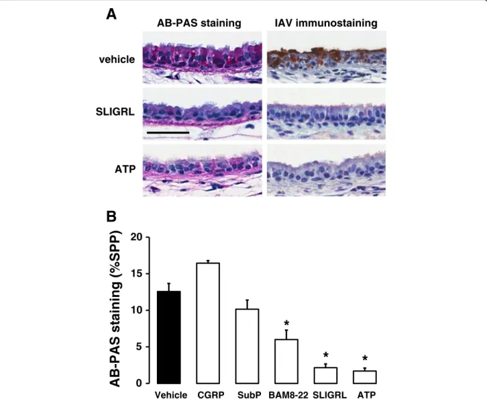
of substances with anti-IAV activity directly from the air-way epithelium. Consistent with this, exposure of tra-cheal segments to SLIGRL-amide was associated with a marked reduction in the levels of AB-PAS staining within the tracheal epithelium, indicative of mucin release (left-hand panels of Fig. 7a and 7b). Levels of AB-PAS staining were also reduced by the established secretagogue ATP and by BAM8-22, but not by either substance P or CGRP (Fig. 7b). Of particular interest, ATP also inhibited IAV infectivity in mouse isolated tra-cheal segments (right hand panels of Fig. 7a), similar to the effect produced by SLIGRL-amide.
Effect of SLIGRL-amide on IAV infectivity in airways of human isolated airways
As indicated above, an advantage of theex vivoperfused airway system is that it can be readily adapted to enable the study of human airways. A 15 min exposure of hu-man isolated bronchioles to IAV, followed by two days of perfusion with IAV-free cRPMI medium was associ-ated with the development of high levels of staining for immunoreactive IAV within the epithelium, consistent with the replication of IAV (Fig. 8a). Co-incubation of IAV with SLIGRL-amide during the initial 15 min expos-ure period was associated with significantly suppressed the levels of epithelial IAV immunoreactivity in ex vivo segments of human bronchioles two days later (P = 0.05; Fig. 8b), consistent with findings obtained from in vivo andex vivostudies using mice.
Discussion
SLIGRL-amide activates murine PAR-2, and has been used for over twenty years to evaluate the role of PAR-2 activation in physiological and pathological processes [42–45]. SLIGRL-amide has also recently been shown to activate MRGPRC11, a G protein-coupled receptor expressed exclusively on sensory nerves [8], and to in-hibit IAV infection in mice that lack PAR-2 [7]. The current study sought to determine whether the antiviral activity of SLIGRL-amide in mouse airways involved 0.0
0.5 1.0 1.5 2.0 2.5
BAL Leukocytes (x10
6)
0
30
0
30
pretreated with NK1R antagonist vehicle
IAV plus SLIGRL
(nmol/mouse) IAV plus SLIGRL(nmol/mouse)
*
*
Fig. 4Effect ofin vivoadministration of an NK1 receptor antagonist on the anti-IAV activity of SLIGRL-amide in mice. Groups of mice were injected with L-703,606 (8.5 mg/kg, i.p.) or vehicle (control), and 30 mins later intranasally inoculated with IAV in the presence or absence (saline) of SLIGRL-amide. A further four days later, BAL was performed on each euthanased mouse and the total numbers of leukocytes recovered was determined, as shown (mean ± s.e.mean of 4–5 mice per group). *, indicatesP<0.05 compared to respective IAV + saline control (one-way ANOVA)
IAV+ SLIGRL
IAV+ saline
NoIAV
IAV+ BAM8
-
22
50 m
B
A
*
*
*
0 5 10 15
IAV+ saline
IAV+ SLIGRL
IAV+ BAM8-22
NoIAV
ir-IA
V
staining (%SPP)
μμ
Fig. 5Semiquantitative measurement of anti-IAV activities of SLIGRL-amide and BAM8-22 in mouse isolated tracheal segments by immunohistochemical staining of IAV nucleoprotein.
activation of MRGPRC11 and/or sensory nerves, and to determine whether SLIGRL-amide also inhibited IAV in-fectivity in human airways.
In the current study, IAV infectivity in intact mice and in mouse isolated tracheal segments was inhibited by BAM8-22, an established activator of MRGPRC11 [8, 13]. Indeed, the observed 10-fold greater anti-IAV potency of BAM8-22 compared to SLIGRL-amide in mice was con-sistent with an action on MRGPRC11 [8, 10]. However, both SLIGRL-amide and BAM8-22 were effective in pre-venting IAV infection in Mrgpr-clusterΔ−/−mice that do not express MRGPRC11 [8]. These latter studies con-ducted inMrgpr-clusterΔ−/−mice provide compelling evi-dence that the anti-IAV activities of SLIGRL-amide and BAM8-22 were mediated independently of MRGPRC11. Additional pharmacologic studies were then undertaken to determine whether the anti-IAV properties of SLIGRL-amide and BAM8-22 involved activation of a popula-tion of capsaicin-sensitive, peptidergic airway sensory nerves.
We hypothesised that if SLIGRL-amide inhibits IAV infection via activation of capsaicin-sensitive sensory nerves, then the anti-IAV activity of SLIGRL-amide should be suppressed following the chemical ablation of sensory nerves by repeated exposure of rodents to capsa-icin [27, 29, 36–38]. However, the anti-IAV effects pro-duced by SLIGRL-amide were preserved in mice that had been exposed to capsaicin on three consecutive days 10 days prior to inoculation with IAV, despite tracheal preparations obtained from these mice failing to produce a characteristic relaxation response to capsaicin [32]. Consistent with the findings of these in vivo studies, in vitro pre-treatment of mouse isolated tracheal segments to capsaicin rendered them unresponsive to subsequent challenges to capsaicin, but not to the anti-IAV actions of SLIGRL-amide. Furthermore, although NK1 receptors have previously been implicated in SLIGRL-amide-induced actions in mouse trachea [46] and small intes-tine [47], neither NK1 nor CGRP receptor antagonists blocked the anti-IAV activities of SLIGRL-amide,in vivo orex vivo. Thus, our data do not support the explicit hy-pothesis that SLIGRL-amide inhibits IAV via activation of capsaicin-sensitive sensory nerves, or via the actions of the neuropeptides substance P or CGRP.
In a complementary series of experiments, we tested the postulate that if SLIGRL-amide inhibits IAV infection via activation of sensory nerves and local release of neuropep-tides, then other agents that stimulate sensory nerves would mimic the anti-IAV actions of SLIGRL-amide. Cap-saicin directly activates sensory nerve C-fibres via stimula-tion of the highly expressed TRPV1 calcium ion channel. However, exposing mouse isolated tracheal segments to capsaicin together with IAV did not mimic the anti-IAV effects produced by SLIGRL-amide. In these experiments,
B
A
*
*
0 5 10 15
IAV + saline
IAV + SubP
IAV + SLIGRL
IAV + SLIGRL +
NK1-RA
ir-IAV staining (%SPP)
0 5 10 15 20 25
IAV + saline
IAV + CGRP
IAV + SLIGRL
IAV + SLIGRL + CGRP-RA
ir-IAV staining (%SPP)
*
*
C
0 2 4 6
IAV + saline
IAV + capsaicin
(acute)
ir-IAV staining (%SPP)
capsaicin was used at a concentration (10 μM) that in-duced large relaxation responses in mouse isolated tra-cheal preparations [32]. These responses were blocked by a TRPV1 antagonist (capsazepine) and an NK1 receptor antagonist (L-733060), consistent with sensory nerve acti-vation [32]. In addition, if the anti-IAV effects of SLIGRL-amide were mediated via the actions of substance P and/ or CGRP released from activated sensory nerves, then the exogenous application of these neuropeptides should also inhibit IAV infectivity. However, neither substance P nor
CGRP, at concentrations known to induce maximal recep-tor activation, inhibited IAV infectivity in mouse isolated tracheal preparations. Together, these findings do not sup-port the postulate that activation of capsaicin-sensitive, neuropeptide-releasing sensory nerves at the time of IAV exposure significantly inhibits IAV infectivity in the epi-thelium of mouse isolated trachea.
Sensory nerve C fibre function has been reported to influence the response of the host to respiratory tract in-sults including bacterial endotoxin, mycoplasma and
SLIGRL vehicle
ATP
AB-PAS staining IAV immunostaining
B
A
0 5 10 15 20
Vehicle CGRP SubP BAM8-22 SLIGRL ATP
AB-PAS staining (%SPP)
*
*
*
Fig. 7Comparative effects of SLIGRL-amide and ATP on promoting mucin release and inhibiting IAV infectivity in mouse isolated tracheal segments.
environmental toxicants [27, 37, 38, 48–50], however lit-tle is known of the consequence of sensory nerve activa-tion or sensory nerve dysfuncactiva-tion on influenza infecactiva-tion. In the current study, stimulation of sensory nerves with capsaicin during an initial period of IAV exposure did not prevent the subsequent propagation of IAV within the epithelium of mouse isolated airways. These findings indicate that local release of neuropeptides following sensory nerve activation does not markedly impact on the capacity of IAV to enter and replicate in airway epi-thelial cells.
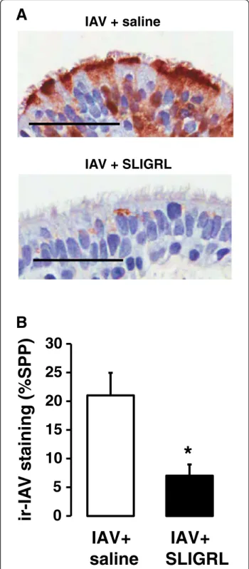
The current study has established that SLIGRL-amide inhibits IAV infectivity in vivo and ex vivo via mecha-nisms that are independent of MRGPRC11 and of capsaicin-sensitive sensory nerves, and also independent of the sensory neuropeptides substance P and CGRP and their receptors. However, it is important to note that vagal bronchopulmonary C-fibers in the mouse are het-erogeneous – a substantial fraction of C-fiber afferent nerves in the mouse respiratory system are capsaicin-insensitive [51], and that a fraction of capsaicin-sensitive C-fibers are non-peptidergic [20]. Thus, we cannot ex-clude the possibility that SLIGRL-amide may exert its antiviral activities through actions on sub-populations of capsaicin-insensitive, nonpeptidergic neurons. Neverthe-less, the current investigation builds on our previous studies that demonstrate SLIGRL-amide does not inhibit IAV infectivity via PAR-2, by direct antiviral or by im-munomodulatory processes [7]. Of particular interest, the current study has shown that the anti-IAV effects of SLIGRL-amide observed in murine airways extend to human airways, although further studies are required to identify its specific molecular target.
SLIGRL-amide and related peptides have been re-ported to directly stimulate secretory pathways in the airways [46, 52–54]. Importantly, components of airway secretions, including the mucin MUC5AC exert protect-ive roles against influenza A virus [55]. Thus, the release of endogenous antiviral substances in mucus may con-tribute to the anti-IAV activity of SLIGRL-amide. In the current study, SLIGRL-amide promoted the release of intracellular mucins from mouse tracheal epithelium, as
IAV + saline
IAV + SLIGRL
B
A
0
5
10
15
20
25
30
IAV+
saline
IAV+
SLIGRL
ir-IA
V
s
taining (%SPP)
*
determined from microscopic imaging of AB-PAS-stained airway sections. The purinergic receptor agonist ATP, an established key secretagogue for airway epithe-lium [56–58], also promoted the release of intracellular mucins from mouse isolated tracheal epithelium, and moreover, inhibited IAV infectivity. These findings pro-vide preliminary epro-vidence that SLIGRL-amide may in-hibit IAV infectivity by stimulating the secretion of endogenous substances with anti-IAV activity. Neverthe-less, it remains to be determined whether the anti-IAV activity of SLIGRL-amide involves the secretion of sialic acid-rich mucins [55], or other anti-IAV molecules such as cationic host defence peptides (β-defensins, cathelici-dins) [59] or phospholipids such as palmitoyl-oleoyl-phosphatidylglycerol (POPG) [60].
Conclusion
The current study using a combination ofin vivoandex vivo approaches provides compelling evidence that the anti-IAV activity of SLIGRL-amide in murine airways occurs independently of the sensory nerve receptor MRGPRC11 and of neuropeptide release from TRPV1-expressing sensory nerves. Preliminary data show that both SLIGRL-amide and ATP release airway secretions and inhibit IAV infection, raising the possibility that SLIGRL-amide inhibits IAV infectivity via the release of antiviral substances from the airway epithelium. Additional release and intervention studies, particularly in human airways, will provide invaluable insight into the molecular target and cellular signalling processes through which SLIGRL-amide inhibits IAV infectivity.
Acknowledgements
The authors acknowledge the facilities, and the scientific and technical assistance of the Australian Microscopy & Microanalysis Research Facility at the Centre for Microscopy, Characterisation & Analysis, The University of Western Australia, a facility funded by The University of Western Australia, and the State and Commonwealth Governments of Australia.
Authors’contributions
Participated in research design: AC, TM, PM, LH, XD, PH. Conducted experiments: AC, TM, LH. Performed data analysis: AC, TM, PH. Wrote or contributed to writing of manuscript: AC, TM, PM, XD, PH. All authors read and approved the final manuscript.
Competing interests
The authors declare that they have no competing interests.
Author details
1
School of Medicine and Pharmacology, University of Western Australia, Crawley, WA 6009, Australia.2School of Anatomy, Physiology & Human
Biology, University of Western Australia, Crawley 6009, WA, Australia.
3Howard Hughes Medical Institute, Johns Hopkins University School of
Medicine, Baltimore, MD 21205, USA.
Received: 5 February 2016 Accepted: 15 May 2016
References
1. Influenza (Seasonal) Fact Sheet No.211 (2014) [http://www.who.int/ mediacentre/factsheets/fs211/en/#] Accessed on 27 Jan 2016.
2. Beigel J, Bray M. Current and future antiviral therapy of severe seasonal and avian influenza. Antiviral Res. 2008;78:91–102.
3. Hayden F. Developing new antiviral agents for influenza treatment: what does the future hold? Clin Infect Dis. 2009;48 Suppl 1:S3–S13.
4. Bright RA, Medina MJ, Xu X, Perez-Oronoz G, Wallis TR, Davis XM, Povinelli L, Cox NJ, Klimov AI. Incidence of adamantane resistance among influenza A (H3N2) viruses isolated worldwide from 1994 to 2005: a cause for concern. Lancet. 2005;366:1175–81.
5. Li TC, Chan MC, Lee N. Clinical Implications of Antiviral Resistance in Influenza. Viruses. 2015;7:4929–44.
6. Spanakis N, Pitiriga V, Gennimata V, Tsakris A. A review of neuraminidase inhibitor susceptibility in influenza strains. Expert Rev Anti Infect Ther. 2014; 12:1325–36.
7. Betts RJ, Mann TS, Henry PJ. Inhibitory influence of the hexapeptidic sequence SLIGRL on influenza A virus infection in mice. J Pharmacol Exp Ther. 2012;343:725–35.
8. Liu Q, Weng HJ, Patel KN, Tang Z, Bai H, Steinhoff M, Dong X. The distinct roles of two GPCRs, MrgprC11 and PAR2, in itch and hyperalgesia. Sci Signal. 2011;4:ra45.
9. Lee MG, Dong X, Liu Q, Patel KN, Choi OH, Vonakis B, Undem BJ. Agonists of the MAS-related gene (Mrgs) orphan receptors as novel mediators of mast cell-sensory nerve interactions. J Immunol. 2008;180:2251–5. 10. Solinski HJ, Gudermann T, Breit A. Pharmacology and signaling of
MAS-related G protein-coupled receptors. Pharmacol Rev. 2014;66:570–97. 11. Dong X, Han S, Zylka MJ, Simon MI, Anderson DJ. A diverse family of GPCRs
expressed in specific subsets of nociceptive sensory neurons. Cell. 2001;106:619–32. 12. Grazzini E, Puma C, Roy MO, Yu XH, O'Donnell D, Schmidt R, Dautrey S,
Ducharme J, Perkins M, Panetta R. Sensory neuron-specific receptor activation elicits central and peripheral nociceptive effects in rats. Proc Natl Acad Sci U S A. 2004;101:7175–80.
13. Han SK, Dong X, Hwang JI, Zylka MJ, Anderson DJ, Simon MI. Orphan G protein-coupled receptors MrgA1 and MrgC11 are distinctively activated by RF-amide-related peptides through the Galpha q/11 pathway. Proc Natl Acad Sci U S A. 2002;99:14740–5.
14. Lembo PM, Grazzini E, Groblewski T, O'Donnell D, Roy MO, Zhang J, Hoffert C, Cao J, Schmidt R, Pelletier M, et al. Proenkephalin A gene products activate a new family of sensory neuron–specific GPCRs. Nat Neurosci. 2002;5:201–9. 15. Burstein ES, Ott TR, Feddock M, Ma JN, Fuhs S, Wong S, Schiffer HH, Brann MR,
Nash NR. Characterization of the Mas-related gene family: structural and functional conservation of human and rhesus MrgX receptors. Br J Pharmacol. 2006;147:73–82.
16. Guan Y, Liu Q, Tang Z, Raja SN, Anderson DJ, Dong X. Mas-related G-protein-coupled receptors inhibit pathological pain in mice. Proc Natl Acad Sci U S A. 2010;107:15933–8.
17. Verhein KC, Fryer AD, Jacoby DB. Neural control of airway inflammation. Curr Allergy Asthma Rep. 2009;9:484–90.
18. Kollarik M, Ru F, Brozmanova M. Vagal afferent nerves with the properties of nociceptors. Auton Neurosci. 2010;153:12–20.
19. Potenzieri C, Meeker S, Undem BJ. Activation of mouse bronchopulmonary C-fibres by serotonin and allergen-ovalbumin challenge. J Physiol. 2012;590:5449–59. 20. Nassenstein C, Taylor-Clark TE, Myers AC, Ru F, Nandigama R, Bettner W,
Undem BJ. Phenotypic distinctions between neural crest and placodal derived vagal C-fibres in mouse lungs. J Physiol. 2010;588:4769–83. 21. Canning BJ, Spina D: Sensory nerves and airway irritability. Handb Exp
Pharmacol. 2009:194;139–183.
22. Lamb JP, Sparrow MP. Three-dimensional mapping of sensory innervation with substance p in porcine bronchial mucosa: comparison with human airways. Am J Respir Crit Care Med. 2002;166:1269–81.
23. White MR, Helmerhorst EJ, Ligtenberg A, Karpel M, Tecle T, Siqueira WL, Oppenheim FG, Hartshorn KL. Multiple components contribute to ability of saliva to inhibit influenza viruses. Oral Microbiol Immunol. 2009;24:18–24. 24. Liu Q, Tang Z, Surdenikova L, Kim S, Patel KN, Kim A, Ru F, Guan Y, Weng HJ, Geng Y, et al. Sensory neuron-specific GPCR Mrgprs are itch receptors mediating chloroquine-induced pruritus. Cell. 2009;139:1353–65. 25. Williams K, Mackenzie JS. Influenza infections during pregnancy in the
mouse. J Hyg (Lond). 1977;79:249–57.
26. Fazekas De St Groth S, White DO. An improved assay for the infectivity of in influenza viruses. J Hyg (Lond). 1958;56:151–62.
28. Kawabata A, Oono Y, Yonezawa D, Hiramatsu K, Inoi N, Sekiguchi F, Honjo M, Hirofuchi M, Kanke T, Ishiwata H. 2-Furoyl-LIGRL-NH2, a potent agonist for proteinase-activated receptor-2, as a gastric mucosal cytoprotective agent in mice. Br J Pharmacol. 2005;144:212–9.
29. Morris JB, Symanowicz PT, Olsen JE, Thrall RS, Cloutier MM, Hubbard AK. Immediate sensory nerve-mediated respiratory responses to irritants in healthy and allergic airway-diseased mice. J Appl Physiol (1985). 2003;94:1563–71. 30. Scheerens H, Buckley TL, Muis T, Van Loveren H, Nijkamp FP. The involvement
of sensory neuropeptides in toluene diisocyanate-induced tracheal hyperreactivity in the mouse airways. Br J Pharmacol. 1996;119:1665–71. 31. Cheah EY, Burcham PC, Mann TS, Henry PJ. Acrolein relaxes mouse isolated
tracheal smooth muscle via a TRPA1-dependent mechanism. Biochem Pharmacol. 2014;89:148–56.
32. Taylor SJ, Mann TS, Henry PJ. Influence of influenza A infection on capsaicin-induced responses in murine airways. J Pharmacol Exp Ther. 2012;340:377–85. 33. Agrawal A, Rengarajan S, Adler KB, Ram A, Ghosh B, Fahim M, Dickey BF.
Inhibition of mucin secretion with MARCKS-related peptide improves airway obstruction in a mouse model of asthma. J Appl Physiol (1985). 2007;102:399–405.
34. Evans CM, Williams OW, Tuvim MJ, Nigam R, Mixides GP, Blackburn MR, DeMayo FJ, Burns AR, Smith C, Reynolds SD, et al. Mucin is produced by clara cells in the proximal airways of antigen-challenged mice. Am J Respir Cell Mol Biol. 2004;31:382–94.
35. Hollenberg MD, Saifeddine M. al-Ani B: Proteinase-activated receptor-2 in rat aorta: structural requirements for agonist activity of receptor-activating peptides. Mol Pharmacol. 1996;49:229–33.
36. Cattaruzza F, Cenac N, Barocelli E, Impicciatore M, Hyun E, Vergnolle N, Sternini C. Protective effect of proteinase-activated receptor 2 activation on motility impairment and tissue damage induced by intestinal ischemia/ reperfusion in rodents. Am J Pathol. 2006;169:177–88.
37. Elekes K, Helyes Z, Nemeth J, Sandor K, Pozsgai G, Kereskai L, Borzsei R, Pinter E, Szabo A, Szolcsanyi J. Role of capsaicin-sensitive afferents and sensory neuropeptides in endotoxin-induced airway inflammation and consequent bronchial hyperreactivity in the mouse. Regul Pept. 2007;141: 44–54.
38. Morris JB, Wilkie WS, Shusterman DJ. Acute respiratory responses of the mouse to chlorine. Toxicol Sci. 2005;83:380–7.
39. Steinhoff MS, von Mentzer B, Geppetti P, Pothoulakis C, Bunnett NW. Tachykinins and their receptors: contributions to physiological control and the mechanisms of disease. Physiol Rev. 2014;94:265–301.
40. Chauhan VS, Kluttz JM, Bost KL, Marriott I. Prophylactic and therapeutic targeting of the neurokinin-1 receptor limits neuroinflammation in a murine model of pneumococcal meningitis. J Immunol. 2011;186:7255–63. 41. Yang Y, Yan M, Zhang H, Wang X. Substance P participates in
immune-mediated hepatic injury induced by concanavalin A in mice and stimulates cytokine synthesis in Kupffer cells. Exp Ther Med. 2013;6:459–64. 42. Nystedt S, Emilsson K, Wahlestedt C, Sundelin J. Molecular cloning of a
potential proteinase activated receptor. Proc Natl Acad Sci U S A. 1994;91: 9208–12.
43. Ossovskaya VS, Bunnett NW. Protease-activated receptors: contribution to physiology and disease. Physiol Rev. 2004;84:579–621.
44. Ramachandran R, Noorbakhsh F, Defea K, Hollenberg MD. Targeting proteinase-activated receptors: therapeutic potential and challenges. Nat Rev Drug Discov. 2012;11:69–86.
45. Rothmeier AS, Ruf W. Protease-activated receptor 2 signaling in inflammation. Semin Immunopathol. 2012;34:133–49.
46. Abey HT, Fairlie DP, Moffatt JD, Balzary RW, Cocks TM. Protease-activated receptor-2 peptides activate neurokinin-1 receptors in the mouse isolated trachea. J Pharmacol Exp Ther. 2006;317:598–605.
47. Zhao A, Shea-Donohue T. PAR-2 agonists induce contraction of murine small intestine through neurokinin receptors. Am J Physiol Gastrointest Liver Physiol. 2003;285:G696–703.
48. Bowden JJ, Baluk P, Lefevre PM, Schoeb TR, Lindsey JR, McDonald DM. Sensory denervation by neonatal capsaicin treatment exacerbates Mycoplasma pulmonis infection in rat airways. Am J Physiol. 1996;270:L393–403.
49. Raemdonck K, de Alba J, Birrell MA, Grace M, Maher SA, Irvin CG, Fozard JR, O'Byrne PM, Belvisi MG. A role for sensory nerves in the late asthmatic response. Thorax. 2012;67:19–25.
50. Schelegle ES, Walby WF. Vagal afferents contribute to exacerbated airway responses following ozone and allergen challenge. Respir Physiol Neurobiol. 2012;181:277–85.
51. Kollarik M, Dinh QT, Fischer A, Undem BJ. Capsaicin-sensitive and -insensitive vagal bronchopulmonary C-fibres in the mouse. J Physiol. 2003;551:869–79. 52. Kunzelmann K, Sun J, Markovich D, Konig J, Murle B, Mall M, Schreiber R.
Control of ion transport in mammalian airways by protease activated receptors type 2 (PAR-2). FASEB J. 2005;19:969–70.
53. Lee HJ, Yang YM, Kim K, Shin DM, Yoon JH, Cho HJ, Choi JY. Protease-activated receptor 2 mediates mucus secretion in the airway submucosal gland. PLoS One. 2012;7, e43188.
54. Lin KW, Park J, Crews AL, Li Y, Adler KB. Protease-activated receptor-2 (PAR-2) is a weak enhancer of mucin secretion by human bronchial epithelial cells in vitro. Int J Biochem Cell Biol. 2008;40:1379–88.
55. Ehre C, Worthington EN, Liesman RM, Grubb BR, Barbier D, O'Neal WK, Sallenave JM, Pickles RJ, Boucher RC. Overexpressing mouse model demonstrates the protective role of Muc5ac in the lungs. Proc Natl Acad Sci U S A. 2012;109:16528–33.
56. Adler KB, Tuvim MJ, Dickey BF. Regulated mucin secretion from airway epithelial cells. Front Endocrinol (Lausanne). 2013;4:129.
57. Fahy JV, Dickey BF. Airway mucus function and dysfunction. N Engl J Med. 2010;363:2233–47.
58. Kreda SM, Okada SF, van Heusden CA, O'Neal W, Gabriel S, Abdullah L, Davis CW, Boucher RC, Lazarowski ER. Coordinated release of nucleotides and mucin from human airway epithelial Calu-3 cells. J Physiol. 2007;584: 245–59.
59. Gwyer Findlay E, Currie SM, Davidson DJ. Cationic host defence peptides: potential as antiviral therapeutics. Biodrugs. 2013;27:479–93.
60. Numata M, Kandasamy P, Nagashima Y, Posey J, Hartshorn K, Woodland D, Voelker DR. Phosphatidylglycerol suppresses influenza A virus infection. Am J Respir Cell Mol Biol. 2012;46:479–87.
• We accept pre-submission inquiries
• Our selector tool helps you to find the most relevant journal
• We provide round the clock customer support
• Convenient online submission
• Thorough peer review
• Inclusion in PubMed and all major indexing services
• Maximum visibility for your research
Submit your manuscript at www.biomedcentral.com/submit