Original Research Article
Incidence of congenital anomalies in newborn in tertiary care hospital
V. Narmadha, M. Nirmala*
INTRODUCTION
Malformations and dysplasia both affect intrinsic structures. A malformation is a primary structural defect arising from a localized error in morphogenesis, resulting in the abnormal formation of a tissue or organ.1 The
present consensus emerged that approximately 3% of newborn children are affected by significant congenital malformations. Hospital-based prospective studies of live and still births from different parts of India, the incidence
of congenital malformations has varied from 2.5-40 / 1000 births. Etiological agents could be broadly divided into 4 categories specific teratogenic agents (8-10%), monogenic (15-25%), chromosomal (15.28%) and unknown (including multifactorial) (40-65%).2 A
teratogen is an agent, applied during prenatal life produces a permanent postnatal damage, change in morphology or function. Such agents can be chemicals, drugs, virus, or physical or deficiency states.3 Majority of
the malformations are due to complex involvement of genetic and environmental factors. As regards prenatal Department of Paediatrics, Government Mohan Kumaramangalam Medical College Hospital, Salem, Tamil Nadu, India
Received: 30 April 2019
Accepted: 08 May 2019
*Correspondence:
Dr. M. Nirmala,
E-mail: drnirmala94@gmail.com
Copyright: © the author(s), publisher and licensee Medip Academy. This is an open-access article distributed under the terms of the Creative Commons Attribution Non-Commercial License, which permits unrestricted non-commercial use, distribution, and reproduction in any medium, provided the original work is properly cited.
ABSTRACT
Background: Just about three decades ago (1976) congenital malformations comprised 8% of perinatal deaths, from available data and ranked fifth as a cause of perinatal mortality. But the trend is rapidly changing over the years. perinatal death was due to congenital malformation, is the second commonest cause. This changing trend over years warns us that with the control of nutritional and infectious diseases, congenital malformations will come to the forefront as it is in India. To find out the incidence of congenital anomalies in stillbirth. And the probable etiology of congenital anomalies.
Methods: The study was conducted at Government Mohan Kumaramangalam Medical College Hospital, Salem in the year 2017 August- September 2018. Totally 5000 babies born of consecutive deliveries were taken for the study, over the period of one year. All mothers were interrogated within 48 Hours of delivery as per the proforma prepared, which contains the following particulars like, maternal and paternal age, consanguinity, detailed antenatal history with reference to exposure to teratogens, especially during 1st Trimester.
Results: Of the five thousand consecutive deliveries 48 deliveries were multiple delivers and a number of stillbirths were 108. The incidence of congenital anomalies was 30.4 per 1000 live birth (152 cases). Major malformations were present in 20.8 per 1000 (104 cases) while minor malformations were 9.6 per 1000 (48 cases).
Conclusions: Incidence of malformation were higher in preterm babies 6.31%. Incidence of malformations were higher in male babies, especially genitourinary system anomalies. Antenatal events in the 1st trimester like fever, drug intake could be implicated in the etiology of malformations especially neural tube defects in our study.
Keywords: Cardiovascular abnormality, Central nervous system malfunction, Congenital anomalies, Diabetes mellitus
infections, as a cause of malformations, very little information is available in our country.4 As most of the
Indian studies are hospital-based and hence do not represent the problem in the community as hospital records are based by inclusion of high-risk mothers, further many of births in India especially in rural areas occur at home and infants with abnormalities are not brought to the hospital for various reasons, and there are many difficulties in the evaluation of stillbirths in the community.5 There is no standard protocol in the study of
malformation in India. Excellent normal standards for physical features are available for Western populations and there is an urgent need to develop our own standards for Indian population by the multicentric collaborative study.6 Centers with good laboratory facilities like
chromosomal study, biochemical screening for inborn errors of metabolism, ultrasonography and radiological services should develop standard protocols for collection of specimens like blood and skin biopsy, their preservation and transportation to regional centers from the periphery. These centers should take part in training the pathologist in autopsy studies of fetuses and newborns.7 Malformations due to single gene mutation
and chromosomal disorders and the only way these could be limited are by genetic counselling and induced abortions in areas where the genetic factors operate as a cause of malformations through the practice of consanguineous marriages, education of people against such practice could reduce the incidence of malformations.8
METHODS
The study was conducted at Government Mohan Kumaramangalam Medical College Hospital, Salem in the year 2017 August-September 2018. Totally 5000 babies born of consecutive deliveries were taken for the study, over the period of one year. All mothers were interrogated within 48 Hours of delivery as per the proforma prepared, which contains the following particulars like, maternal and paternal age, consanguinity, detailed antenatal history with reference to exposure to teratogens, especially during 1st Trimester. and medical disease complicating pregnancy like Diabetes, Rheumatic heart disease, Hypertension, detailed obstetric history
with reference to previous abortions and stillbirth. Routine investigations like hemoglobin, urine analysis, blood grouping, and Rh typing, VDRL, and HIV were done for all cases and blood sugar, renal function, and liver function tests were done when indicated for mothers. Every newborn was subjected to detailed examination from head to toe within 48 hours of birth. Assessment of the newborn included birth weight, sex, live born/stillborn, gestational age and details of congenital malformations. All were recorded in a pre-designed proforma. A gavage tube was used to check choanal and esophageal atresia, Anorectal anomaly in suspected cases. All the newborns were followed up every day until the time of discharge from the hospital. Necessary investigations were done wherever required. Cardiovascular anomalies were subjected to ECG, X-ray chest AP view and ECHO cardiography. The umbilical cord stump was examined to note down the anomalies of arteries and vein. The placenta was examined in Detail.
Statistical analysis
Data were entered using Microsoft Excel and analyzed using STATA software. A continuous variable was analyzed using the student ‘t’ test which was used to determine the significant difference.
RESULTS
Of the five thousand consecutive deliveries 48 deliveries were multiple delivers and a number of stillbirths were 108. The incidence of congenital anomalies was 30.4 per 1000 live birth (152 cases). Major malformations were present in 20.8 per 1000 (104 cases) while minor malformations were 9.6 per 1000 (48 cases).
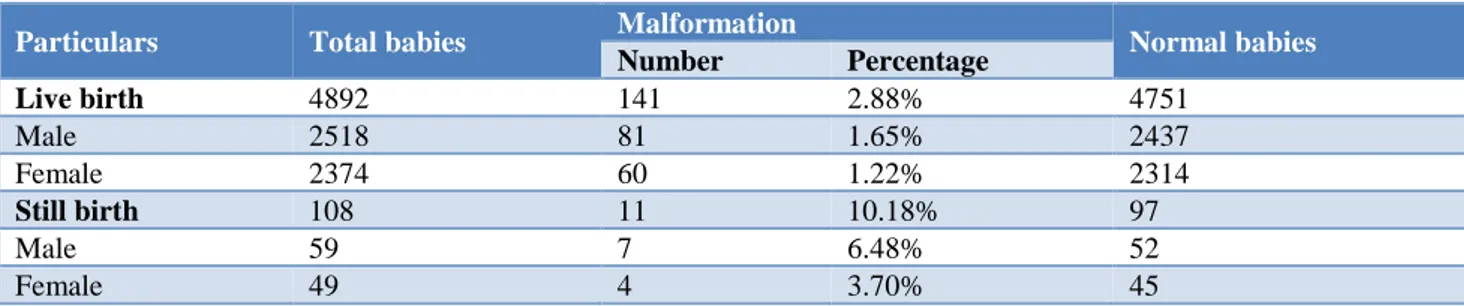
Table 1 shows Congenital malformations were seen more in stillbirths as compared to the live birth, the frequency being 10.18% and 2.88% respectively and was found to be statistically significant (p <0.05).88 babies were males and 64 were females. Male to female ratio was 1.35:1. In our study, there was an increased incidence of congenital malformations in male babies and it was statistically significant (p<0.001).
Table 1: Congenital malformation: frequency and distribution in relation to sex and live and stillbirth.
Particulars Total babies Malformation Normal babies
Number Percentage
Live birth 4892 141 2.88% 4751
Male 2518 81 1.65% 2437
Female 2374 60 1.22% 2314
Still birth 108 11 10.18% 97
Male 59 7 6.48% 52
Table 2 shows of 7 cases of hydramnios 2 mothers delivered babies with esophageal atresia/ tracheoesophageal fistula, 3 babies with hydrocephalus and 2 babies with anencephaly. Four mothers gave a history of diabetes during the antenatal period, 1 baby had congenital cyanotic heart disease and died before doing ECHO. Other 2 baby had bilateral CTEV and 4th baby had ASD.
Table 3 shows in the study it was observed that majority of anomalies were found in the central nervous system.
The other systems where major anomalies recorded were the gastrointestinal system and the skeletal system.
Table 4 shows one baby with agenesis of corpus callosum born of 2nd-degree consanguinity, it was the 3rd child. Previous two babies died in the neonatal period and this child presented with status epilepticus and diagnosis was established by cranial ultrasonogram. The baby died on the first postnatal day.
Table 2: Correlation of complication during pregnancy with congenital malformations.
Complication Total babies Malformation Normal Babies
Number Percentage
Diabetes 12 4 33.33% 8
Hydramnios 22 7 31.81% 15
PIH 46 3 6.52% 43
Table 3: Systemic distribution of congenital malformation.
Systems Malformation No. of
Live Birth
No. of Still Birth
Neonatal Death
MB Per 1000 Birth
No. %
Central nervous system 28 18.42% 23 5 6 5.6 Gastro intestinal system 23 15.13% 19 4 3 4.6 Skeletal system 20 13.15% 20 Nil Nil 4
Oro facial 12 7.89% 12 Nil Nil 2.4
Cardio Vascular 11 7.23% 11 Nil 2 2.2
Genito Urinary 10 6.57% 8 2 Nil 2
Minor malformation 48 31.57% 48 Nil Nil 9.6
Table 4: Analysis of central nervous system malformation.
Malformation No % of MB LB SB M F SGA AGA MB/1000 birth
Meningomyelocele 7 4.60% 7 Nil 5 2 5 2 1.4 Anencephaly 5 3.28% 3 2 3 2 5 Nil 1 Spina bifida 4 2.63% 4 Nil 2 2 2 2 0.8 Microcephaly 4 2.63% 4 Nil 3 1 4 Nil 0.8 Hydrocephalus 3 1.97% 2 1 2 1 2 1 0.6 Hydrocephalus with
meningomyelocele 3 1.97% 1 2 2 1 2 1 0.6 Craniosynostosis 1 0.65% 1 Nil 1 Nil 1 Nil 0.2 Agenesis of corpus callosum 1 0.65% 1 Nil 1 Nil 1 Nil 0.2
Table 5: Analysis of cardiovascular system.
Malformation No % of MB Live Birth Still Birth M F SGA AGA MB/1000 birth
VSD 5 3.28% 5 Nil 3 2 3 2 1
VSD with PS 2 0.4% 2 Nil 2 Nil 1 1 0.4 PS 1 0.65% 1 Nil 1 Nil Nil 1 0.2
ASD 2 0.4% 2 Nil 1 1 1 1 0.4
Table 5 shows most common congenital heart disease observed in the study is VSD (3.28%). Out of the 2 babies with congenital malformations in a cardiovascular system born to a diabetic mother, one baby presented with congestive cardiac failure and cyanosis at birth. Chest X-ray showed cardiomegaly and ECG showed biventricular hypertrophy. Echocardiogram was not done
due to the very poor general condition and the baby died after 36 hrs of life. The other baby had ASD. Antenatally two mothers gave a history of drug intake during the first trimester. In one case mother had taken anticonvulsants phenytoin and phenobarbitone and delivered with VSD. 5 babies born of consanguineous marriage, 3 of them were 2nd-degree consanguinity.
Table 6: Analysis of gastrointestinal system.
Malformation No % of MB Live Birth Still Birth M F SGA AGA MB/1000 birth
Cleft lip and cleft
palate 13 8.55% 11 2 6 7 8 5 2.6 Cleft lip 5 3.28% 5 Nil 4 1 3 2 1 Ano rectal anomaly 5 3.28% 5 Nil 2 3 1 4 1 Cleft palate 3 1.97% 3 Nil 2 1 Nil 3 0.6
TEF 3 1.97% 3 Nil 2 1 2 1 0.6
Megacolon 2 1.31% 2 Nil 1 1 Nil 2 0.4 Exomphalus 2 1.31% 1 1 1 1 Nil 2 0.4 Pierre Robin
syndrome 1 0.65% Nil 1 Nil 1 Nil 1 0.2 Omphalocele 1 0.65% 1 Nil 1 Nil 1 Nil 0.2
Table 7: Analysis of genitourinary system.
Malformation No % of MB Live Birth Still Birth M F SGA AGA MB/1000 birth
Undescended testis 4 2.63% 4 Nil 4 Nil 2 2 0.8 Hypospadias 3 1.97% 2 1 3 Nil 1 2 0.6 Epispadias 1 0.65% 1 Nil 1 Nil Nil 1 0.2 Hydronephrosis 1 0.65% 1 Nil 1 Nil 1 Nil 0.2 Micropenis 1 0.65% Nil 1 1 Nil Nil 1 0.2
Table 8: Skeletal anomalies.
Malformation No % of MB Live Birth Still Birth M F SGA AGA MB/1000 birth
CTEV 10 6.57% 10 Nil 3 7 4 6 2
Polydactyly 6 3.94% 6 Nil 2 4 2 4 1.2 Osteogenesis
Imperfect
1 0.65% 1 Nil Nil 1 Nil 1 0.2
Polysyndactyly 1 0.65% 1 Nil Nil 1 Nil 1 0.2 Arthrogryphosis 1 0.65% 1 Nil Nil 1 1 Nil 0.2 Achondroplasia 1 0.65% 1 Nil 1 Nil Nil 1 0.2
Table 6 shows cleft lip with palate formed the major group. 3 of them had multiple anomalies - CTEV in one case, microcephaly in two. Mothers gave a history of drug intake in early pregnancy. Next major group is anorectal malformation. Total of 5 cases, 2 cases were of low anorectal anomalies. Three of them were female children. In this group, we had two male children with high anomaly. Three cases of tracheal esophageal fistulae, all the cases were diagnosed and underwent surgery.
Table 7 shows Analysis of the genitourinary system showed 4 out of 10 cases were undescended testis and 3 cases showed hypospadias and one among them was stillborn with abdominal distension. The autopsy of the stillborn showed bilateral hydronephrosis with posterior urethral valve.
delivered by the breech, 2 babies were preterm and 1 had a cleft lip. We had a case of osteogenesis imperfecta diagnosed immediately after birth.
DISCUSSION
Congenital malformations present at birth have been known since earliest times. Primitive man’s interest and fascination with these bizarre phenomena have found expression in drawings, carvings, and scriptures.9 During
the last 25 years numerous publications of experimental teratology and of many clinical genetic studies the elucidation of the multiple and intertwined causes of human malformation is still in an early state. Nevertheless, only through such understanding will prevention become a reality.10 The frequency of
congenital malformation in the present study is 30.4 per 1000 birth. This rate in the study is comparable to the incidence of congenital malformation in other hospital-based studies of life and stillbirths. Analysis of 22,434 births from various part of India in different studies varies from 8.6-20.2 per 1000 births. Population-based studies of live births, study by J.S. Anand et al is of great interest as it involved a prospective survey of a population of 1,00,000 in Delhi.11 The incidence was 26.2
/1000. A survey of these infants after an interval of years was carried out, wherein 3816 children were re-examined. Among this new malformation was detected in 54 children, even though the examination at birth was normal. On adding the later diagnosed malformations 14.15 per 1000 to those detected at birth, a total frequency of 40.37 per 1000 was obtained in the birth cohort. The incidence of major malformations increased from 11.07 to 16.5 per 1000. Most of the increase was accounted for by cardiovascular and central nervous system malformations.12 Central nervous system and
Gastrointestinal system including orofacial group were the most commonly involved in the present study. The skeletal system is the third commonest and 4th one is the cardiovascular system. Our results are also comparable with other studies. The commonest system involved in other studies where the central nervous system.13 In
contrast Kesavan P, et al in a community-based study found musculoskeletal anomalies as the commonest.
Terry et al, found gastrointestinal anomalies to occur more commonly among mothers of Indian origin.in the present study, out of central nervous system malformations, neural tube defects were the commonest 3.2 per/1000.14 Reports of neural tube defects from
various parts of India have given the incidence ranging from 0.5 to 8/1000. It has been suggested that a high incidence of neural tube defects and that the high incidence is maintained even after migration to the U.K. Khanna K.K and colleagues reported such a high incidence of neural tube defects not only in people of Punjab but also in those of Rajasthan. In most parts of the country musculoskeletal disorders are the commonest malformations. It is likely that many of the musculoskeletal disorders are deformations rather than
malformations, representing an aberrant form or position of a formed structure, Minor anomalies are defined as unusual morphologic features that are of no serious medical or cosmetic consequences to the patient. The value of this recognition is that they may serve as indicators, of altered morphogenesis or may constitute valuable clue in the diagnosis of a specific pattern of malformations.15 Most of the babies have a single minor
anomaly, but several minor anomalies in the same individual indicate a more serious problem. In our study 9.6 per 1000 births (48 cases) had minor anomalies. Five of these children had birth Asphyxia. Incidence of minor anomalies reported by Kulshrestha et al, was 4 per 1000 births. The present study shows 67.11% of malformed babies were born of consanguineous marriages.
There is a definite increase in the rate of malformations in the offspring of consanguineous marriages. 32.89% among nonconsanguineous, 38.81% among second-degree consanguinity and 28.28% among 3rd-second-degree consanguinity.16 M.L Kulkarni et al from Trivandrum
who reported that consanguineous parents had 3.59% malformed offspring while nonconsanguineous parents had only 1.69% offspring with malformations. In the global study of congenital malformations, the rate of neural tube defect was observed to be higher in the offspring of consanguineous marriages in Alexandria and Egypt. However, the data from India on this aspect are equivocal.17 Congenital malformations were responsible
for 12.3% perinatal deaths in the present study and fifth as the cause of perinatal mortality after prematurity, birth asphyxia, septicemia, and respiratory distress.18
Mathur B.C et al, have attributed to 17.8% and 15.7% of perinatal deaths to congenital malformations. Congenital malformations compromise 8% of the perinatal mortality in India as estimated from the data available from hospital-based studies76 and fifth as a cause of perinatal mortality after asphyxia, respiratory problems, infections, and birth trauma.19,20
CONCLUSION
Congenital malformations especially neural tube defects were common in babies born of second degree consanguineous marriage. (Significant contribution of second-degree consanguinity was noted in all major malformations 38.81%. Antenatal events in the 1st trimester like fever, drug intake could be implicated in the etiology of malformations especially neural tube defects in our study. Prenatal diagnosis using ultrasound is accurate but performed late. So, a high index of suspicion and early scanning in high-risk mothers are necessary.
Funding: No funding sources Conflict of interest: None declared
REFERENCES
1. Singh M. Hospital based data on perinatal and neonatal mortality in Indian Paediatrics. 1986;23:579-84.
2. Choudhury AR, Mukherjee M, Sharma A, Talukder G, Ghosh PK. Study of 1,26,266 consecutive births for major congenital defects. Indian J.Pediatr. 1989,56:493-499.
3. Chinara PK, Singh S. East-West differentials in congenital malformations in India. Indian J Pediatr. 1982 May-Jun;49(398):325-9.
4. Singh DN. Fetal environment and congenital malformations. Indian J. Pediatr. 1989,56:575-584. 5. Saiffulah S, Chandra RK, Pathak IC, Dhall GI.
Congenital malformations in new born. Prospective longitudinal study. Indian Pediatrics. 1957;4:251. 6. Fraser FC, Fainstat TD. Causes of congenital
defects: A Review. AMA Am J Dis Child. 1951;82(5):593-603.
7. Funderburk SJ, Guthrie D, Meldrum D. Outcome of pregnancies complicated by early vaginal bleeding. Br J Obstet Gynaecol. 1980 Feb;87(2):100-5. 8. Ghosh S, Bali L. Congenital malformations in the
newborn. Indian J Child Health. 1963 Jul;12:448-52.
9. Ghosh S, Ramanujacharyulu TK, Hooja V, Madhavan S. Mortality pattern in an urban birth cohort. Indian J Med Res. 1979 Apr;69:616-23. 10. Goravalingappa JP, Nashi HK. Congenital
malformations in a study of 2398 consecutive births. Indian J Med Res. 1979 Jan;69:140-6.
11. Kalter H, Warkany J. Medical progress. Congenital malformations: etiologic factors and their role in prevention (first of two parts). N Engl J Med. 1983 Feb 24;308(8):424-31.
12. Anand JS, Javadekar BB, Belani M. Congenital malformations in 2000 consecutive births. Indian Pediatr. 1988 Sep;25(9):845-51.
13. Dadhwal V, Kochhar S, Mittal S, Kumar S, Agarwal S, Arora V, et al. Fetal gastrointestinal malformations. Indian J Ped. 2001;68(1):27-30. 14. Kesavan P, Nataraja U, Murugesan K,
Ramakrishnan MS. Congenital malformations and consanguinity. In: I.C. Verma. Medical genetics in India. 1st ed. 1978:65.
15. Khanna KK, Prasad N, congenital malformations in newborn. Indian J Pediatr. 1967;34:63.
16. Kulshrestha R, Nath LM, Upadhyaya P. Congenital malformations in live born infants in a rural community. Indian Pediatr. 1983 Jan;20(1):45-9. 17. Kulkarni ML, Mathew MA, Reddy V. The range of
neural tube defects in Southern India. Arch dis childhood. 1989;64:201-4.
18. Verma M, Chhatwal J, Singh D. Congenital malformations- a retrospective study of 10,000 cases. Indian J Pediatr. 1991 Mar-Apr;58(2):245-52.
19. Mathur BC, Karan S, Vijyadevi KK. Congenital malformations in newborns. Indian paediatr.1975,12:179-81.
20. Mcintosh R, Merritt KK, Richards MR, Samuels MH, Bellows MT. The incidence of congenital malformations: a study of 5,964 pregnancies. Pediatrics. 1954 Nov;14(5):505-22.