R E V I E W
Open Access
Neuropeptides in asthma, chronic
obstructive pulmonary disease and cystic
fibrosis
Kalina R. Atanasova and Leah R. Reznikov
*Abstract
The nervous system mediates key airway protective behaviors, including cough, mucus secretion, and airway
smooth muscle contraction. Thus, its involvement and potential involvement in several airway diseases has become
increasingly recognized. In the current review, we focus on the contribution of select neuropeptides in three
distinct airway diseases: asthma, chronic obstructive pulmonary disease (COPD), and cystic fibrosis. We present data
on some well-studied neuropeptides, as well as call attention to a few that have not received much consideration.
Because mucus hypersecretion and mucus obstruction are common features of many airway diseases, we place
special emphasis on the contribution of neuropeptides to mucus secretion. Finally, we highlight evidence
implicating involvement of neuropeptides in mucus phenotypes in asthma, COPD and cystic fibrosis, as well as
bring to light knowledge that is still lacking in the field.
Keywords:
Neuropeptides, Lung diseases, Mucus, Cystic fibrosis, Asthma, COPD
Background
Lung diseases are among the top 5 mortality-causing
diseases in the World [
1
]. As with many organs, lung
diseases vary significantly in their etiology and
path-ology, ranging from inherited genetic diseases like cystic
fibrosis (CF), to allergic and inflammatory disorder
dis-eases, such as asthma and chronic obstructive
pulmon-ary disease (COPD). Although hallmark features of
asthma, COPD, and CF differ, mucus obstruction is a
common attribute among all [
2
]. Studies examining
asthma, COPD and CF have focused on the role of the
immune system [
3–7
] and epithelial ion channels in
mucus secretion [
8–12
], but the involvement of the
ner-vous system, and in particular neuropeptide signaling,
remains a field of much unknowns [
13
]. This is
import-ant given the rich innervation of the airway and its
mod-ulatory role in mucus secretion [
14–19
]. The goal of this
review is to summarize the current knowledge on the
ef-fects of select neuropeptides in the lungs, with an
em-phasis on asthma, COPD and CF. A specific interest is
placed on neuropeptide-mediated regulation of mucus
secretion and their effects on the expression of the two
major secreted gel-forming mucins in the airway,
muci-n5AC (muc5AC) and mucin5B (muc5B) [
20
].
Neuropeptides production and secretion
Neuropeptides, by definition, are peptides that are
formed by the enzymatic processing of gene-encoded
precursor molecules [
21
]. They are produced, stored,
and secreted upon demand via regulated secretory
path-ways. Due to different enzyme cleaving and processing
of the precursor molecules, current neuropeptides are
classified into families based upon the genes encoding
those precursors [
21
,
22
]. The molecules that fit this
def-inition are called
“
classic neuropeptides
”
. However, with
increased interest and evolving research, new members
are constantly emerging, and the definition continues to
expand as new molecules that have some neuropeptide
features, but lack others, are discovered [
21
]. Expression
of precursor molecules occurs predominantly in neurons
where they are stored in large granular vesicles in the
cytoplasm and released upon stimulation [
21
,
23
]. After
their release, classical neuropeptides exert their specific
actions upon a variety of target cells via G-protein
* Correspondence:leahreznikov@ufl.eduDepartment of Physiological Sciences, College of Veterinary Medicine, University of Florida, 1333 Center Drive, PO Box 100144, Gainesville, FL 32610, USA
coupled receptors [
21
,
23
]. Their actions can be exerted
on other neurons as modulators of signaling, or on
non-neuronal cells as signaling molecules. Therefore in
many organs, neuropeptides can exert effects through
direct innervation of the end organ (synaptic contact),
but also through non-synaptic contact and paracrine
ac-tivity on neighboring cells [
21
]. Additionally, more than
one receptor type and different G-protein coupling of
neuropeptide receptors in different tissues lead to
vari-able effects of the same neuropeptides in different
tis-sues/cell types [
24–36
]. This also adds to the complexity
observed in the effects of neuropeptides.
Many neuropeptides co-exist in the same neurons,
where they influence production and secretion of one
an-other, thus exerting a neuromodulatory role [
21
,
23
,
37
].
Because of this, predicting the consequences of
neuropep-tide release and/or activation can be difficult. Moreover, in
the lung, non-neuronal cells, known as neuroendocrine
cells, synthesize and secrete neuropeptides. These cells
add an additional layer of regulation and have recently
gained interest in asthma and CF [
38–44
].
Neuropeptides in asthma, COPD and cystic fibrosis
Asthma and COPD are common, chronic, and
heteroge-neous pulmonary diseases that have a significant impact
on quality of life [
45
]. Asthma is primarily viewed as an
in-flammatory disorder of the airways and often is diagnosed
at young age [
37
]. It is characterized by wheezing, cough,
chest tightness and variable airflow limitation that is
par-tially reversible [
37
,
46
]. Key features of asthma include
airway hyperreactivity, as well as alterations in the
quan-tity and quality of airway mucus [
18
,
47
,
48
]. Although
in-flammation is a cornerstone of asthma [
49
], several
studies have shown that the nervous system plays a
funda-mental role in its pathogenesis [
50–53
].
Unlike asthma, COPD almost exclusively affects adult
populations and is often related to long-term exposure
to tobacco smoke or other chemicals [
54–56
].
Inflamma-tion, persistent airflow limitaInflamma-tion, and mucus obstruction
are also salient features [
57
]. The role of the nervous
system in COPD is still being elucidated [
58–61
].
In contrast to both asthma and COPD [
62
], CF is a
single gene disorder that arises from mutations in the
cystic fibrosis conductance regulator (CFTR)
gene [
63
].
Mutations in CFTR result in faulty ion transport,
which in the airway impairs several key airway host
defenses (reviewed here [
64
,
65
]. Features of CF
in-clude mucus obstruction and recurrent airway
infec-tions. Neural involvement in CF has been proposed
[
66–68
].
Here we review a few select neuropeptides that have
either been shown to impact or have the potential to
im-pact the pathogenesis and progression of either asthma,
COPD,
or
CF.
Special
emphasis
is
placed
on
neuropeptides that have not received much attention,
and/or those that have been recently discovered. We
summarize the involvement of these neuropeptides in
mucus secretion in Table
1
, and their effects on mucus
secretion in asthma, COPD, and CF in Fig.
1
. Further, a
summary of the expression and/or release of these
neu-ropeptides in people with asthma, COPD, or CF is
pro-vided in Table
2
. Fig.
2
and Table
3
provide a summary
of the known/proposed G-protein coupled receptors
me-diating the effects of the select neuropeptides.
Tachykinins
Tachykinins are a group of neuropeptides that are
enzy-matically cleaved from precursor proteins to form 10-12aa
biologically active products [
69
]. The two major gene
products that encode tachykinins are the preprotachykinin
A (TAC1)
gene
–
the parent of Substance P (SubP) and
neurokinin A (NKA), and preprotachykinin B (TAC3)
gene
–
encoding the precursor of neurokinin B (NKB)
[
21
,
69
,
70
]. Of these, Sub P is the best studied. SubP and
NKA, but not NKB, are localized to sensory nerves that
are situated beneath and within the airway epithelium
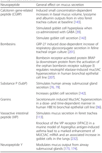
Table 1
General overview of neuropeptides and their effect on
airway mucus secretion
Neuropeptide General effect on mucus secretion
Calcitonin gene-related peptide (CGRP)
Induced small concentration-dependent increases in basal mucus volume, lysozyme and albumin outputs from in vitro ferret trachea culture at baseline [143].
Stimulated goblet cell hyperplasia when co-administered with GABA [39].
Stimulate goblet cell secretion [142]
Bombesins GRP-27 induced dose-dependent increase of respiratory glycoconjugate secretion in feline tracheal organ culture [201].
Bombesin receptor-activated protein BRAP (a downstream protein from the activation of the orphan bombesin receptor subtype-3) regulates neutrophil elastase-induced muc5AC hypersecretion in human bronchial epithelial cell line [207].
Substance P (SubP) Stimulates human airway submucosal gland secretion [76,79].
Increases goblet cell secretion [142].
Granins Secretoneurin induced Muc5AC hypersecretion in a dose- and time-dependent manner in human HBE16 bronchial epithelial cell line [36].
Vasoactive intestinal peptide (VIP)
Stimulates mucus secretion in ferret trachea [113].
Knockout of the VIP receptor (VPAC2) in a murine model of Aspergillus antigen-induced asthma lead to a marked enhancement of MUC5AC mRNA and an associated increase in goblet cells in the lungs [232].
(C-fibers), around blood vessels, and to a lesser degree
within airway smooth muscle [
37
,
71
]. Their effects are
mediated via NK1 receptors (mainly for SubP) and NK2
receptors (activated by NKA). Activation of these
recep-tors induces airway smooth muscle constriction in
humans, although the effects of SubP are variable [
72–75
].
SubP also has a direct effect on airway goblet cells and
submucosal glands through NK1 receptor, causing mucus
secretion [
76–79
]. Using receptor antagonists against NK1
and NK3 receptors in porcine tracheal explants, Philips
and colleagues showed that SubP and NKB induced
sub-mucosal gland fluid flux, while NKA had no effect on the
gland flux [
80
]. Their results also suggested that NK1 and
NK3 receptors may induce glandular effects by different
mechanisms
–
NK3 receptors are likely inducing
activa-tion of parasympathetic nerves, while NK1 may have
dir-ect effdir-ect on the glands [
80
].
Asthma
In asthmatic airways, mucus content positively correlated
with SubP expression; NK1 receptors were also elevated with
strongest expression detected on goblet cells [
81
]. The
au-thors of that study concluded that neurogenic mechanisms
contributed to asthma. Consistent with that, Tomaki and
colleagues found that SubP levels in sputum correlated with
airway obstruction in asthma [
82
]. In an experimental
mur-ine model of allergic asthma, increased bronchoalveolar
lav-age fluid concentrations of SubP were associated with
induction of muc5AC mRNA [
83
], further suggesting a
po-tential pathogenic role for tachykinins in asthma. Other
stud-ies have found a positive correlation between muc5AC and
NKA protein expression in the sputum of asthmatics [
84
].
There are currently no studies available that provide
infor-mation regarding the role of NKB in regulating muc5AC or
muc5B expression, nor of SubP or NKA in the regulation of
muc5B, in asthma. However, given that muc5AC is increased
and muc5B decreased in the sputum obtained from
asth-matics [
48
], it is possible (although speculative) that
tachyki-nins have stimulatory roles on muc5AC, but inhibitory roles
on muc5B.
Therapeutically, tachykinin receptor antagonists have
been explored in asthma, with some reports suggesting
beneficial effects (reviewed here [
85
]). For such studies,
bronchoconstriction, inflammation and lung function
were primary endpoints. For example, Van Schoor and
Fig. 2Simplified schematic of select neuropeptide and their proposed receptor mechanisms mediating mucus secretion. Receptors coupled to Gsincrease cAMP through adenylyl cyclase (not shown). cAMP then increases intracellular Ca
2+
through downstream mediators, such as protein kinase A (not shown). Receptors coupled to Gqlead to breakdown to inositol 1,4,5-phosphate (IP3) and subsequent mobilization of Ca
2+
from intracellular stores. Ca2+serves as a common mediator of mucus granule discharge and exocytosis [231]. Additional details regarding receptor and effector mechanisms are shown in Table3. Abbreviations: SubP, substance P; NKA, neurokinin A; VIP, vasoactive intestinal peptide; CGRP, calcitonin gene-related peptide; NPY, neuropeptide Y; GRP, gastrin-releasing peptide; NMB, neuromedin B; IP3, inositol 1,4,5-trisphosphate; VPAC2, vasoactive intestinal peptide receptor 2; bombesin subtype-1 receptor (BB1); bombesin subtype-2 receptor (BB2)
Table 3
Receptor mechanisms of select neuropeptides
Neuropeptide Receptor Effectors & signaling molecules involved
Substance P/ Neurokinin A neurokinin-1,2 & 3 receptors (NK-1,2 &3) (discussed and reviewed in [239] and [240]).
Gsand Gq[241-243].
↑intracellular Ca2+, DAG, cAMP, IP3 [79,239,244-246]. CFTR has been implicated [100].
VIP VPAC1, VPAC2 and PAC1 [30,247]. Gs, Giand Gq[248,249].
↑intracellular Ca2+, DAG, cAMP, IP3, [249]. CFTR has been implicated [132].
CGRP CGRP-αsubtype and CGRP-βsubtype [31,138,250]. Gqand Gs[251].
↑intracellular Ca2+, DAG, IP3, cAMP [251,252]. NPY Y-1, Y-2, Y-4 and Y-5 receptors [32,34,182]. Giand Gq[182,253].
↑intracellular Ca2+, DAG, cAMP, IP3, [182,253-256].
Bombesins (GRP-27; NMB) NMB = subtype 1 receptor (BB1);
GRP-27 = subtype 2 receptor (BB2);
subtype-3 (BB3) = orphan [35].
Giand Gq[35].
↑intracellular Ca2+, DAG, cAMP, IP3 [35,257].
Chromogranins (Secretoneurin) Currently suspected, but not identified G-protein coupled receptor.
colleagues found that SR 48969, a NK2 receptor
antagon-ist, prevented bronchoconstriction provoked by NKA in
mild asthmatics [
86
]. However, more recently, the NK1/
NK2 receptor antagonist AVE5883 has provided mixed
re-sults, in which it mitigated NKA-mediated
bronchocon-striction, but augmented allergen-induced decreases in
forced expiratory volume (FEV
1) in people with asthma
[
87
]. Because decreases in FEV
1could be due to either
bronchoconstriction and/or mucus obstruction, it is
pos-sible that dual blockade of NK1 and NK2 receptors in
asthmatics augmented mucus secretion and/or
broncho-constriction to allergen. Therefore, examining expression
of muc5AC and muc5B in the sputum of people with
asthma provided tachykinin receptor antagonists might be
especially important. Finally, despite some evidence that
tachykinin receptor antagonists might be beneficial, they
have failed to reach the market for asthma [
88
].
COPD
One report suggests that the concentration of SubP is
el-evated in the sputum of COPD patients [
89
], and it was
suggested that neurogenic inflammation might
contrib-ute to the airway narrowing in COPD. Interestingly,
however, in individuals with COPD, sputum SubP and
NKA decreased during exacerbation [
90
]. The authors
proposed that continual stimulation of sensory nerves
during an exacerbation might have led to neuropeptide
depletion, thus accounting for the decreased content.
Additional studies have shown that cigarette smoke
in-duces mucus secretion of goblet cells through activation
of sensory nerve fibers [
91
], with SubP being the
pro-posed mediator. Thus, since smoking is a major risk
fac-tor for COPD [
92
,
93
], these findings suggested that
tachykinin antagonists might be beneficial for COPD
[
29
]. Furthermore, if SubP does mediate cigarette-induced
secretion of goblet cells, then it is possible that it
might also contribute to increased expulsion of mucus
in COPD airways and/or regulate muc5AC and
muc5B expression.
Therapeutically, tachykinin receptor antagonists have
been shown to decrease inflammation in animal models
of COPD [
94
]. Specifically, the number of macrophages
and dendritic cells was decreased in the lung lavage fluid
of mice exposed to cigarette smoke and provided the
tachykinin receptor antagonist AVE5883. It is also
in-teresting to note that several studies have found a
pro-relaxing effect of SubP and NKA human blood
vessels [
95
]. From this perspective, tachykinin
antago-nists might be of detriment in subpopulations of
people with COPD (e.g., those that have pulmonary
hypertension). This might explain why none of the
tachykinin receptor antagonists that were in
develop-ment for COPD [
96
] have exhibited a clear
thera-peutic benefit.
Cystic fibrosis
Although SubP stimulates gland secretion in
“
normal
”
submucosal glands, reports suggest that it is ineffective
in people with CF [
76
]. This finding has been
repro-duced in pigs with CF [
97
]. An implication from those
studies was that defective responses to SubP might
con-tribute to airway pathology in CF. Given that glandular
secretion in response to SubP is defective in CF, a
specu-lation is that SubP-mediated secretion in CF might be
associated with enhanced muc5AC to muc5B secretion
ratios, effectively mimicking asthma [
98
]. Moreover, in
contrast to asthma and COPD, where enhanced SubP
and/or tachykinin signaling is potentially pathogenic, a
decrease in SubP-mediated secretion in CF might be of
detriment [
99
]. However, restoring SubP-mediated
sig-naling through tachykinin agonists seems like an
inef-fective strategy in CF because evidence suggests that the
defect in SubP-mediated glandular secretion in CF is
due to loss of CFTR [
100
]; thus, CFTR correctors and/or
modulators would be required. Even if such a strategy
was pursued, the pro-inflammatory effects [
101
] and
po-tential bronchoconstricting effects [
72–75
] of
tachyki-nins make this a less appealing option.
Other Kinins
Recently a new gene and associated peptides have been
added to the kinin group, although they do not entirely
fulfill the requirements to be classical neuropeptides [
69
,
102
]. These peptides have structural similarity with the
known tachykinins but are present in a large variety of
tissues and organs and are synthesized mainly by cells of
the hematopoietic lineage [
69
,
102
]. In humans,
hemoki-nin 1 (HK-1), together with endokihemoki-nins (EK) A, B, C and
D, which originate from the different splice variants of
the TAC4 gene (preprotachykinin C), have been found
[
102
]. Due to the wide spread of HK-1 in the body, and
its preferential affinity to the NK-1 tachykinin receptor,
it has been implicated in many diseases (reviewed
else-where, [
103–105
]) including asthma and possibly COPD
[
106–108
]. HK-1, EKA and B pro-contractile effects have
been shown in ex vivo bronchi both in humans and
guinea pigs, although seemingly these effects are
medi-ated through different receptors [
109
]. Very recently
HK-1 has also been shown to cause degranulation of the
human mast cell line leukocyte adhesion deficiency-2
(LAD2) [
110
]. These findings suggest that HK-1 might
play an important role in the pathogenesis and
sympto-mology of asthma and COPD.
Vasoactive intestinal peptide (VIP)
histidine methionine 42 (PHV-42) [
111
]. Major functions
of VIP include airway smooth muscle relaxation [
112
],
stimulation of mucus secretion from airway glands and
goblet cells [
113
], and vasodilation [
112
,
114
]. The
ef-fects of VIP on mucus secretion in humans are complex
as an inhibitory effect of VIP on cholinergic-mediated
mucus secretion in human submucosal glands has been
reported [
115
].
Asthma
Some evidence suggests a functional loss of VIP-innervation
to the airways in asthma [
116
], although this may be
sec-ondary to inflammation. It was speculated that the loss of
VIP-innervation to the airway diminished the amount
of bronchodilation mediated by the nervous system.
Athari et al. also found that mucus hypersecretion
and muc5AC mRNA were significantly decreased
when enzyme-degradation resistant VIP was delivered
to the airways of mice with experimentally induced
asthma [
117
]. Although speculative, this finding might
suggest that VIP has an inhibitory role on muc5AC
expression in asthma. However, perhaps inconsistent
with this speculation is data from the pancreas
sug-gesting that VIP increases muc5AC expression [
118
].
Although we were not able to find any studies
con-cerning the effects of VIP on muc5B regulation and/
or expression in asthma, since muc5B might be
de-creased in asthma [
119
], and VIP might also be
de-creased, then one speculation is that VIP directly
stimulates production of muc5B.
In 2003, Linden and colleagues examined the effects of
a VIP agonist in asthma and found a short, but effective,
bronchodilatory effect [
120
]. Others have shown that
VIP possesses potent anti-inflammatory effects and
in-hibits eosinophil migration [
121
]. These properties make
it an appealing therapeutic candidate for asthma.
How-ever, due to short plasma half-lives, VIP analogs have
been met with limited enthusiasm clinically [
122
].
Ef-forts to modify formulation and delivery have been
on-going. Very recently, one agonist with sustained release
(PB1046) has undergone further development for
pul-monary hypertension [
123
]. Perhaps a renewed interest
in VIP analogs will reinvigorate efforts to examine their
therapeutic potential in asthma.
COPD
Increased serum levels of VIP might be a marker of
acute exacerbations in COPD [
124
], although whether
they are a cause or consequence is unknown. An
argu-ment for consequence is derived from studies suggesting
a beneficial effect of inhaled VIP on quality of life in
COPD [
125
], as well as evidence indicating a protective
role of VIP against pulmonary hypertension in COPD.
However, an argument for causal relationship might be
inferred from studies demonstrating that chronic
bron-chitis, which is a common feature of COPD [
126
], is
as-sociated with increased VIP innervation to the mucus
glands [
127
]. The authors suggested that increased VIP
innervation to the mucus glands was linked to increased
sputum production.
No studies were available that described VIP-mediated
activation of goblet cells or glands in either humans or
animal models of COPD. Similarly, we were unable to
identify any studies that examined the effects of VIP on
muc5AC or muc5B expression in COPD airways.
How-ever, receptors for VIP (vasoactive intestinal peptide
re-ceptor 1 (VPAC), vasoactive intestinal peptide rere-ceptor 2
(VPAC2)) were elevated in the epithelium and glands in
biopsies from smokers with chronic bronchitis [
128
].
This finding might suggest that regulation of mucus
se-cretion by VIP is altered in COPD. Additionally, since
CFTR dysfunction is involved in COPD pathogenesis
[
56
], one speculation is that VIP-mediated submucosal
gland secretion, which is dependent upon CFTR [
129
], is
impaired in COPD.
Very few studies have examined the therapeutic
poten-tial of VIP in COPD. As highlighted above, PB1046 is a
VIP agonist with sustained release that is being
devel-oped for pulmonary hypertension [
123
]. Because
pul-monary hypertension is common in COPD [
130
], it is
possible that PB1046 will be of clinical value in COPD.
However, given that increased VIP innervation to the
mucus glands in COPD is proposed to contribute to
in-creased sputum production [
126
], VIP agonists might be
of mixed benefit.
Cystic fibrosis
Studies suggest that VIP nerve distribution and density
are decreased in the airway epithelium, submucosal
glands, alveolar walls and blood vessels of people with
CF [
131
]. The decrease in VIP distribution was proposed
to be secondary to infection and inflammation. It has
also been shown that CF airway glands do not respond
to VIP stimulation [
129
]. This lack of response has been
interpreted to indicate that VIP stimulates gland
secre-tion through CFTR-dependent mechanisms [
132
]. The
authors suggested that the lack of VIP-mediated gland
secretion might promote a hyper-inflammatory airway
environment that contributes to CF lung disease.
Inter-estingly, in mice, lack of VIP evokes CFTR dysfunction,
creating a CF-like disease [
133
]. Together, these finding
suggests a potential reciprocal relationship between VIP
signaling and CFTR function.
Since VIP is ineffective at stimulating gland secretion
in CF, then it seems an unlikely candidate to mediate the
increased muc5B concentrations (which is largely
expressed in the glands and to a lesser extent in surface
exacerbations [
135
]. It is also unlikely that VIP agonists
would be therapeutically beneficial for restoring
glandu-lar secretion in CF, since VIP-mediated secretion is
dependent upon CFTR [
132
]. Thus, co-administration
with CF correctors or potentiators would be necessary.
However, the proposed anti-inflammatory and
broncho-dilator properties of VIP [
121
] make it a therapeutic
op-tion worth exploring.
Calcitonin gene-related peptide (CGRP)
CGRP is a member of the calcitonin gene family related
neuropeptides and its precursors are encoded by the
cal-citonin II gene [
136
,
137
]. CGRP-alpha and -beta
precur-sors are cleaved into two 37aa-long isoforms:
α−
CGRP
and
β−
CGRP respectively [
21
,
114
,
138
]. CGRP is
pre-dominantly expressed in the central and peripheral
ner-vous systems [
139
,
140
]. CGRP-positive nerve fibers that
innervate the airways originate from the trigeminal,
nodose-jugular and dorsal root ganglia [
138
]. CGRP is
also expressed in pulmonary neuroendocrine cells
throughout the airway tree and in the alveoli [
37
,
141
].
CGRP can induce mucus secretion in the airways, from
both glands and goblet cells [
142
,
143
]. CGRP also
amp-lifies the pro-contractile effects of capsaicin [
144
] and
electrical field stimulation [
145
].
Asthma
CGRP has long been suspected for having important
modulatory role in asthma, due to its airway constricting
capacity [
146
,
147
]. Indeed, reports suggest that CGRP is
increased in the bronchoalveolar lavage fluid of
asth-matics and might contribute to the late phase asthmatic
reactions following provocation by allergen inhalation
[
148
]. CGRP-positive nerve fibers are also increased in
animal models given viral infections, which are risk
fac-tors for asthma [
149
]. Interestingly, Larson and
col-leagues
demonstrated
that
CGRP
is
expressed
ectopically in mucus cells of ovalbumin
(OVA)-sensi-tized Brown-Norway rats [
150
]. They suggested that this
accumulation of CGRP might represent an additional
leasing mechanism involved in quick hypersensitivity
re-sponses and mucus secretion. A recent investigation by
Sui and colleagues found that a combination of CGRP
and gamma-aminobutyric acid (GABA) are responsible
for goblet cell hyperplasia and muc5AC induction in a
murine model of asthma [
39
]. Using the same model,
they also found that elimination of pulmonary
neuroen-docrine cells, which express CGRP, decreased the
ex-pression of goblet cells and muc5B.
There are currently several small molecules, as well as
a monoclonal antibodies [
151
], that inhibit CGRP
signal-ing [
152
]. Studies exploring the therapeutic potential of
inhibiting CGRP signaling have focused largely on the
cardiovascular system [
153–156
], with an emphasis on
migraines. Many of those studies demonstrated an acute
beneficial effect, however, liver toxicity associate with
frequent use of CGRP small molecule inhibitors has
hampered progress. To the best of our knowledge, there
are no clinical studies that have examined the potential
of CGRP receptor antagonists in clinical populations of
asthmatics. However, with several tools available,
exam-ining inhibition of CGRP in the context of asthma
should be achievable, even if only on a small scale. Based
upon studies in animals, it is expected that inhibition of
CGRP would alleviate some of the mucus phenotypes in
allergic asthma [
39
].
COPD
Increased concentrations of CGRP have been identified
in the sputum of people with COPD [
157
], where they
have been speculated to play a role in promoting airway
inflammation. Similarly, Gu et al. found increased
fre-quency of CGRP-positive cells in the airways of people
with COPD but decreased epithelial expression of CGRP
receptors [
158
]. Although there are no studies that have
specifically examined the effects of CGRP on muc5AC
or muc5B expression in COPD, finding altered CGRP
receptor expression in COPD airway epithelial cells
sug-gests a possible influence of CGRP on muc5AC and
muc5B expression. Correlative data also demonstrates a
relationship between COPD, notch signaling, muc5AC
mRNA, and pulmonary neuroendocrine cells (which
synthesize CGRP among many other neuropeptides)
[
159
].
We carefully examined the literature but were unable
to find any studies that assessed the therapeutic
poten-tial of CGRP antagonists/blockers in preclinical or
clin-ical COPD studies. However, given that CGRP can
stimulate smooth muscle contraction in human airways
[
160
], and has been implicated in mucus secretion in
normal airways [
142
,
143
], it is predicted that inhibition
of CGRP might be of benefit in COPD. Yet, studies
indi-cating
that
CGRP
agonists
possess
specific
anti-inflammatory properties, as well as vasodilatory
properties [
138
], might suggest that inhibiting CGRP
could be of potential negative consequence in COPD.
Thus, clinical and/or preclinical studies that address the
potential benefits of blocking CGRP (or the potential
negative consequences) in COPD are necessary.
Cystic fibrosis
Although CGRP has also been shown to increase
sub-mucosal gland secretions in non-CF airways, it is an
in-effective secretagogue in CF airways [
161
]. Of note,
might be a compensatory mechanism to counter the lack
of CGRP-mediated submucosal gland secretion. The
au-thors also demonstrated an important role for CGRP in
maintaining airway progenitor cells, which they
specu-lated might be important for lung injury and repair in
CF airways. Since CGRP participates in goblet cell
hyperplasia and muc5AC induction in other disease
models [
39
], it is possible that it is also involved in CF
exacerbations, in which increased muc5AC has been
noted [
135
].
With that said, it is difficult to predict whether
inhibit-ing CGRP would be of significant benefit in CF, as one
might anticipate mixed results (e.g. anti-inflammatory
and vasodilatory properties versus involvement in mucus
secretion). However, in the specific context of mucus,
in-hibition of CGRP might be of benefit in CF. Given the
numerous CF animal models [
162–165
], preclinical
stud-ies examining this possibility are feasible.
Neuropeptide Y (NPY)
Neuropeptide Y (NPY) is a 36 aa peptide that belongs to
the F- and Y-amide gene family [
21
,
166
,
167
]. NPY is
synthesized in both the peripheral and central nervous
systems [
168
]. NPY has been implicated in a wide variety
of autoimmune and inflammatory diseases, including
asthma and COPD [
37
,
167
,
169–171
]. It plays an
im-portant role in regulation of airway blood flow [
37
],
smooth muscle contraction [
172
], immune cell/mast cell
modulation [
167
,
169
] and secretion output from
sub-mucosal glands [
173
,
174
].
Asthma
The role of NPY in asthma remains unclear. For example,
NPY has been associated with stress-induced exacerbation
in asthma [
175–177
]. From those studies, it was suggested
that NPY facilitated inflammation, as its concentrations
correlated with the number of leukocytes and eosinophils
in the bronchoalveolar lavage fluid. Similarly, NPY
through activation of its Y1 receptor, was demonstrated to
be critical for the development of allergic airway
inflam-mation in mice [
178
]. Specifically, NPY and Y1 receptor
knock-out mice showed decreased eosinophils in the
bronchoalveolar lavage fluid, as well as decreased
circulat-ing immunoglobulin E (IgE) levels, compared to wild-type
mice under allergic conditions [
178
]. Additional studies
have found that loss of
forkhead box protein P1
(Foxp)1
and
forkhead box protein P4 (Foxp4)
in mice can induce
ectopic expression of NPY in airway epithelia, resulting in
airway hyperresponsiveness [
172
]. Loss of
Foxp1
and
Foxp4
are also associated with ectopic expression of
muc5AC in the airway [
179
]. Thus, although a direct
regulation of muc5AC by NPY has not been established,
the two parallel ectopic expressions, suggest an
associ-ation. Consistent with that, elimination of pulmonary
neuroendocrine cells in a murine model of asthma,
de-creased
Npy
gene expression, which was associated with
decreased goblet cell hyperplasia [
39
]. However, in
con-trast to the aforementioned studies, Chanez and
col-leagues found decreased expression of NPY in the airway
smooth muscle, but not in the epithelium, of people with
asthma [
180
]. The authors proposed that NPY might be
protective in the airway, and that loss of NPY might
con-tribute to mucus hypersecretion. Similarly, Lacroix and
Mosimann reported that pretreatment with NPY
de-creased nasal obstruction and mucus secretion in allergic
rhinitis [
181
].
Although the NPY modulation has been explored in
obesity, alcoholism, anxiety, depression, epilepsy and
pain (reviewed in [
34
,
182
,
183
]), its potential benefit in
asthma is largely unknown and unexplored. The
numer-ous small molecules that have been developed to either
potentiate or inhibit NPY receptor offer promise for
in-vestigating NPY signaling in asthma [
182
].
COPD
In COPD, it has been reported that NPY expression is
de-creased in the lung epithelium, glands and smooth muscle
tissue [
171
], and the authors speculated that such changes
might contribute to COPD pathogenesis. In mice,
second-ary tobacco smoke exposure, which is considered a risk
factor for COPD [
184
], increases the density of the NPY
nerve fibers in the tracheal smooth muscle [
185
]. The
in-crease in NPY nerve fibers was associated with airway
hyperresponsiveness, suggesting a potential pathological
role for NPY. Others have shown that NPY stimulates
se-cretion of interleukin 6 from airway fibroblasts [
186
]. An
implication therefore was that NPY might contribute to
diseases like COPD and asthma, in which elevated
inter-leukin 6 has been found in the sputum [
187
]. Interestingly,
reports suggest that the concentration of muc5AC is
in-creased in the sputum of smokers [
188
]; thus, one
specu-lation is that NPY expression is positively correlated with
muc5AC. Unfortunately, no studies have directly
exam-ined the effects of NPY of submucosal gland secretion or
goblet cell degranulation, in COPD.
To address the role of NPY in COPD, studies utilizing
antagonists or agonists in animal models or humans
with COPD are required. Additionally, although no
stud-ies have examined the effects of modulating NPY in
COPD, the NPY antagonist BIBO 3304 has purported
beneficial effects in pulmonary hypertension [
189
].
Therefore, it is possible that NPY modulation might be
of benefit in some people with COPD, such as those that
have pulmonary hypertension [
130
].
Cystic fibrosis
loci of CFTR and NPY was excluded, and thus it was
concluded that NPY likely played little role in CF
patho-genesis. The only other study that has examined NPY in
CF was conducted in the olfactory system, in which it
was found that NPY was increased in the olfactory
epi-thelium of CF mice [
191
]. The increased NPY was
asso-ciated altered expression of specific microvilli proteins,
and it was suggested that olfactory function might be
af-fected. Thus, whether NPY is involved in CF
pathogen-esis, or modulates mucus secretion in CF, remains
unknown. The numerous CF animal models that develop
airway disease similar to humans [
162–165
], as well as
the multiple small molecules available that inhibit or
po-tentiate NPY signaling [
182
,
189
] offer ample
opportun-ities to investigate the potential role of NPY in CF.
Bombesins
The bombesin-like peptide gene family is currently
com-prised of 5 peptides, originating from two genes
–
the
gastrin-releasing peptide gene and the Neuromedin-B
gene [
21
]. Bombesins are synthesized by both the
ner-vous system [
192
] and by pulmonary neuroendocrine
cells [
193
], as well as other tissues [
194
]. In mammals,
two of the peptides have been cloned and characterized
–
the 27 aa gastrin-releasing peptide (GRP) and the 10
aa neuromedin B (NMB), which act through their
spe-cific G-coupled receptors. For NMB, it is the subtype-1
receptor, and for GRP-27, it is the subtype-2 receptor
[
195–197
]. Similar to the above described groups of
neu-ropeptides, in humans, bombesin-like peptides (BLPs)
have roles in many different physiological functions and
pathological conditions
–
from glucose homeostasis to
malignancies [
195
,
196
,
198
]. In the lungs, bombesins
promote fetal lung development and maturation, as well
as epithelial cell differentiation [
37
,
196
]. They also play
important roles in modulating airway physiology,
includ-ing bronchoconstriction [
199
] and mucus secretion [
200
,
201
], which we explore in more detail below.
Asthma
Recently, work by Sun and colleagues showed that the
num-ber of bombesin positive cells was increased in the airways
of asthmatics [
39
]. Similarly, there have been several studies
suggesting that exposure to tobacco products, which are
triggers for asthma [
202
], increase components of BLP
sig-naling in the lungs [
203
,
204
]. Interestingly, the increased
expression of BLP signaling components persisted after
smoking ceased, suggesting a long-term sensitivity to the
proliferative effects of BLPs. Additionally,
bronchopulmon-ary dysplasia, in which bombesins have been implicated
[
205
], is considered a risk factor for asthma [
206
]. Evidence
also suggests that bombesin receptor-activated protein
regu-lates muc5AC hypersecretion through neutrophil elastase
[
207
]; neutrophil elastase been implicated in goblet cell
degranulation and mucus hypersecretion in a rodent model
of asthma [
208
]. While we found no studies describing the
effects of bombesins on submucosal gland activation and/or
regulation of muc5B in asthma, its ability to modulate
glyco-protein secretion from whole tracheas under normal
physio-logical conditions [
201
] suggests a potential role for it in
pathophysiological conditions. Finally, bombesin antagonists
have not been explored in asthma; however, several small
peptide antagonists have been developed [
209
]. For example,
monoclonal antibodies targeted against GRP have shown
promise for disrupting bombesin signaling in human
air-ways. Thus, using these tools to disrupt bombesin signaling
might shed new light onto the role that they play in asthma.
COPD
Bombesins have been implicated in COPD [
193
], with
evidence suggesting that there is greater release of
bom-besins from COPD airways. Consistent with that, greater
numbers of pulmonary neuroendocrine cells, which
ex-press bombesins, have been observed in the airways of
people with COPD [
158
]. Potential consequences of
in-creased bombesin signaling in COPD include
inflamma-tion [
210
], bronchoconstriction [
211
], and altered lung
injury repair [
212
]. Additionally, bronchopulmonary
dys-plasia, in which bombesins have been implicated, may
increase the risk for development of COPD later in life
[
213
]. Although the role of bombesins in modulating
mucus secretion or muc5AC/muc5B regulation in
COPD is unknown, given that bombesins are increased
in COPD [
193
] and muc5B is also increased [
188
], then
it is possible that either a direct or indirect relationship
between bombesin signaling and muc5B expression
ex-ists. Bombesin antagonists have not been explored in
COPD, and therefore preclinical studies focused on
inhi-biting bombesins might be of particular interest.
Cystic fibrosis
Granins
Granins are a large family of putative neuropeptides
found in the secretory granules of the chromaffin
adre-nomedullary cells in adrenal glands [
216
]. They display
structural features close to the classical neuropeptides.
They are also synthesized by neurons and are co-stored
and/or released together with other neuropeptides;
how-ever, currently they are not considered to be among the
classical neuropeptides [
21
,
216
].
There are 6 genes, 7 prepro-peptides and 14 active
cleaved peptides in the granin family [
21
,
22
]. The most
characterized of these peptides are chromogranin A and
its derivative
–
vasostatin; chromogranin B,
secretogra-nin II, and secretoneurin. Although the functions of
granins are still being elucidated, secretoneurin has been
shown to induce mucus hypersecretion in human airway
epithelial cell lines (16HBE and NCI-H292) and induce
expression of muc5AC [
36
], suggesting a modulatory
role in mucus secretion. Consistent with that,
chromo-granin A is more commonly found in epithelial
mucin-ous tumors compared to nonmucinmucin-ous tumors [
217
].
Asthma
Very few studies have examined granins in asthma. One
study reported increased serum levels of chromogranin A
in a mixed population of individuals, some of which had
asthma [
218
]. The premise of that study was to identify
biomarkers of pulmonary neuroendocrine tumors.
Re-cently, Sui and colleagues reported increased
chromogra-nin A in the airways of mice sensitized to ovalbumin [
39
].
Elshafie and colleagues also reported that increased
wheezing in a patient with asthma was associated with
in-creased serum levels of chromogranin A [
219
]. It has also
been reported that secretogranin II (known as
chromogra-nin C), is a potent chemoattractant for eosinophils [
220
],
which play a critical role in asthma [
221
]. The proposed
signaling molecule mediating eosinophil chemotaxis in
re-sponse to secretogranin II was cyclic AMP [
220
].
While we were not able to find any studies examining
the role of granins in mucus hypersecretion in asthma,
secretoneurin
induces
mucus
hypersecretion
in
non-asthmatic human airway cells [
36
]. Moreover,
stud-ies suggesting that secretoneurin is co-released with
SubP and CGRP raise the possibility that secretoneurin
is pro-inflammatory [
222
]. If true, then inhibiting
secre-toneurin might be particularly useful in asthma. Since
the cell surface receptor secretoneurin and other granins
are currently unknown (Fig.
2
), future studies focused
on identifying the receptor responsible for granin
signal-ing are important [
223
].
COPD
Elevations in serum levels of chromogranin A have been
reported in people with COPD, and interestingly, the
degree of obstruction correlated with chromogranin A
concentrations [
224
]. The authors concluded that elevated
chromogranin A reflected ongoing inflammatory
pro-cesses in the airway. Circulating secretoneurin
concentra-tions were also measured in people with COPD;
compared to those with acute heart failure, people with
COPD had decreased levels of secretoneurin [
225
]. It was
proposed that secretoneurin was a
“
protective mediator
”
in people with heart failure. To the best of our knowledge,
no studies have examined the potential contribution of
granins in mucus-related phenotypes in COPD. However,
it is possible that granins contribute to airway obstruction
in COPD [
224
] through modulation of inflammation
[
222
] and/or mucus secretion [
36
]. Additional studies are
required to explore these possibilities.
Cystic fibrosis
Data indicate that pulmonary neuroendocrine cells
ex-pressing chromogranin A also express CFTR [
226
]. In
neuroendocrine cells with diminished CFTR,
neurose-cretory properties were altered [
226
]. In follow-up study,
Pan and colleagues determined that the number of
pul-monary neuroendocrine cells were decreased in a mouse
model of CF, resulting in diminished oxygen sensing
[
67
]. An implication from those studies was that loss of
CFTR might impair the function of neuroendocrine cells
that express chromogranin A, and possibly alter
chro-mogranin A expression and/or signaling. While we were
unable to find any additional studies that investigated
granins in CF, their potential for modulating
inflamma-tion [
222
] and mucus secretion [
36
] suggest that these
neuropeptides might be of interest in CF airway disease.
Conclusion
Neuropeptides continue to be a source of insight and
com-plexity when it comes to airway disease. Although significant
progress has been made in understanding the contributions
of some neuropeptides to asthma, COPD, and CF, their
spe-cific contributions to mucus obstruction and/or
hypersecre-tion is, in many ways, unknown. Therefore, the field of
neuropeptides and mucus secretion is fertile with questions.
Below we highlight a few potential future directions that can
be expanded upon to advance the field.
A greater emphasis needs to be placed on
understand-ing the role that neuropeptides play in modulatunderstand-ing
mucus secretion in
“
normal
”
airways. More specifically,
studies that delineate the effects of neuropeptides on
submucosal gland secretion versus surface epithelial cell
secretion need to be addressed. Techniques including
(but not limited to) epithelial cells cultured at the
air-liquid interface [
227
], as well as methods developed
by Wine and colleagues [
228
] and/or Welsh and
Additionally, given that muc5AC and muc5B
expres-sion profiles are affected in asthma [
98
], COPD [
188
],
and CF [
135
], it is of interest to understand whether
neuropeptides
preferentially
regulate
expulsion
of
muc5AC and/or muc5B. Similarly, studies focused on
determining whether neuropeptides influence the
ex-pression of muc5AC and/or muc5B at the protein or
transcriptional levels will provide new insight regarding
the regulation of mucins in both health and disease.
We found it surprising that there is relatively limited
information available regarding the role that
neuropep-tides assume in regulating mucus secretion in asthma,
COPD, and CF. Based upon our review of the literature
available, more progress has been made in asthma
com-pared to COPD or CF, but much work still remains. A
few simple studies could narrow this knowledge gap. For
example, using animal models or human samples
de-rived from people with asthma, COPD, or CF, one can
determine whether mucus secretion to a given
neuro-peptide is blunted or exaggerated. Similarly, examining
how acute and/or repeated applications of neuropeptides
affect expression of mucins (e.g., muc5AC, muc5B) in
asthma, COPD, and CF would reveal a potentially
causa-tive role for neuropeptides in airway mucus phenotypes.
Additional studies might include the use of knockout
or pharmacological approaches to assess the
contribu-tion of specific neuropeptides and/or their receptors to
regulation of mucus secretion and expression. Moreover,
a thorough investigation of the expression (both spatially
and temporally) of neuropeptides and their receptors in
the airway in both health and disease would provide
additional insight regarding potential causative and/or
modulatory roles in asthma, COPD, and CF.
Although it is difficult to select which neuropeptides are
the most important and/or interesting to pursue, our
re-view provides a road map to help guide researchers and
clinicians. Clearly, species of interest will influence which
neuropeptides can be readily studied, as the tools, models,
and neuropeptides available vary according to species. In
addition, there is less information regarding the signaling
mechanisms of bombesins and granins. Thus, for these
peptides, progress may require further efforts.
Mucus is a critical defensive mechanism [
230
].
En-hanced mucus secretion/production and/or abnormal
mucus properties are associated with several airway
dis-eases, as highlighted in this review. In order to find more
specific targets and additional treatment options for
people with asthma, COPD, or CF, a greater
understand-ing of the regulation of mucus secretion by
neuropep-tides is undeniably important. We anticipate that future
research aimed at investigating neuropeptide-mediated
regulation of mucus secretion and expression will
pro-vide new insight into airway pathogenesis, as well as
un-cover new targets for therapeutic disun-covery.
Abbreviations
aa:Amino acid; BB1: Bombesin subtype-1 receptor; BB2: Bombesin subtype-2
receptor; BLPs: Bombesin-like peptides; cAMP: Cyclic adenosine monophosphate; CF: Cystic fibrosis; CFTR: Cystic fibrosis conductance regulator; CGRP: Calcitonin gene-related peptide; DAG: Diacylglycerol; EK: Endokinins; FEV1: Forced expiratory volume; Foxp1: Forkhead box protein
P1; Foxp4: Forkhead box protein P4; GABA: Gamma-Aminobutyric acid; GRP: Gastrin-releasing peptide; HK-1: Hemokinin 1; IgE: Immunoglobulin E; IP3: Inositol 1,4,5-trisphosphate; LAD2: Leukocyte adhesion deficiency-2; muc5AC: Mucin5AC; muc5B: Mucin5B; NK1: Neurokinin 1; NK2: Neurokinin 2; NK3: Neurokinin 3; NKA: Neurokinin A; NKB: Neurokinin B; NMB: Neuromedin B; NPY: Neuropeptide Y; OVA: Ovalbumin; PHM-27: Peptide histidine methionine 27; PHV-42: Peptide histidine methionine 42; RAMP1: Receptor activity modifying protein 1; RAMP2: Receptor activity modifying protein 2; SubP: Substance P; TAC1: Preprotachykinin A; TAC3: Preprotachykinin B; TAC4: Preprotachykinin C; VIP: Vasoactive intestinal Peptide;
VPAC1: Vasoactive intestinal peptide receptor 1; VPAC2: Vasoactive intestinal peptide receptor 2
Acknowledgements
We thank Kevin Vogt for helpful assistance in the preparation of this manuscript.
Funding
R00HL119560–03 (PI, LRR) and 10T2TR001983–01 (Co-I, LRR).
Availability of data and materials
All data generated or analysed during this study are included in this published article.
Authors’contributions
LRR and KRA read and interpreted available literature for the review. LRR and KRA generated tables and figures. LRR and KRA wrote and edited the manuscript. Both authors read and approved the final manuscript.
Ethics approval and consent to participate
Not applicable.
Consent for publication
Not applicable.
Competing interests
The authors declare that they have no competing interests.
Publisher
’
s Note
Springer Nature remains neutral with regard to jurisdictional claims in published maps and institutional affiliations.
Received: 13 May 2018 Accepted: 13 July 2018
References
1. Summary tables of mortality estimates by cause, age and sex, globally and by region, 2000–2015 [http://www.who.int/entity/healthinfo/global_ burden_disease/GHE2015_Deaths_Global_2000_2015.xls?ua=1]. 2. Cohn L. Mucus in chronic airway diseases: sorting out the sticky details. J
Clin Invest. 2006;116:306–8.
3. Lai H, Rogers DF. New pharmacotherapy for airway mucus hypersecretion in asthma and COPD: targeting intracellular signaling pathways. J Aerosol Med Pulm Drug Deliv. 2010;23:219–31.
4. Livraghi-Butrico A, Grubb BR, Wilkinson KJ, Volmer AS, Burns KA, Evans CM, O'Neal WK, Boucher RC. Contribution of mucus concentration and secreted mucins Muc5ac and Muc5b to the pathogenesis of muco-obstructive lung disease. Mucosal Immunol. 2017;10:829.
5. Dunican EM, Elicker BM, Gierada DS, Nagle SK, Schiebler ML, Newell JD, Raymond WW, Lachowicz-Scroggins ME, Di Maio S, Hoffman EA, et al. Mucus plugs in patients with asthma linked to eosinophilia and airflow obstruction. J Clin Invest. 2018;128:997–1009.
7. Mall MA. Unplugging mucus in cystic fibrosis and chronic obstructive pulmonary disease. Ann Am Thorac Soc. 2016;13(2):S177–85.
8. Kang JW, Lee YH, Kang MJ, Lee HJ, Oh R, Min HJ, Namkung W, Choi JY, Lee SN, Kim CH, et al. Synergistic mucus secretion by histamine and IL-4 through TMEM16A in airway epithelium. Am J Physiol Lung Cell Mol Physiol. 2017;313: L466–76.
9. Gorrieri G, Scudieri P, Caci E, Schiavon M, Tomati V, Sirci F, Napolitano F, Carrella D, Gianotti A, Musante I, et al. Goblet cell hyperplasia requires high bicarbonate transport to support mucin release. Sci Rep. 2016;6:36016. 10. Zhou-Suckow Z, Duerr J, Hagner M, Agrawal R, Mall MA. Airway mucus,
inflammation and remodeling: emerging links in the pathogenesis of chronic lung diseases. Cell Tissue Res. 2017;367:537–50.
11. Quinton PM. Role of epithelial HCO3(−) transport in mucin secretion: lessons from cystic fibrosis. Am J Physiol Cell Physiol. 2010;299:C1222–33. 12. Ghosh A, Boucher RC, Tarran R. Airway hydration and COPD. Cell Mol Life
Sci. 2015;72:3637–52.
13. Reznikov LR. Cystic fibrosis and the nervous system. Chest. 2017;151:1147–55. 14. Tokuyama K, Kuo HP, Rohde JA, Barnes PJ, Rogers DF. Neural control of
goblet cell secretion in Guinea pig airways. Am J Phys. 1990;259:L108–15. 15. Rogers DF, Dewar A. Neural control of airway mucus secretion. Biomed
Pharmacother. 1990;44:447–53.
16. Rogers DF, Alton EW, Dewar A, Lethem MI, Barnes PJ. Impaired stimulus-evoked mucus secretion in cystic fibrosis bronchi. Exp Lung Res. 1993;19:37–53. 17. Rogers DF. Motor control of airway goblet cells and glands. Respir Physiol.
2001;125:129–44.
18. Rogers DF. Airway mucus hypersecretion in asthma: an undervalued pathology? Curr Opin Pharmacol. 2004;4:241–50.
19. Wine JJ. Parasympathetic control of airway submucosal glands: central reflexes and the airway intrinsic nervous system. Auton Neurosci. 2007;133:35–54. 20. Williams OW, Sharafkhaneh A, Kim V, Dickey BF, Evans CM. Airway mucus:
from production to secretion. Am J Respir Cell Mol Biol. 2006;34:527–36. 21. Burbach JP. What are neuropeptides? Methods Mol Biol. 2011;789:1–36. 22. Burbach JP. Neuropeptides from concept to online database www.
neuropeptides.nl. Eur J Pharmacol. 2010;626:27–48.
23. Merighi A, Salio C, Ferrini F, Lossi L. Neuromodulatory function of neuropeptides in the normal CNS. J Chem Neuroanat. 2011;42:276–87. 24. Coles SJ, Bhaskar KR, O'Sullivan DD, Neill KH, Reid LM. Airway mucus:
composition and regulation of its secretion by neuropeptides in vitro. CIBA Found Symp. 1984;109:40–60.
25. Billington CK, Penn RB. Signaling and regulation of G protein-coupled receptors in airway smooth muscle. Respir Res. 2003;4:2.
26. Deshpande DA, Penn RB. Targeting G protein-coupled receptor signaling in asthma. Cell Signal. 2006;18:2105–20.
27. Bradley SJ, Wiegman CH, Iglesias MM, Kong KC, Butcher AJ, Plouffe B, Goupil E, Bourgognon JM, Macedo-Hatch T, LeGouill C, et al. Mapping
physiological G protein-coupled receptor signaling pathways reveals a role for receptor phosphorylation in airway contraction. Proc Natl Acad Sci U S A. 2016;113:4524–9.
28. De Swert KO, Joos GF. Extending the understanding of sensory neuropeptides. Eur J Pharmacol. 2006;533:171–81.
29. Joos GF, De Swert KO, Pauwels RA. Airway inflammation and tachykinins: prospects for the development of tachykinin receptor antagonists. Eur J Pharmacol. 2001;429:239–50.
30. Groneberg DA, Rabe KF, Fischer A. Novel concepts of neuropeptide-based drug therapy: vasoactive intestinal polypeptide and its receptors. Eur J Pharmacol. 2006;533:182–94.
31. McLatchie LM, Fraser NJ, Main MJ, Wise A, Brown J, Thompson N, Solari R, Lee MG, Foord SM. RAMPs regulate the transport and ligand specificity of the calcitonin-receptor-like receptor. Nature. 1998;393:333–9.
32. Balasubramaniam AA. Neuropeptide Y family of hormones: receptor subtypes and antagonists. Peptides. 1997;18:445–57.
33. Pedragosa-Badia X, Stichel J, Beck-Sickinger AG. Neuropeptide Y receptors: how to get subtype selectivity. Front Endocrinol (Lausanne). 2013;4:5. 34. Yi M, Li H, Wu Z, Yan J, Liu Q, Ou C, Chen M. A promising therapeutic target
for metabolic diseases: neuropeptide Y receptors in humans. Cell Physiol Biochem. 2018;45:88–107.
35. Jensen RT, Battey JF, Spindel ER, Benya RV. International Union of Pharmacology. LXVIII. Mammalian bombesin receptors: nomenclature, distribution, pharmacology, signaling, and functions in normal and disease states. Pharmacol Rev. 2008;60:1–42.
36. Xu R, Li Q, Zhou J, Zhou X, Perelman JM, Kolosov VP. Secretoneurin induces airway mucus hypersecretion by enhancing the binding of EGF to NRP1. Cell Physiol Biochem. 2014;33:446–56.
37. Barnes PJ, Rodger IW, Thomson NC. Asthma, basic mechanisms and clinical management. 3rd ed. London: San Diego Academic Press; 1998. 38. Branchfield K, Nantie L, Verheyden JM, Sui P, Wienhold MD, Sun X.
Pulmonary neuroendocrine cells function as airway sensors to control lung immune response. Science. 2016;351:707–10.
39. Sui P, Wiesner DL, Xu J, Zhang Y, Lee J, Van Dyken S, Lashua A, Yu C, Klein BS, Locksley RM, et al. Pulmonary neuroendocrine cells amplify allergic asthma responses. Science. 2018;360(6393):eaan8546.
40. Pilmane M, Luts A, Sundler F. Changes in neuroendocrine elements in bronchial mucosa in chronic lung disease in adults. Thorax. 1995;50:551–4. 41. Cutz E, Yeger H, Pan J. Pulmonary neuroendocrine cell system in pediatric
lung disease-recent advances. Pediatr Dev Pathol. 2007;10:419–35. 42. Pan J, Copland I, Post M, Yeger H, Cutz E. Mechanical stretch-induced
serotonin release from pulmonary neuroendocrine cells: implications for lung development. Am J Physiol Lung Cell Mol Physiol. 2006;290:L185–93. 43. Yeger H, Pan J, Fu XW, Bear C, Cutz E. Expression of CFTR and cl(−)
conductances in cells of pulmonary neuroepithelial bodies. Am J Physiol Lung Cell Mol Physiol. 2001;281:L713–21.
44. Weichselbaum M, Sparrow MP, Hamilton EJ, Thompson PJ, Knight DA. A confocal microscopic study of solitary pulmonary neuroendocrine cells in human airway epithelium. Respir Res. 2005;6:115.
45. Soler X, Ramsdell JW. Are asthma and COPD a continuum of the same disease? J Allergy Clin Immunol Pract. 2015;3:489–95. quiz 496-487 46. GINA. Global Strategy for Asthma Management and Prevention. Global
Initiative for Asthma (GINA); 2017. p. 2017.
47. McCracken JL, Veeranki SP, Ameredes BT, Calhoun WJ. Diagnosis and Management of Asthma in adults: a review. JAMA. 2017;318:279–90. 48. Lachowicz-Scroggins ME, Yuan S, Kerr SC, Dunican EM, Yu M, Carrington SD,
Fahy JV. Abnormalities in MUC5AC and MUC5B protein in airway mucus in asthma. Am J Respir Crit Care Med. 2016;194:1296–9.
49. Wills-Karp M, Luyimbazi J, Xu X, Schofield B, Neben TY, Karp CL, Donaldson DD. Interleukin-13: central mediator of allergic asthma. Science. 1998;282:2258–61. 50. Mazzone SB, Undem BJ. Vagal afferent innervation of the Airways in Health
and Disease. Physiol Rev. 2016;96:975–1024.
51. Talbot S, Abdulnour RE, Burkett PR, Lee S, Cronin SJ, Pascal MA, Laedermann C, Foster SL, Tran JV, Lai N, et al. Silencing nociceptor neurons reduces allergic airway inflammation. Neuron. 2015;87:341–54.
52. Fryer AD, Jacoby DB. Parainfluenza virus infection damages inhibitory M2 muscarinic receptors on pulmonary parasympathetic nerves in the Guinea-pig. Br J Pharmacol. 1991;102:267–71.
53. Aven L, Paez-Cortez J, Achey R, Krishnan R, Ram-Mohan S, Cruikshank WW, Fine A, Ai X. An NT4/TrkB-dependent increase in innervation links early-life allergen exposure to persistent airway hyperreactivity. FASEB J. 2014;28:897–907.
54. Hikichi M, Hashimoto S, Gon Y. Asthma and COPD overlap pathophysiology of ACO. Allergol Int. 2018;67:179–86.
55. Shaikh M, Sood RG, Sarkar M, Thakur V. Quantitative computed tomography (CT) assessment of emphysema in patients with severe chronic obstructive pulmonary disease (COPD) and its correlation with age, sex, pulmonary function tests, BMI, smoking, and biomass exposure. Pol J Radiol. 2017;82:760–6.
56. Dransfield MT, Wilhelm AM, Flanagan B, Courville C, Tidwell SL, Raju SV, Gaggar A, Steele C, Tang LP, Liu B, Rowe SM. Acquired cystic fibrosis transmembrane conductance regulator dysfunction in the lower airways in COPD. Chest. 2013;144:498–506.
57. Ramos FL, Krahnke JS, Kim V. Clinical issues of mucus accumulation in COPD. Int J Chron Obstruct Pulmon Dis. 2014;9:139–50.
58. Yamada M, Ichinose M. The cholinergic anti-inflammatory pathway: an innovative treatment strategy for respiratory diseases and their comorbidities. Curr Opin Pharmacol. 2018;40:18–25.
59. van Gestel AJ, Steier J. Autonomic dysfunction in patients with chronic obstructive pulmonary disease (COPD). J Thorac Dis. 2010;2:215–22. 60. Stewart AG, Marsh F, Waterhouse JC, Howard P. Autonomic nerve
dysfunction in COPD as assessed by the acetylcholine sweat-spot test. Eur Respir J. 1994;7:1090–5.