The Roles of the Subicula of the Hippocampus in Producing Heroin-Induced Conditioned
Immunosuppression to a Context
By
Lynde M. Wangler
Senior Honors Thesis Psychology & Neuroscience
University of North Carolina at Chapel Hill
Spring 2018
Approved:
Acknowledgements
I would like to thank my advisor, Dr. Donald Lysle, and my mentor, Christina
Lebonville, for their willingness and enthusiasm in instructing and supporting me throughout the
duration of my thesis. Their mentorship has remarkably enhanced my capabilities as a scientist
and has prepared me for the rigors of post-baccalaureate training and graduate school. I would
also like to thank Dr. Beth Kurtz-Costes and Dr. Peter Ornstein for their invaluable expertise and
undying support. This project was supported by a David Bray Peele Memorial Research Award
(LW), a Lindquist Undergraduate Research Fund (LW), and a King’s Research Excellence
Award (CL) from the Department of Psychology and Neuroscience, at the University of North
Abstract
Background & Significance
The opioid epidemic is an enduring and ever-growing problem in the United States and
contributes to increasing rates of drug abuse and overdose-related deaths (Rudd, Seth, David, &
Scholl, 2016). Opioids are a category of drugs that includes the illegal drug heroin, prescription
pain relievers, such as oxycodone, hydrocodone, codeine, morphine, etc., and synthetic opioids
such as fentanyl (Opioid Crisis, 2017). Though most opioids can be acquired legally and are
generally benign when taken for short amounts of time, their use is being increasingly
discouraged. The potent reinforcing qualities of opioid drugs (mainly, pain relief and euphoric
high) place chronic opioid users at a high risk of developing drug abuse tendencies. Not only are
drug users vulnerable to becoming dependent, they are also at an increased risk of developing
infections, ranging from lung, GI tract, and skin infections to infections in the skeletal and
nervous systems (Risdahla, Khannaa, Peterson, & Molitora, 1998).
Increased infections in opioid users stem not only from increased exposure to pathogens
due to non-sterile needle use, but also from exposure to the drug itself. Opioids, especially
heroin, have been shown to produce deleterious immunological effects unrelated to needle use
(Horsburgh, Anderson, & Boyko, 1989). Furthermore, the negative immunological effects of
opioids can become conditioned in a Pavlovian manner in which the drug becomes associated
with a predictive cue that then acquires an immunomodulatory function. In this way, opioid users
may become more vulnerable to infection when exposed to drug-related contextual cues.
Behaviorally conditioned immunosuppression has been shown in humans but these processes are
usually studied within the context of the placebo effect (Goebel, 2002), and much remains
Animal models using immunosuppressive drugs have been increasingly used to study the
neurological mechanisms and potency of conditioned immune effects. Researchers utilize a
Pavlovian conditioning paradigm with rats in which an unconditioned stimulus (US) is paired
with a conditioned stimulus (CS) to produce a conditioned response (CR). In this case, the US is
a drug substance that causes immunosuppression, and the CS is an initially inert substance that,
through repeated pairings with the US, gains the capacity to affect the animal’s immune
functioning when presented on its own. Early studies paired an immunosuppressant, such as
cyclophosphamide, with saccharine and found that after sufficient pairings, rats showed
immunosuppression, measured by levels of antibody production in response to an immune
challenge, after re-exposure to saccharine alone (Ader & Cohen, 1975). Early studies also found
that immunomodulation could be achieved in rats through re-exposure to a non-aversive stimulus
that had been previously paired with an aversive, immunosuppressive foot shock (Lysle,
Cunnick, Fowler, & Rabin, 1988). Furthermore, researchers found that immunosuppression
could also be induced by exposure to a previously cocaine-associated cue (Kubera et al., 2008).
Our interest has turned to studying heroin-induced conditioned immunosuppression because the
illegal use of heroin as a replacement for prescribed pain medication continues to rise
exponentially (Muhuri, Gfroerer, & Davies, 2013). With these increasing rates of opioid
addiction, there is an unprecedented need to build a more comprehensive understanding of the
physiological underpinnings of this phenomenon, especially as it relates to drugs of abuse like
heroin.
Studies from our laboratory have confirmed that heroin possesses immunosuppressive
properties (Lysle & How, 2000), and that heroin-associated environmental stimuli are capable of
infer changes in immune functioning is through indirect measures of Nitric Oxide (NO), which is
a substance known to be important in immunological defense processes. Previous work in our
laboratory has found that nitrate/nitrite (the product of NO reacting with oxygen) production in
blood plasma is one such measure (Lysle & How, 2000). Quantification of inducible nitric oxide
synthase (iNOS) production, which is an enzyme that produces nitric oxide, has also been used
to infer changes in immune function in an animal (Coleman, 2001; Lysle & How, 2000). By
using an immune challenge, such as injection of lipopolysaccharide (LPS), immediately after
re-exposure to the CS, one can measure conditioned effects on nitrate/nitrite and iNOS mRNA
production in heroin-conditioned immunosuppressed rats (Lysle & Ijames, 2002). In rats that are
re-exposed to the heroin-paired context, production of iNOS mRNA in response to LPS was
significantly decreased, as opposed to control animals, which were not re-exposed to the
conditioned context and did not experience immunosuppression.
Following these discoveries, research has been aimed at using these measures to uncover
the neural mechanisms involved in producing a conditioned immune response to
opioid-associated places. Regions that have been shown to have a role in producing this effect include
the nucleus accumbens shell (NAc shell; Saurer, Ijames, & Lysle, 2009) and the basolateral
amygdala (BLA; Szczytkowski & Lysle, 2008), which are regions also known to be heavily
involved in other contextual drug-related behaviors (Crombag et al., 2008). Studies have also
demonstrated critical involvement of the hippocampus (Szczytkowski et al., 2013; Lebonville et
al., 2016), a region known to be heavily involved in contextual memory retrieval (Maren & Holt,
2000) and to be functionally connected to both the NAc shell and the BLA (e.g., Naber & Witter,
1998). The hippocampus is a uniquely complex and heterogeneous structure, with much
primary interests is in determining whether or not the dorsal and ventral aspects of the
hippocampus are dissociable within the context of heroin-induced conditioned
immunosuppression. The functions of the hippocampus have been heavily researched in the
context of drug- and reward-related behaviors, and its well-known involvement in these
behaviors, specifically those that rely on contextual processing, provides cogent support for our
interest in investigating its neural outputs.
Extensive evidence suggests that the dorsal hippocampus (dHPC) is heavily involved in
spatial memory (Bannerman et al., 2002; Broadbent, N.J., Squire, L., Clark, R.E., 2004; Moser et
al., 1995) and spatial associations (Piekema et al., 2006; Gilbert, P.E. & Kesner, R.P., 2002), as
well as navigation (Miyoshi et al., 2012).Relating this function to drug behaviors, studies found
that during the acquisition of cocaine and nicotine conditioned place preference (CPP), dHPC
neuronal ensembles form that encode the reward-paired context (Trouche et al., 2016; Xia et al.,
2017). Several researchers have also found that the dHPC performs a role in context-induced
reinstatement of drug seeking. For example, dHPC inactivation decreases context-induced (but
not cue-induced) reinstatement of cocaine seeking (Fuchs et al., 2005). Furthermore, the dorsal
subiculum (dSub), a prominent neural output of the dHPC, has been shown to have importance
in mediating the acquisition of conditioned reinstatement of cocaine-seeking behavior.
Researchers found that bilateral infusion of tetrodotoxin (TTX) – a potent neurotoxin that blocks
sodium channels, effectively stopping the function of the cell – in the dorsal, but not ventral,
subiculum of rats blocked re-exposure to a associated cue from reinstating
cocaine-seeking behavior (Martin-Fardon, Ciccocioppo, Aujla, & Weiss, 2008). Most critically to the
current study, functionally inhibiting the dHPC with an infusion of a GABA agonist prior to CS
dHPC is necessary for producing this effect (Szczytkowski, Lebonville, Hutson, Fuchs, & Lysle,
2013). However, much remains unknown about the involvement of the dHPC in conditioned
immunosuppression, as most of the research investigating this brain region outside of our lab
focuses heavily on drug-related context-reward associations and does not characterize
contextually conditioned immune functioning mediated by this region. Our lab is specifically
interested in characterizing the neurocircuitry that produces this effect and in contributing to a
nascent line of research that may one day determine the relationship between underlying factors
that mediate drug-related contextual-reward and/or contextual-immune responses. Accordingly,
recent efforts have been oriented towards characterizing the role of the dSub of the hippocampus,
as the neural output of the dHPC to cortical and subcortical areas, to determine its influences on
the expression of conditioned immune effects.
In contrast to the dHPC’s pronounced role in contextual processing, the ventral
hippocampus (vHPC) and it’s corresponding output the ventral subiculum (vSub), has been
shown to be more involved in other distinct processes, most notably motivated behaviors and
reward learning (Riaz et al., 2017). The vSub has been shown to modulate the activity of the
NAc (Blaha et al., 1997) and the BLA (French, Hailstone, & Totterdell, 2003), both of which
have been shown to be involved in conditioned immunosuppression (noted previously). For
example, in vivo stimulation of rat vSub evoked glutamate-receptor-mediated changes in release of dopamine in the NAc (Blaha et al., 1997). Additionally, excitotoxic lesioning of the vSub
significantly attenuated rats’ probability of approaching a conditioned stimulus predictive of
positive reinforcement and on the acquisition of a new conditioned response to a reinforcing
stimulus potentiated by intra-accumbens infusions of D-amphetamine (Burns, Robbins, &
limbic regions found that vSub stimulation affected plasticity in the BLA and the NAc (Horovitz
& Richter-Levin, 2015). The vSub not only plays a role in appetitive behaviors, it has also been
shown to play a critical role in context-induced relapse to cocaine- (Sun & Rebec, 2003) and
heroin-seeking (Bossert & Stern, 2014), as well as contributes to context-induced relapse of
alcohol seeking (Marchant et al., 2016) via its projections to the NAc. There is strong evidence
of drug-specific roles of the vSub, which make it a primary region of interest in investigating
conditioned immunosuppression to a heroin-paired context. Additionally, there is substantial data
to suggest that the contextual processing abilities of the dSub may intimate its involvement in
expression of conditioned immunosuppression in our model. It has long been theorized that
declarative, including contextual memory, recall is dependent upon a hippocampal-cortical
system (Eichenbaum, 2000), but the exact role of the subicula in facilitating contextual memory
retrieval is unclear. As the main output structures of the hippocampus (O’Mara, 2005), we
suspect that the dorsal and ventral subiculum are involved in producing conditioned
immunological effects. Therefore, this study sought to characterize the roles of the dorsal and
ventral subicula of the hippocampus in opioid-conditioned immunosuppression to a context.
In this paradigm, we paired heroin (US) with exposure to a specific context (CS) and then
use an immune challenge (LPS) to assess immunosuppression by measuring nitrate/nitrite in
blood plasma and iNOS mRNA in spleen tissue. To determine whether or not the subicula are
involved, we used a designer receptor exclusively activated by a designer drug (DREADD) to
inactivate the dSub (Experiment 1) or vSub (Experiment 2) immediately prior to context
re-exposure. DREADDs are a widely used chemo-genetic tool that allow researchers to engineer
receptors in specific regions of the brain that will only be activated by designer drugs, such as
which can be infused directly into a brain region of interest. In this study, we used a Gi-coupled
receptor, which is an inhibitory G-protein coupled receptor. G-protein coupled receptors
(GPCRs) are abundant transmembrane proteins that initiate molecular cascades within cells.
Inhibitory G-protein coupled receptors, therefore, are able to inhibit functional activation in
specific brain regions through molecular mechanisms (Urban & Roth, 2015). Given all of the
evidence that the vSub is involved in context-induced relapse of drug-seeking and appetitive
behaviors, as well as its direct functional connections with the NAc and BLA, we hypothesize
that inhibition of this brain region will block expression of a conditioned immune effect,
resulting in immune measures that are comparable to controls in animals that are exposed to a
heroin-paired context. Though fewer studies have implicated the dSub in this process, there is
still evidence to suggest that, through its well-described role in contextual processing, it may also
mediate this effect, and as such, we predict that inhibition of the dSub will also block expression
of conditioned immunosuppression to a heroin-paired context, such that immune functioning will
be comparable to controls in animals that are exposed to a heroin-paired context.
Methods
Animals
This experiment utilized adult male Lewis rats (N = 70), weighing 225-250 g, which were
acquired from Charles River Laboratories (Kingston, NY, USA). Rats were individually housed
in a room that maintained a reverse light-dark cycle (dark from 7 am – 7 pm). Food and water
was provided ad libitum throughout the experimental period. All experimental and housing conditions are in accordance with the university’s Institutional Animal Care and Use Committee
Drugs & delivery
Heroin, acquired from NIDA’s Drug Supply System (Bethesda, MD, USA), was
dissolved in 0.9 % sterile saline at a final concentration of 1.0 mg/mL and stored at 4 °C. Heroin
was allowed time to warm to room temperature before use. Rats received subcutaneous
injections of 1.0 mg/kg heroin during each conditioning session. This dosage has been shown to
modulate LPS-induced iNOS mRNA expression in the spleen (Lysle & How, 2000; Lysle &
Ijames, 2002; Szczytkowski, 2007). Lipopolysaccharide (LPS; derived from Escherischia coli, serotype 055:B5, Cat# L2880, MilliporeSigma, St. Louis, MO, USA) dissolved in sterile,
pyrogen-free saline was administered at a concentration of 1.0 mg/kg via subcutaneous injections
immediately following final exposure to the conditioned context (home cage exposure in control
animals). This dosage produces sickness behavior and reliable induction of nitric oxide
production. Clozapine-N-oxide (CNO) from NIDA’s Drug Supply System (Bethesda, MD, USA)
was first dissolved in 100% DMSO and was then brought up to volume with 0.9% sterile saline
at a final concentration of 3.0 mg/mL and 0.5% DMSO. CNO was administered at a dose of 3.0
mg/kg subcutaneously.
An Adeno-associated virus (AAV) construct, serotype 5, was used to deliver the
inhibitory DREADD, CAMKIIa-hM4D(Gi), which was tagged with mCherry (titer: 4.4 x 10¹²
GC/mL); the full construct was CAMKIIa-hM4D(Gi)-mCherry. Calcium/calmodulin-dependent
protein kinase II alpha (CAMKIIa) is a promoter that preferentially drives the expression of
DREADDs in excitatory neurons (Liu & Jones, 1996; Tsien et al., 1998; Guo et al., 2010;
Johansen et al., 2010), and AAVs using this promoter within the hippocampus have been used to
successfully manipulate hippocampal LTP and accompanying memory (Lopez et al., 2016).
muscarinic receptor so that it is only activated by the otherwise physiologically inert substance
clozapine-N-oxide (CNO) and not its native agonist acetylcholine. The mCherry tag within the
viral construct was visualized for later viral location verification.
Surgical procedure
Animals were deeply anaesthetized with 1.0 mL/kg 9:1 (vol: vol) ketamine hydrochloride
(100 mg/mL) and xylazine (100 mg/mL) via intraperitoneal injection. Hair was removed from
the top of their heads using an electric razor. Animals were then placed on a stereotaxic
apparatus, and the shaven area was then cleaned with three alternating applications of 70%
ethanol and iodine surgical scrub. After letting the iodine sit for 10 minutes on the skin to fully
disinfect, a vertical incision was then made (approximately 2 cm in length) and a stereotaxic drill
was used to make holes in the skull so that injectors could be directed bilaterally to the dorsal or
ventral subiculum (Experiment 1 or Experiment 2, respectively) at the following stereotaxic
coordinates relative to Bregma: dSub – AP -6.0 mm, ML ± 2.8 mm, DV -3.5 mm at 0° angle;
vSub – AP -6.0 mm, ML ± 4.6 mm, DV -8.5 mm at 0° angle. From these coordinates, injectors
were then raised 0.1 mm DV to create a pocket to house the viral infusion. Rats received bilateral
intracranial infusions of 0.7 μL per hemisphere into the dorsal subiculum at a rate of 0.05 μL per
minute. Injectors were left to sit for a ten-minute period before raising them out of the brain to
allow virus to diffuse away from the injection site. Surgical holes in the skull were then filled
with bone wax, and 0.25 % bupivacaine (200 μL) was administered in and around the wound
using the splash method. Incisions were then sutured with a 4-0 nylon monofilament
non-absorbable suture (MV-662-V, MedVet, Mettawa, IL, USA). Rats were monitored closely during
the surgery by checking reflexes and breathing rates to ensure deep anesthetization. Rats
the wound site with EMLA cream (a mixture of 2.5 % lidocaine and 2.5 % prilocaine) and triple
antibiotic while being monitored closely to ensure weight gain and recovery. Viral incubation
occurred in the following two weeks after which conditioning procedures began.
Conditioning procedure & CS re-exposure
Rats underwent five, 60-minute conditioning sessions every other day for ten consecutive
days. Rats received heroin injections (US) immediately before placement in the conditioning
chamber (BRS/LVE, Laurel, MD, USA; W 30.5 cm x D 24.1 cm x H 26.7 cm), which served as
the conditioned stimulus (CS). The conditioning chambers were placed within sound and light
attenuating chambers (W 50.8 cm x D 34.3 cm x H 36.8 cm) and were located in a room away
from the animal colony (refer to Szczytkowski, 2013). Importantly, cedar bedding was used to
enhance the contextual distinction of the conditioning chamber from the home cage by providing
a distinct olfactory stimulus. Chambers were cleaned between animals with Roccal-D Plus
disinfectant (Zoetis, Kalamazoo, MI, USA). All rats (N = 70) were subjected to these
conditioning procedures.
On the sixth day following the final conditioning session (day 15), rats underwent a test
session in which they were either re-exposed to the conditioning chamber (CS) or left in their
home cages as a control (HC). Half of the rats that were re-exposed to the context received an
intraperitoneal injection of3.0 mg/kg CNO directly prior (30 minutes) to CS exposure and the
other half received an intraperitoneal injection of an equivalent volume of vehicle. Rats that were
left in the home-cage on test day were subjected to the same injections, with half receiving CNO
and the other half vehicle. This procedure produced four conditions, enabling a comparison of
between conditions. All rats were injected with LPS immediately following the test session and
sacrificed six hours later for blood and tissue collection
Blood plasma & tissue collection
Six hours after the testing session on day 15, each rat was sacrificed by cervical
dislocation without anesthesia. A vertical abdominal incision was made and blood from the
abdominal aorta was collected for later analysis. Additionally, spleen tissue was harvested and
analyzed because immune cells found in the spleen, including macrophages, lymphocytes, and
neutrophils, have been shown to harbor LPS-induced iNOS enzyme. Harvested spleen tissue was
then cut into approximately 100 mg-sized pieces in preparation for quantitative reverse
transcription PCR (RT-qPCR). For RT-qPCR analysis, tissue samples were stored in RNAlater (Invitrogen/Thermo Fisher Scientific, Waltham, MA, USA) for two days at 4°C and then -80°C
until processing.
Nitrate/nitrite assay
Greiss reagent assay was used to assess nitrate/nitrite concentration in plasma samples.
Nitrate and nitrite are formed when NO is exposed to oxygen, and levels of NO production can
be assessed by converting nitrate to nitrite (using nitrate reductase in the presence of NADPH
and flavin adenine dinucleotide) and treating with Greiss reagent. Our procedure has been
described in more detail previously (Szczytkowski & Lysle, 2007; Lebonville et al., 2016).
Briefly, after allowing color to develop for ten minutes at room temperature, absorbance at 550
nm was measured using a spectrophotometer. Total micromolar concentrations of nitrite were
determined for each sample based on a known standard curve and each reaction was carried out
of the triplicate average was excluded), the remaining replicates were averaged for each subject.
Nitrate recovery is greater than 95% using this assay (Szczytkowski & Lysle, 2007).
RNA extraction & RT-qPCR
Two-step reverse transcription quantitative polymerase chain reaction (RT-qPCR) was
performed on spleen tissue to ascertain iNOS mRNA expression. Spleen tissue was kept on ice
and homogenized using a bead mill homogenizer (Precellys Evolution, Bertin Instruments,
Montigny-le-Bretonneux, France). TRI-Reagent (Molecular Research Center, Cincinnati, OH,
USA) was used to extract total RNA according to the manufacturer protocol with the following
modifications: performed the optional centrifugation step after homogenization, added 100 μL of
DEPC H2O with 100 μL BCP to improve phase separation of dense homogenate in PhaseLock
Gel Tubes (Heavy formulation, 5Prime/Quantabio, Beverly, MA, USA), and performed three
75% ethanol washes instead of one. The RNA pellet was dissolved in 150 μL DEPC H2O.
Extracted RNA was then quantified spectrophotometrically (Epoch Take3 microdot plate,
BioTek, Winooski, VT, USA). RNA purity was also assessed by looking at the A260nm/280nm
ratio and all samples exhibited ratios close to 2.1, indicating high purity. Reverse transcriptase
was performed using Oligo(dT)18 primer and Maloney Murine Leukemia Virus-Reverse
transcriptase following the Advantage RT-for-PCR Kit (Clontech, Palo Alto, CA, USA)
protocol. Applied Biosystems TaqMan Gene Expression Assays for iNOS and L13A were used
in combination with TaqMan Fast Advanced Master Mix (iNOS: Rn00561646_m1, L13A:
Rn01475911_g1, Thermo Fisher Scientific, Waltham, MA, USA) to detect specific products of
the PCR reaction. Each amplification was run in triplicate on a QuantStudio 6 Flex thermocycler
(AP Biosystems/Thermo Fisher Scientific, Waltham, MA, USA) under the following reaction
cycles of 1 sec. at 95°C (denature) and 20 sec. at 60°C (anneal and extend). RT-qPCR results
were analyzed using the delta-delta Ct method (Livak & Schmittgen, 2001). Briefly, raw Ct
results were surveyed for significant triplicate variation; any individual replicate that differed
from the other two values in the triplicate by 0.5 Ct or more was removed from analysis. Cleaned
triplicate averages for iNOS were normalized to cleaned triplicate Ct averages for the reference
gene L13A to control for the variation in the amount of starting cDNA template. These values
were then normalized again to the average of reference normalized Ct for all groups since there
are two independent control groups for this experiment. The resulting delta delta Ct values were
linearly transformed for graphical representation.
Histology
Brains were harvested and suspended 10% formalin for 24-48 hours, and later transferred
to a 30% sucrose solution for approximately 3-4 days or until brains sunk. Brains were
embedded in frozen section compound (VWR, Radnor, PA, USA) and frozen slowly at
-25°C within a freezing microtome (cryostat). Coronal sections (40μm) were taken using a
cryostat (Leica CM 3050 S, Leica-Microsystems, Germany), mounted onto charged glass slides
(FisherBrand Superfrost, Thermo Fisher Scientific, Waltham, MA, USA), and coverslip mounted
with HardSet VECTASHIELD mounting medium with DAPI (Vector Laboratories, Burlingame,
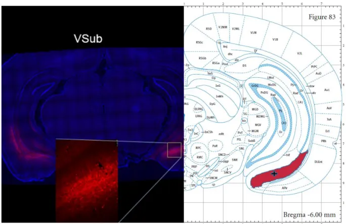
CA, USA). Fluorescent microscopy using Leica DM6000 B widefield light microscope (Leica
Microsystems, Wetzlar, Germany) was utilized to verify virus placement (See Figure A. for
representative dSub viral expression and Figure B. for representative vSub viral expression;
shaded regions indicate the structures as labeled in the atlas( dSub and vSub), and the + symbol
indicates the coordinates that we used to inject virus). Animals were removed from analysis if
Statistical analysis
A 2x2 analysis of variance (ANOVA) was performed on data sets, and for all tests,
significance was set at p < 0.05. Fisher’s Least Significant Difference (LSD) post-hoc test was
used to determine differences between CS-exposed and home cage control groups after a
statistically significant 2x2 ANOVA result. RT-qPCR values were statistically analyzed using
delta delta Ct values, which tend to better meet the assumptions of ANOVA, rather than the
linear transform values, which were only used for graphical presentation. The assumptions of an
ANOVA, namely normality of errors and equal variance across treatments, were each tested and
deemed to satisfy the requirements, making ANOVA a statistically appropriate measure for our
study. Specifically, normality was tested using the Shapiro-Wilk test and equality of variance
was tested using Levene’s Test of Equality of Error Variances. One violation of an assumption is
reported in the results for Experiment 2, but all other assumptions were supported. The presence
of statistical outliers was probed using Grubb’s test. Any statistically significant outliers within
groups were removed from final analysis.
Results
Experiment 1: Inhibition of dorsal subiculum
Experiment 1 investigated the effects of inhibiting the dorsal subiculum of the
hippocampus on the expression of heroin-induced conditioned immunosuppression by activating
an inhibitory DREADD expressed in this region with a systemic injection of CNO directly prior
to re-exposure to the heroin-paired context. There were two factors: treatment (CNO or vehicle,
Veh) x exposure (context, CS, or home cage, HC), resulting in four groups. In the final analyses,
(N = 7; one animal was dropped from the nitrate/nitrite analysis due to an error in blood
collection). Descriptive statistics for Experiment 1 are shown in Table 1.
A 2x2 ANOVA revealed significant differences between groups for plasma nitrate/nitrite
levels (F(3, 27) = 14.264, p < 0.001, Figure 1). There were significant main effects of treatment
(F(1, 27) = 8.002, p = 0.009) and context exposure (F(1, 27) = 29.321, p < 0.001) on plasma
nitrate/nitrite levels. There was also a statistically significant interaction effect between context
exposure and treatment (F(1, 27) = 4.725, p = 0.039). Post-hoc analysis, using LSD, (summarized
in Table 2) revealed that nitrate/nitrite levels were statistically significantly reduced in animals
that received a vehicle injection prior to re-exposure to the heroin-paired context (Veh-CS) in
comparison to the corresponding vehicle control group that remained in home cages (Veh-HC, p
< 0.001). These data indicate that conditioning resulted in a conditioned immune response in the
form of suppressed nitrate/nitrite levels in blood plasma with exposure to the heroin-paired
context. No statistically significant difference in nitrate/nitrite levels was observed between
animals that received the vehicle treatment prior to home-cage exposure (Veh-HC) in
comparison to animals that received an injection of CNO prior to home-cage exposure
(CNO-HC, p = 0.652), indicating that there was no effect of CNO administration alone on this measure.
However, there was a statistically significant difference in plasma nitrate/nitrite levels between
animals that received CNO before home-cage exposure (CNO-HC) and animals that received
CNO prior to context re-exposure (CNO-CS, p = 0.027), as well as between animals that
received the vehicle treatment prior to context re-exposure (Veh-CS) and animals that received
an injection of CNO prior to context re-exposure (CS, p < 0.001), indicating that
CNO-treated animals exposed to the heroin-paired environment exhibited partial attenuation of
CNO-mediated Gi signaling activation prior to re-exposure to the context partially blocked the
expression of conditioned suppression of nitrate/nitrite levels in plasma.
A 2x2 ANOVA revealed significant differences between groups for splenic iNOS mRNA
expression as well (F(3, 28) = 8.060, p = 0.001, Figure 2). There were significant main effects of
treatment (F(1, 28) = 7.346, p = 0.011) and exposure (F(1, 28) = 13.718, p = 0.001) on iNOS mRNA
expression. LSD post-hoc analysis, summarized in Table 2, revealed that iNOS mRNA
expression was statistically significantly reduced in animals that received a vehicle injection
prior to re-exposure to the context (Veh-CS) in comparison to the corresponding vehicle control
group that remained in home cages (Veh-HC, p = 0.001). No statistically significant difference
was observed between vehicle-treated animals that were left in home cages (Veh-HC) and
animals that received CNO and remained in home cages (CNO-HC; p = 0.510). No statistically
significant suppression of iNOS mRNA expression after re-exposure to the context was observed
in animals that received CNO before re-exposure to the heroin-paired context (CNO-CS) when
compared to the corresponding home cage control group that was not re-exposed to the context
(CNO-HC, p = 0.181). However, there was a significant difference between vehicle-treated
(Veh-CS) animals and CNO-treated (CNO-CS) animals that were exposed to the context (p =
0.004). These data indicate that inhibition of the dorsal subiculum prior to context re-exposure
blocks the expression of conditioned suppression of iNOS mRNA expression.
These data, taken together, replicate previous findings from our laboratory that
heroin-induced conditioned immunosuppression reduces nitrate/nitrite levels in blood plasma, as well as
iNOS mRNA expression. Chemogenetic inhibition of the dorsal subiculum of the hippocampus
partially or fully blocks expression of this immunosuppressive effect. Specifically, animals
context differ significantly on these measures of immune functioning from control animals that
receive a vehicle injection prior to re-exposure, and in splenic iNOS mRNA expression at least,
they do not differ from home cage controls.
Experiment 2: Inhibition of ventral subiculum
Experiment 2 investigated the effects of inhibiting the ventral subiculum of the
hippocampus on the expression of heroin-induced conditioned immunosuppression by activating
an inhibitory DREADD with a systemic injection of CNO directly prior to re-exposure to the
heroin-paired context. As in experiment 1, there were two factors: treatment (CNO or vehicle,
Veh) x exposure (context, CS or home cage, HC), resulting in four groups. In the final analyses,
group sizes were as follows: CNO-CS (N = 6; four animals were dropped due to unilateral viral
expression in vSub), CNO-HC (N = 7; three animals were dropped due to unilateral viral
expression in vSub), Veh-CS (N = 9; one animal was dropped due to unilateral viral expression
in vSub), and Veh-HC (N = 7; one animal was dropped as a statistical outlier in the data and one
for unilateral viral expression in vSub). Descriptive statistics for Experiment 2 are given in Table
3.
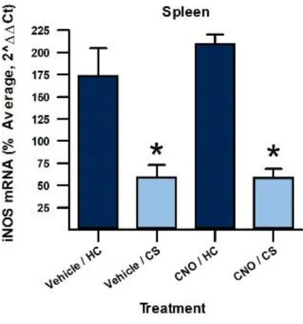
A 2x2 ANOVA revealed a significant difference between groups (plasma nitrate/nitrite:
F(3, 24) = 16.091, p < 0.001, Figure 4; iNOS mRNA: F(3, 23) = 9.556, p < 0.001, Figure 5).The one
violation of assumptions (mentioned earlier in the statistical analysis section) was in the vSub
qPCR data for which Levene’s test reported a value of 0.014, indicating that variances were not
equal across groups. There was a significant main effect of exposure to the context versus home
cage for plasma nitrate/nitrite levels (F(1, 24) = 38.893, p < 0.001) and iNOS mRNA expression
(F(1, 23) = 26.310, p < 0.001). There was a non-significant main effect of treatment on
0.296). There was a significant interaction effect between context exposure and treatment (F(1, 24)
= 5.361, p = 0.029) for plasma nitrate/nitrite levels, but a significant interaction between these
factors was not seen in iNOS mRNA expression (F(1,23) = 0.036, p = 0.851). LSD post-hoc
analysis, summarized in Table 4, revealed that vehicle-treated context re-exposed groups
CS) showed significantly decreased immune activation compared to home cage controls
(Veh-HC; nitrate/nitrite: p = 0.008, iNOS mRNA: p < 0.001). Additionally, vehicle-treated animals
that were left in home cages (Veh-HC) were statistically different from CNO-treated animals that
were left in home cages (CNO-HC) in nitrate/nitrite levels (p = 0.006), but this was not reflected
in iNOS mRNA expression for these two groups (p = 0.370). CNO-treated animals that were
re-exposed to the context (CNO-CS) showed statistically significantly decreased immune activation
compared to CNO-treated animals left in home cages (CNO-HC; nitrate/nitrite levels: p < 0.001,
iNOS mRNA expression: p = 0.002), indicating that animals with functionally inactivated vSub
still showed significant immune suppression to the heroin-paired context. Additionally, animals
that were treated with either vehicle or CNO that were re-exposed to the context (Veh-CS vs.
CNO-CS) were not significantly different in immune measures (nitrate/nitrite: p = 0.804; iNOS
mRNA: p = 0.551), indicating that there was comparable immune suppression in both groups.
These data indicate that inhibition of the ventral subiculum of the hippocampus does not
block the expression of heroin-induced conditioned immunosuppression of blood plasma
nitrate/nitrite levels or splenic iNOS mRNA expression. Taken together, the data from
Experiment 1 and Experiment 2 suggest that the dorsal, but not the ventral, subiculum of the
Discussion
Heroin is a substance that is known to possess immunosuppressive qualities. Opioid users
have an increased susceptibility to a range of infections and this vulnerability could be partially
mediated by a conditioned immune response. Previous studies have shown that pairing an
immunosuppressive substance (such as heroin) with a conditioned stimulus (such as a behavioral
chamber) can induce a conditioned immune response, such that, after sufficient pairings, the
previously neutral conditioned stimulus can elicit suppression of several immune measures. The
hippocampus is known to be involved in both the acquisition and expression of this effect. The
hippocampus is also well-known to be important in contextual memory and has diffuse
connections to both cortical and subcortical regions known to also be involved in heroin
conditioned immunosuppression to a context. A sub-region of the hippocampus called the
subiculum is implicated in this effect, as it is the main output region of the hippocampus and
might therefore mediate the communication of the hippocampus with other required brain
regions. Here, we investigated the roles of both the dorsal and ventral subicula of the
hippocampus in producing a conditioned immune effect. We functionally inactivated these
regions to investigate their respective roles by expressing and activating an inhibitory Gi-coupled
DREADD in either of these regions in animals conditioned to associate heroin with a context just
prior to re-exposure to this context and administering an immune challenge (LPS). Our data
indicate that heroin-induced conditioned immunosuppression of plasma nitrate/nitrite levels and
iNOS mRNA expression do not occur with inhibition of the dorsal subiculum. However,
inhibition of the ventral subiculum failed to block this effect, as the aforementioned immune
measures were suppressed for the CNO-CS group in a comparable way to the group that received
that the dorsal, but not the ventral, subiculum of the hippocampus plays a critical role in the
expression of heroin conditioned immune effects to a context.
This finding is rather surprising given the abundance of evidence that the ventral
subiculum plays a critical role in mediating cue-induced reinstatement of drug seeking with
cocaine (Sun & Rebec, 2003), heroin (Bossert & Stern, 2014), and alcohol (Marchant et al.,
2016). Furthermore, researchers have found that stimulation of the vHPC after training and
extinction reinstated cocaine seeking (Vorel et al., 2001). Additionally, the vHPC is thought to
be capable of encoding context under some conditions, as it has been found to contain place cells
(Poucet, Thinus-Blanc, & Muller, 1994) and to be required for contextual bidirectional
discrimination using a reward (Riaz et al., 2017). Based on existing evidence of the functional
capabilities of the vHPC and vSub, we hypothesized that it would be involved in expression of a
conditioned immune effect. However, it was somewhat surprising that the dorsal subiculum was
found to play a role instead. Our study may indicate a dissociation between the ventral and dorsal
subicula of the hippocampus, such that one may be more involved in processing reward- and
drug-related behaviors and the other more important for producing drug-induced conditioned
immune responses to contexts, respectively.
Unpublished data from our lab indicates that CPP-trained rats that received a cytokine
receptor antagonist in the dHPC prior to testing still demonstrated a strong conditioned
preference for the place that was previously paired with heroin, indicating that this mechanism
within the dHPC does not play a critical role in this paradigm. However, rats that received the
same receptor antagonist in the same region of the dHPC did not express conditioned
immunosuppression to a heroin-paired context, suggesting that this dHPC mechanism is
(unpublished data). These data intimate a more immune-response oriented role for the dHPC,
whereas the vHPC may be more involved in processing conditioned reward-related behaviors.
By investigating the subicula, the most prominent output from the hippocampus, our aim was to
characterize potential neural circuitry mediating this effect. Though these data suggest a
dissociation between conditioned drug-reward and drug-immune behaviors, it is still unclear
whether or not the circuitry underlying these effects is similar, overlapping, or distinct, and to
what degree. Evidence of the dHPC’s involvement in reward-processing and drug-seeking
behavior complicates our understanding of the distinction between the functions of these brain
regions. For example, previous research demonstrated the dSub’s involvement in mediating the
acquisition of conditioned reinstatement of cocaine-seeking behavior (Martin-Fardon,
Ciccocioppo, Aujla, & Weiss, 2008), and it has also been hypothesized that the dHPC may be
involved in the neurocircuitry that encodes rewarding properties of drugs. One study found that
rats learned to self-administer dynorphin A, an endogenous opioid peptide, into dorsal CA3 of
the hippocampus, a behavior that was blocked with co-administration of naloxone (a
non-selective opiate antagonist; Stevens et al., 1991). Furthermore, directly injecting morphine into
dHPC produces a conditioned place preference (CPP) for the morphine-paired side (Corrigall &
Linseman, 1987). It may be of interest to investigate whether the dorsal subiculum is involved in
producing a conditioned immune effect using a non-rewarding immunosuppressive drug, like
cyclophosphamide, which has been used previously (Ader & Cohen, 1975).
In addition to considering the context-paired immunosuppressant used, it might also be
important to probe the involvement of the dSub in a paradigm that does not pair an
immunosuppressant with a context; given that previous research has shown the dHPC’s
to the use of a specific context as our CS. Future research might use a different conditioned
stimulus to better characterize the role of the dSub. It would also be interesting to note whether
or not a different CS could require activation of the vSub in producing a conditioned immune
effect. Furthermore, though visualization of mCherry was used to confirm that the virus was
expressed in the vSub, it is possible that not enough of the cells were affected to produce a
difference in effect. Another possibility is that there are other populations of projection cells in
the vSub that do not express CAMKIIa, but that are involved in communicating with other
regions from the hippocampus to produce conditioned immunosuppression to a heroin-paired
context. Given all of the previous research that indicates that the vSub is important in reward-
and drug-related behaviors, future researchers should conduct a similar study using a global
inactivation method for silencing neuronal activity in the ventral subiculum.
Another factor to consider is the possibility that, by expressing in the cell membrane the
inhibitory G-protein coupled receptor that we utilized in this experiment, we may have modified
the ability of a conditioned immune effect to be expressed; infecting cells with foreign genetic
constructs can have unforeseen effects, and as such, stringent measures should be taken to
corroborate any data that has been collected by implementing these methods. CAMKIIa-EGFP, a
construct consisting of solely a promoter and a fluorescent tag, can be used as a control to ensure
that insertion of a viral construct does not affect behavior in and of itself. Another, more
rigorous, option might be to use a construct with a CAMKIIa promoter, but a different receptor
with an mCherry tag. In this way, cells that express CAMKIIa would drive the expression of a
different receptor that can be used as a control to ensure that our construct specifically did not
affect conditioned immunosuppression in and of itself. One such measure would be to use a
template (KORD), which is activated by the pharmacologically inert ligand salvinorin B, and has
been shown to significantly attenuate neuronal activity (Vardy et al., 2015). Using a different
DREADD would also eliminate the possibility that treatment with CNO mediated an immune
effect, as recent research suggested that CNO might not be entirely inert and that DREADD
experiments should include a CNO-no DREADD control group (MacLaren et al., 2016).
However, it should be noted that this study used a significantly lower dosage than that shown to
have a behavioral effect and that a CNO-treated non-CS-exposed group served as a control for
comparisons to CNO-treated CS-exposed animals.
Previous literature has shown that heroin users are susceptible to a wide range of
infections (Risdahla, Khannaa, Peterson, & Molitora, 1998) and other deleterious immunological
effects unrelated to needle use (Horsburgh, Anderson, & Boyko, 1989). It has also been shown
that negative immunological effects of opioids can be conditioned in a Pavlovian manner in
animals (Ader & Cohen, 1975; Lysle, Cunnick, Fowler, & Rabin, 1988; Kubera et al., 2008), and
though these effects are generally studied within the context of the placebo effect in humans, it is
reasonable to assume that humans may express conditioned immunosuppression to a context as
well. This study sheds light on the neural circuitry that contributes to the expression of
conditioned immunosuppression to a heroin-paired context, and future research should aim to
elaborate on this characterization of hippocampal outputs and their contributions to producing
Works Cited
Ader, R., & Cohen, N. (1975). Behaviorally Conditioned Immunosuppression. Psychosomatic Medicine,37(4), 333-340. doi:10.1097/00006842-197507000-00007
Bannerman, D.M., Deacon, R.M.J., Offen, S., Friswell, J., Grubb, M., Rawlins, J.N.P. (2002). Double dissociation of function within the hippocampus: spatial memory and
hyponeophagia. Behavioral Neuroscience, 116(5), 884-901.
Blaha, C.D., Yang, C.R., Floresco, S.B., Barr, A.M., & Phillips, A.G. (1997). Stimulation of the ventral subiculum of the hippocampus evokes glutamate receptor-mediated changes in dopamine efflux in the rat nucleus accumbens. European Journal of Neuroscience, 9(5), 902-911. doi: 10.1111/j.1460-9568.1997.tb01441.x
Bossert, J.M., Stern, A.L. (2014). Role of ventral subiculum in context-induced reinstatement of heroin seeking in rats. National Institutes of Health, 19(3), 338-342. doi:
10.1111/adb.12015.
Broadbent, N.J., Squire, L.R., Clark, R.E. (2004). Spatial memory, recognition memory, and the hippocampus. Proceedings of the National Academy of Sciences of the United States of America, 101(40), 14515-14520. doi: 10.1073/pnas.0406344101
Burns, L.H., Robbins, T.W., & Everitt, B.J. (1993). Differential effects of excitotoxic lesions of the basolateral amygdala, ventral subiculum and medial prefrontal cortex on responding with conditioned reinforcement and locomotor activity potentiated by intra-accumbens infusions ofD-amphetamine. Behavioural Brain Research, 55(2), 167-183. doi:
10.1016/0166-4328(93)90113-5
Coleman, J.W. (2001). Nitric oxide in immunity and inflammation. International Immunopharmacology, 1(8), 1397-1406.
Corrigall, W.A. & Linseman, M.A. (1988). Conditioned place preference produced by intra-hippocampal morphine. Pharmacology, Biochemistry, and Behavior, 30(3), 787-9. doi: 10.1016/0091-3057(88)90100-1
Crombag, H.S., Bossert, J.M., Koya, E., & Shaham, Y. (2008). Context-induced relapse to drug seeking: a review. Phil. Trans. R. Soc. B, 363, 3233-3243. doi: 10.1098/rstb.2008.0090 Eichenbaum, H. (2002). A brain system for declarative memory. The Cognitive Neuroscience of
Memory, 213-236. doi: 10.1093/acprof:oso/9780195141740.003.0009
French, S.J., Hailstone, J.C., & Totterdell, S. (2003). Basolateral amygdala efferents to the ventral subiculum preferentially innervate pyramidal cell dendritic spines. Brain Research, 981(1-2), 160-1967. doi: 10.1016/S0006-8993(03)03017-8
Fuchs, R.A., Evans, K.A., Ledford, C.C., Parker, M.P., Case, J.M., Mehta, R.H., See, R.E. (2005). The role of the dorsomedial prefrontal cortex, basolateral amygdala, and dorsal hippocampus in contextual reinstatement of cocaine seeking in rats.
Gilbert, P.E., Kesner, R.P. (2002). Role of rodent hippocampus in paired-associate learning involving associations between a stimulus and a spatial location. Behavioral
Neuroscience, 116(1), 63-71. doi: 10.1037/0735-7044.116.1.63
Goebel, M. U. (2002). Behavioral conditioning of immunosuppression is possible in humans. The FASEB Journal,16(14), 1869-1873. doi:10.1096/fj.02-0389com
Guo, S., Chen, S.,, Zhang, Q., Wang, Y., Xu, K., & Zheng, X. (2014). Optogenetic activation of the excitatory neurons expressing CaMKIIa in the ventral tegmental area upregulates the locomotor activity of free behaving rats. BioMed Research International, 1-11. doi: 10.1155/2014/687469
Horovitz, O., Richter-Levin, G. (2015). Dorsal periaqueductal gray simultaneously modulates ventral subiculum induced-plasticity in the basolateral amygdala and the nucleus accumbens. Frontiers in Behavioral Neuroscience, 9(53), 1-8.
Horsburgh, C. R., Anderson, J. R., & Boyko, E. J. (1989). Increased Incidence of Infections in Intravenous Drug Users. Infection Control and Hospital Epidemiology,10(5), 211-215. doi:10.1086/646004
Johansen, J.P., Hamanaka, H., Monfils, M.H., Behnia, R., Deisseroth, K., Blair, H.T., & DeDoux., J.E. (2010). Optical activation of lateral amygdala pyramidal cells instructs associative fear learning. Proceeedings of the National Academy of Sciences, 107(28), 12692-12697. doi: 10.1073/pnas.1002418107.
Kubera, M., Filip, M., Budziszewska, B., Basta-Kaim, A., Wydra, K., Leskiewicz, M.,…Lason, W. (2008). Immunosuppression induced by a conditioned stimulus associated with cocaine self-administration. Journal of Pharmacological Sciences, 107(4), 361-369. doi: 10.1254/jphs.fp0072106.
Liu, X. & Jones, E.G. (1996). Localization of alpha type II calcium calmodulin-dependent protein kinase at glutamatergic but not gamma-aminobutyric acid (GABAergic) synapses in thalamus and cerebral cortex. Proceedings of the National Academy of Sciences, 93(14), 7332-7336.
Livak, K.J., & Schmittgen, T.D. (2001). Analysis of relative gene expression data using real-time quantitative PCR and the 2DDCT method. Methods, 25, 402-408.
Lopez, A.J., Kramar, E., Matheos, D.P., White, A.O., Kwapis, J., Vogel-Ciernia, A., Sakata, K., Espinoza, M., & Wood, M.A. (2016). Promoter-specific effects of DREADD modulation on hippocampal synaptic plasticity and memory formation. The Journal of Neuroscience, 36(12), 3588-3599.
Lysle, D.T., Cunnick, J.E., Fowler, H., & Rabin, B.S. (1988). Pavlovian conditioning of shock-induced suppression of lymphocyte reactivity: acquisition, extinction, and preexposure effects. Life Sciences, 42(22), 2185-2194. doi: 10.1016/0024-3205(88)90369-4
Lysle, DT., & Ijames, SG. (2002). Heroin-associated environmental stimuli modulate the expression of inducible nitric oxide synthase in the rat. Psychopharmacology, 164, 416-422. doi: 10.1007/s00213-002-1208-x.
MacLaren, D.A., Browne, R.W., Shaw, J.K., Radhakrishnan, S.K., Khare, P., Espana, R.A., & Clark, S.D. (2016). Clozapine N-oxide administration produces behavioral effects in long-evans rats: implications for designing DREADD experiments. eNeuro, 3(5), 0219-16. doi: 10.1523/ENEURO.0219-0219-16.200219-16.
Marchant, N.J., Campbell, E.J., Whitaker, L.R., Harvey, B.K., Kaganovksy, K., Adhikary, S., Hope, B.T., Heins, R.C., Prisinzano, T.E., Vardy, E., Bonci, A., Bossert, J.M., & Shaham, Y. (2016). Role of ventral subiculum in context-induced relapse to alcohol seeking after punishment-imposed abstinence. The Journal of Neuroscience, 36(11), 32881-3294.
Maren, S., & Holt, W. (2000). The hippocampus and contextual memory retrieval in Pavlovian conditioning. Behavioural Brain Research,110(1-2), 97-108.
doi:10.1016/s0166-4328(99)00188-6
Martin-Fardon, R., Ciccocioppo, R., Aujla, H., Weiss, F. (2008). The dorsal subiculum mediates the acquisition of conditioned reinstatement of cocaine-seeking.
Neuropsychopharmacology, 33, 1827-1834. doi: 10.1038/sj.npp.1301589
Miyoshi, E., Wietzikoski, E.C., Bortolanza, M., Boschen, S.L., Canteras, N.S., Izquierdo, I., Da Cunha, C. (2012). Both the dorsal hippocampus and the dorsolateral striatum are needed for rat navigation in the Morris water maze. Elselvier 226(1), 171-178.
doi:10.1016/j.bbr.2011.09.011
Moser, M.B., Moser, E.I., Forrest, E., Andersen, P., & Morris, R.G. (1995). Spatial learning with a minislab in the dorsal hippocampus. Proceedings of the National Academy of Sciences, 92(21), 9697-9701.
Muhuri PK, Gfroerer JC, Davies MC. Associations of Nonmedical Pain Reliever Use and Initiation of Heroin Use in the United States. CBHSQ Data Rev. August 2013. Naber, P.A. & Witter, M.P. (1998). Subicular efferents are organized mostly as parallel
projections: a double-labeling, retrograde-tracing study in the rat. The Journal of Comparative Neurology, 393, 284-297.
O’Mara, S. (2005). The subiculum: what it does, what it might do, and what neuroanatomy has yet to tell us. Journal of Anatomy, 207(3), 271-282.
Piekema, C., Kessels, R.P.C., Mars, R.B., Petersson, K.M., Fernandez, G. (2006). The right hippocampus participates in short-term memory maintenance of object-location
associations. Elselvier, 33(1), 374-382. doi: doi.org/10.1016/j.neuroimage.2006.06.035 Poucet, B., Thinus-Blanc, C., & Muller, R.U. (1994). Place cells in the central hippocampus of
Riaz, S., Schumacher, A., Sivagurunathan, S., Van Der Meer, M., Rutsuko, I. (2017). Ventral, but not dorsal, hippocampus inactivation impairs reward memory expression and retrieval in contexts defined by proximal cues. Hippocampus, 27(7), 822-836. doi:
10.1002/hipo.22734
Risdahla, JM., Khannaa, KV., Peterson, PK., & Molitora, TW. (1998). Opiates and infection. Journal of Neuroimmunology, 83(1-2), 4-18. doi: 10.1016/S0165-5728(97)00216-6. Rudd RA, Seth P, David F, Scholl L. Increases in Drug and Opioid-Involved Overdose Deaths
— United States, 2010–2015. MMWR Morb Mortal Wkly Rep. 2016;65. doi:10.15585/mmwr.mm655051e1.
Saurer, T.B., Ijames, S.G., & Lysle, D.T. (2009). Evidence for the nucleus accumbens as a neural substrate of heroin-induced immune alterations. The Journal of Pharmacology and Experimental Therapeutics, 329(3), 1040-1047.
Stevens, K.E., Shiotsu, G., & Stein, L. (1991). Hippocampal μ-receptors mediate opioid reinforcement in the CA3 region. Brain Research, 545(1-2), 8-16. doi: 10.1016/0006-8993(91)91263-Z
Swanson, L.W., & Cowan, W.M. (1977). An autoradiographic study of the organization of the efferent connections of the hippocampal formation in the rat. Journal of Comparative Neurology, 172, 49-84.
Szczytkowski, J.L., Lebonville, C., Hutson, L., Fuchs, R.A., & Lysle, D.T. (2013). Heroin-induced conditioned immunomodulation requires expression of IL-1beta in the dorsal hippocampus. Brain, Behavior, and Immunity, 30, 95-102.
Szczytkowski, J.L. & Lysle, D.T. (2008). Conditioned effects of heroin on proinflammatory mediators require the basolateral amygdala. European Journal of Neuroscience, 28(9), 1867-1876.
Trouche, S., Perestenko, P.V., van de Ven, G.M., Bratley, C.T., McNamara, C.G., Campo-Urriza, N., Black, S.L., Reijmers, L.G., & Dupret, D. (2016). Recoding a cocaine-place memory engram to a neutral engram in the hippocampus. Nature Neuroscience, 19, 564-567. doi: 10.1038/nn.4250
Tsien, J.Z., Chen, D.F., Gerber, D., Tom, C., Mercer, E.H., Anderson, D.J., Mayford, M., Kandel, E.R., & Tonegawa, S. (1996). Subregion- and cell type-restricted gene knockout in mouse brain. Cell, 87, 1317-1326.
Urban, D. J., & Roth, B. L. (2015). DREADDs (Designer Receptors Exclusively Activated by Designer Drugs): Chemogenetic Tools with Therapeutic Utility. Annual Review of Pharmacology and Toxicology,55(1), 399-417. doi:10.1146/annurev-pharmtox-010814-124803
Vorel, S.R., Robert, X.L., Hayes, R.J., Spector, J.A., & Gardner, E.L. Relapse to cocaine-seeking after hippocampal theta burst stimulation. Science, 292(5519), 1175-1178. doi:
10.1126/science.1058043
Sun, W. & Rebec, G.V. (2003). Lidocaine inactivation of ventral subiculum attenuates cocaine-seeking behavior in rats. Journal of Neuroscience, 23(32), 10258-10264.
Tables Experiment 1
Table 1. Descriptive Statistics for Experiment 1 (dSub)
Table 2. Summary of Post-hoc Analyses for Experiment 1 (dSub) Experiment 2
Table 3. Descriptive Statistics for Experiment 2 (vSub)
Table 4. Summary of Post-hoc Analyses for Experiment 2 (vSub)
Figures Experiment 1
Figure 1. Nitrate/Nitrite Production with Inhibition of Dorsal Subiculum Figure 2. iNOS mRNA Production with Inhibition of Dorsal Subiculum Figure A. mCherry Visualization of Virus Expression in Dorsal Subiculum Experiment 2
Table 1. Descriptive Statistics for Experiment 1 (dSub)
A. Nitrate/Nitrite
Descriptive Statistics Mean Standard Error
Veh/HC 78.807 4.516
Veh/CS 23.342 7.157
CNO/HC 83.163 7.949
CNO/CS 61.609 5.915
B. iNOS mRNA
Descriptive Statistics Mean Standard Error
Veh/HC 128.695 17.141
Veh/CS 63.369 12.259
CNO/HC 144.998 15.926
Table 2. Post-Hoc Analyses for Experiment 1 (dSub)
A. Nitrate/Nitrite
Comparison Significance
Veh-CS x Veh-HC p < 0.001
Veh-HC x CNO-HC p = 0.652
CNO-HC x CNO-CS p = 0.027
Veh-CS x CNO-CS p < 0.001
B. iNOS mRNA
Comparison Significance
Veh-CS x Veh-HC p = 0.001
Veh-HC x CNO-HC p = 0.510
CNO-HC x CNO-CS p = 0.181
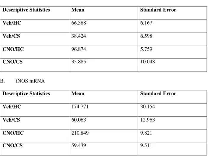
Table 3. Descriptive Statistics for Experiment 2 (vSub)
A. Nitrate/Nitrite
Descriptive Statistics Mean Standard Error
Veh/HC 66.388 6.167
Veh/CS 38.424 6.598
CNO/HC 96.874 5.759
CNO/CS 35.885 10.048
B. iNOS mRNA
Descriptive Statistics Mean Standard Error
Veh/HC 174.771 30.154
Veh/CS 60.063 12.963
CNO/HC 210.849 9.821
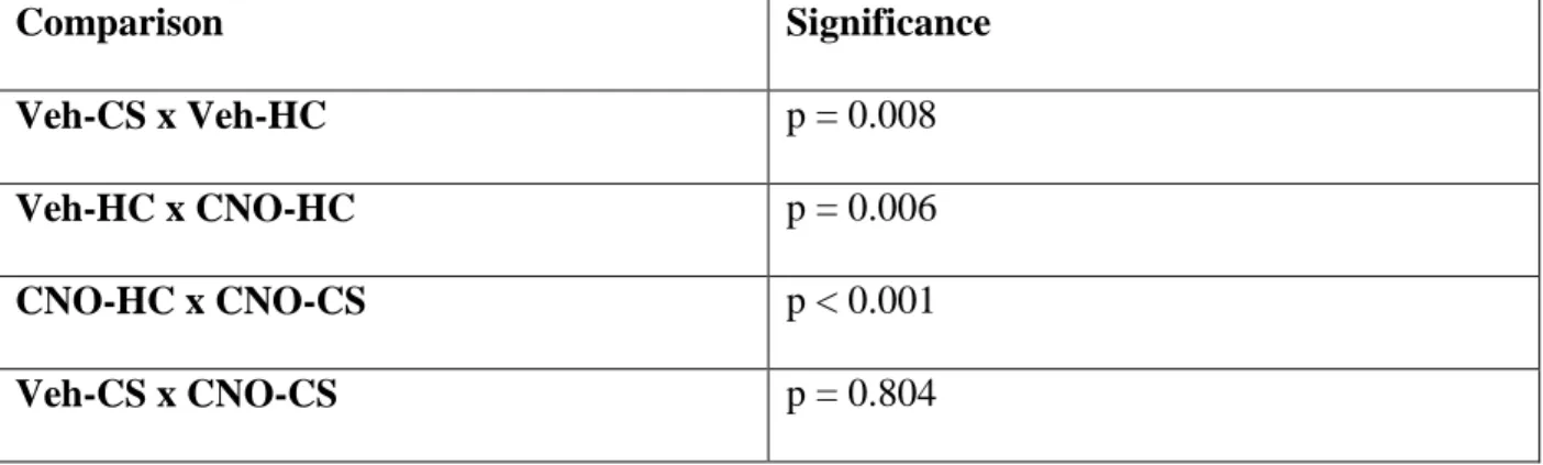
Table 4. Post-Hoc Analyses for Experiment 2 (vSub)
A. Nitrate/Nitrite
Comparison Significance
Veh-CS x Veh-HC p = 0.008
Veh-HC x CNO-HC p = 0.006
CNO-HC x CNO-CS p < 0.001
Veh-CS x CNO-CS p = 0.804
B. iNOS mRNA
Comparison Significance
Veh-CS x Veh-HC p = 0.001
Veh-HC x CNO-HC p = 0.370
CNO-HC x CNO-CS p = 0.002
Veh-CS x CNO-CS p = 0.551