Original Research Article
Role of tissue C4d in differentiation between acute rejection and HCV
recurrence after living donor liver transplantation
Zeinab Salah Eldin Hamza Khalil
1*, Waleed Gad Dandarawy Hassan
2INTRODUCTION
Liver transplantation is a well-established procedure for curative treatment of various liver diseases.1 Inspite of
continuously improving immunosuppressive therapy, acute graft rejection is still a complication after liver transplantation.2 Both acute rejection and HCV
recurrence often display the same clinical alterations in liver specimen.3 Liver biopsy represents the gold standard
for diagnosis of both acute rejection and HCV recurrence; nevertheless, discrimination can be highly difficult due to a quite similar display of alterations in liver specimen.3
Both conditions are characterized by predominantly portal-based inflammation, which may involve bile ducts and portal venous endothelium.4 Correct differentiation
between acute rejection and recurrent hepatitis is very important because of differences in treatment.5
Therefore, a specific marker expressed only in rejection but not in HCV recurrence cases would be a great asset to differentiate patients with clinically suspicious symptoms in order to validate rejection diagnosis.6 An increasing
role, both in acute and chronic rejection processes, has been attributed to humoral response mechanisms, as
ABSTRACT
Background: Liver biopsy represents the gold standard for diagnosis of acute rejection and HCV recurrence after liver transplantation; never the less discrimination can be difficult due to similar display of alterations in liver specimen. Therefore, a specific marker expressed only in rejection but not in HCV recurrence would be a great asset to differentiate between both conditions. The aim of this study was to assess the role of tissue C4d complement fragments in liver biopsy as a marker for differentiating between acute rejection and HCV recurrence in recipients post LDLT.
Methods: A case control study on 25 recipients after liver transplantation with the suspicion of either acute rejection or HCV disease recurrence, patients were classified according to pathological finding into two groups, Group 1: patients with acute rejection (n=13), Group 2: patients with HCV recurrence (n=12), The C4d was evaluated by immunohistochemical staining of the formalin-fixed, paraffin-embedded tissue in different liver compartments.
Results: C4D staining of all the studied tissue compartments (Sinusoids, portal vein endothelium, hepatic vein endothelium, arterial internal elastic lining, portal stroma, bile ducts) had high specificity (100%) and positive predictive value (100%) in diagnosis of rejection cases except portal vein endothelium. (Specificity 91.7%, positive predictive value 88.9%).
Conclusions: Tissue C4d staining was almost present in rejection cases only; further studies on larger cohort are required to stand on standard diagnostic criteria for C4d to be included in diagnosis of acute rejection after liver transplantation as its role in other organ transplantation.
Keywords: Acute rejection, Hepatitis C recurrence, Liver biopsy, Liver transplantation
1Department ofAmbulatory Care, 2Department of Family Medicine, Primary Healthcare Corporation, Qatar
Received: 10 June 2020
Accepted: 03 July 2020
*Correspondence:
Dr. Zeinab Salah Eldin Hamza Khalil, E-mail: prasanthvelekkat@gmail.com
Copyright: © the author(s), publisher and licensee Medip Academy. This is an open-access article distributed under the terms of the Creative Commons Attribution Non-Commercial License, which permits unrestricted non-commercial use, distribution, and reproduction in any medium, provided the original work is properly cited.
rejection dependent on the antibodies directed against class I or II MHC antigens of the donor present in the recipient’s serum.7
The markers of an active humoral process are the selected complement fragments - C4d, and, to a lesser extent C3d - formed as a result of classic complement activation, induced by interaction of complement-binding antibodies with alloantigens (class I and II HLA molecules, present on endothelial cells, polymorphic endothelial antigens) or with autoantigens.8
METHODS
Participant selection criteria A case control study conducted on 25 patients who had received LDLT with clinical suspicion of either acute rejection or HCV disease recurrence at Ain Shams center for organ transplant and Egypt air hospital.
Sample size according to the results that show 67.7% of patients with acute cellular rejection displayed C4d-positive staining in liver biopsy whereas only 11.8% of patients with hepatitis C recurrence tested positive for C4d, the sample size will be 24 patients assuming the power of test = 0.90, α error=0.05.
Inclusion criteria
Patients transplanted for HCV related disease that are presented with elevated liver enzymes and/or hyper-bilirubinemia without active CMV infection with normal Abdominal Doppler ultrasound and MRCP (excluding vascular and biliary disorders).
Patients classified into
• Group 1 (12 patients): patients with HCV recurrence diagnosed by positive serum PCR (HCV RNA) and histopathology suggestive of HCV recurrence.
• Group 2(13 patients): Patients with histopathology suggestive of rejection.
All patients subjected to
• Full history taking and thorough clinical examination
• Laboratory investigations:
• CBC
• Liver profile tests
• Albumin
• Bilirubin (total and direct)
• Transaminases (ALT and AST)
• Alkaline phosphatase
• INR
• PCR (HCV RNA quantitative)
• CMV PCR
• Radiological investigations
• Abdominal Doppler ultrasound
• Histopathological evaluation of liver biopsies from the 25 patients.
Histopathological evaluation
Liver core biopsy tissue were fixed in 10% buffered formalin and 3-4 micrometer thick sections were cut from the paraffin embedded tissue for hematoxylin and eosin (H&E) and trichrome stains for morphological evaluation.
The slides were evaluated and scored on the basis of Macsween’s pathological criteria for acute cellular rejection and recurrent hepatitis. 9
C4d immunostaining
• C4d immunostaining was performed on 3 micrometer thick sections cut from formalin -fixed, paraffin-embedded sections.
• The C4d polyclonal antibody against C4d (CELL MARQUE) was used for immunostaining.
• For antigen retrieval, we used Dako Target Retrieval solution PH 9.
Interpretation of C4d immunostaining
• C4d deposition in the sinsuoids, endothelium of the portal veins or hepatic veins, the arteriolar internal elastic lamina, portal stroma and bile duct epithelium were documented
• The intensity of staining was recorded as follows (0) negative, (1+) weak, (2+) moderate and (3+) strong.
• Regarding sinusoidal staining, the staining of more than 10% of sinusoidal compartment in the liver (intensity of 1+ or greater) was considered as positive. Diffuse staining was defined as staining involving more than 50% of the sinusoids whereas focal staining involved less than 50% of the sinusoids. Staining of the hepatocytes and staining of elastic fibers were regarded as non-specific and considered negative.
Re-evaluation of patients within one month of treatment to determine the response of medical treatment by liver function tests and transaminases.
Statistical methods
The collected data were coded, tabulated, and statistically analyzed using SPSS program (Statistical Package for Social Sciences) software version 18.0.
Descriptive statistics were done for quantitative parametric data as minimum and maximum of the range well as mean±SD and for numerical non-parametric data as median and 1st and 3rd inter-quartile range, while they
groups with parametric data and Mann Whitney U in cases of two independent groups with non-parametric data.
In qualitative data, inferential analysis for independent variables were done using Chi square test for differences between proportions as well as McNemar test for agreement between paired categorical data. ROC curve was used to evaluate the performance of different tests differentiate between certain groups. The level of significance was taken at p value <0.050 is significant, otherwise is non-significant. The p-value is a statistical measure for the probability that the results observed in a study could have occurred by chance.
RESULTS
The demographic features of the studied population showed that there were 22 men (88%) and 3 women (12%) with their age ranging from 19 to 63 years (mean 49.2±9.7). Concerning the pre-transplant Child scoring system; one patient (4%) was child A, seven patients (28%) were child B and seventeen patients were child C (68%), their MELD score ranged from (7-26) mean (16.4±4.4). As regard the pre-transplant clinical presentation, ascites was a common presentation. Post-transplant laboratory findings of the studied cases at time of liver biopsy Table 1.
Table 1: Post transplant laboratory findings of the studied cases at time of liver biopsy (n=25).
Mean±SD Range
WBC 4-11 (10^3/ul) 5.1±1.9 2.2-9.8
HB (13-17 g/dl) 12.0±1.8 9.3-16.9
Platelet (150-450 10^3/ul) 144.7±57.6 36.0-288.0
INR 1.2±0.4 0.8-2.5
Albumin (3.5-5 g/dl) 3.2±0.8 2.0-4.5
Median (IQR) Range
ALT (7-40 IU/L) 127.0 (100.0-239.5) 62.0-792.0 AST (7-37 IU/L) 137.0 (62.0-193.0) 41.0-394.0 Alkaline phosphatase (75-145 IU/L) 185.5 (137.3-355.5) 68.0-830.0 T. Bilirubin (0.3-1.2 mg/dl) 2.5 (0.8-7.1) 0.4-25.0 D. Bilirubin up to 0.2 mg/dl) 1.1 (0.3-4.7) 0.1-20.0
AFP (up to 25ng/mL) 5.0 (3.0-6.5) 1.6-91.0
HCV RNA (x103 IU/mL) 80.7 (0.0-473.9) 0.0-17550.1
N %
CRP (N <6 mg/dl)
Positive 6 24.0
Negative 19 76.0
IQR: Inter-quartile range
Table 2: Pathological findings and diagnosis of the post-transplant liver biopsies of the studied cases (n=25).
Pathological finding N %
Portal tract inflammation 20 80.0
Fibrosis 17 68.0%
Cholestasis 11 44.0
Parenchymal inflammation 13 52.0
Interface hepatitis 11 44.0
Parenchymal ballooning 10 40.0
Bile duct proliferation 9 36.0
Bile duct degenerative changes/ damage 5 20.0
Bile duct inflammation 2 8.0
Endotheliits (venous endothelial inflammation) 6 24.0
Pathologic diagnosis
Recurrence 13 52.0
Rejection 12 48.0
Mean±SD Range
Pathological findings and diagnosis of the post-transplant liver biopsies of the studied cases Table (2).
The bilirubin and alkaline phosphatase were significantly higher in rejection group (5.5mg/dl, 245.5mg/dl respectively) than HCV recurrence group (1.1mg/dl, 145.5mg/dl respectively) Figure 1.
Figure 1: Comparison between rejection and recurrence cases as regards to bilirubin.
Table 3: Diagnostic characteristics of total and direct bilirubin in diagnosis of rejection.
Character
Total bilirubin ≥3.0 mg/dL
Direct bilirubin ≥2.0 mg/dL
Sensitivity 61.5% 58.3% Specificity 91.7% 83.3% Predictive positive
value (PPV) 88.9% 77.8% Predictive negative
value (PNV) 68.8% 66.7% Diagnostic accuracy
(DA) 76.0% 70.9%
Total bilirubin at cut value 3mg\dl and direct bilirubin at cut value 2 mg\dl had good diagnostic characteristics in diagnosis of rejection Table 3.
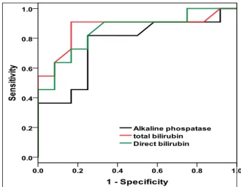
Results of the present work showed high diagnostic performance of alkaline phospatase, total and direct bilirubin in the diagnosis of rejection, total and direct bilirubin had significantly good AUC (0.871, 0.852 respectively), with significant (p value 0.003, 0.004 respectively) and alkaline phosphatase had significant AUC (0.761) (p value 0.034) Figure 2.
Tissue C4d staining was almost present in rejection cases only, this was statistically significant in all studied tissue compartments except hepatic vein endothelium and arterial IEL didn’t reach statistical significance Table 4.
Figure 2: ROC for alkaline phospatase, total and direct bilirubin in diagnosis of rejection.
Table 4: Comparison between rejection and recurrence cases as regards to tissue C4d staining.
Recurrence (N=12)
Rejection
(N=13) #p
Sinusoids 0 (0.0%) 6 (46.2%) 0.007* Portal vein
endothelium 1 (8.3%) 8 (61.5%) 0.006* Hepatic vein
endothelium 0 (0.0%) 1 (7.7%) 0.327 Arterial internal
elastic lining (IEL)
0 (0.0%) 1 (7.7%) 0.327 Portal stroma 0 (0.0%) 5 (38.5%) 0.016* Bile ducts 0 (0.0%) 4 (30.8%) 0.036*
#Chi square test, *Significant
There was agreement between histopathological diagnosis and tissue C4d staining of the examined tissue compartments in prediction of rejection except with hepatic vein endothelium, arterial internal elastic lining (IEL) and bile ducts (Table 5).
Table 5: Difference between histopathological features of rejection in biopsy results and tissue C4d staining in prediction of rejection.
Biopsy C4d staining
Pathological features of Rejection
#p
Positive Negative
Sinsuoids Pos 6 (24.0%) 0 (0.0%) 0.238
Neg. 7 (28.0%) 12 (48.0%)
Portal vein endothelium Pos 8 (32.0%) 1 (4.0%) 0.648
Neg. 5 (20.0%) 11 (44.0%)
Hepatic vein endothelium
Pos 1 (4.0%) 0 (0.0%)
0.003*
Neg. 12 (48.0%) 12 (48.0%)
Neg. 9 (36.0%) 12 (48.0%)
Percentages were calculated from total cases (N=25), #McNemar test, *Significant
The C4d staining of all the studied tissue compartments had high specificity 100% and positive predictive value 100% in diagnosis of rejection except portal vein endothelium (specificity 91.7%, positive predictive value 88.9%) (Figures 3-10).
Figure 3: Liver biopsy, recurrent hepatitis C virus infection in the liver graft portal inflammation.
Figure 4: Liver biopsy, recurrent hepatitis C virus infection in the liver graft showing portal inflammation, focal interface hepatitis and spotty
parenchymal inflammation.
Figure 5: Liver graft, acute (cellular) rejection. (H&E stain x400).
Figure 6: Liver graft, recurrent hepatitis negative staining for C4d (H&E stain x400).
Figure 8: Focal C4d deposits in sinusoidal endothelium and portal stroma.
Figure 9: C4d staining of arteriolar internal lamina.
Figure 10: C4d staining of bile duct epithelium.
DISCUSSION
The discovery of C4d as a clinical marker for AMR in the early 90’s10 marked a turning point in the history of solid organ transplantation. The authors showed that patients with suspected antibody-mediated injury in the renal graft had a linear C4d staining pattern in peritubular capillaries and that the presence of C4d was associated with impaired graft function. At the turn of the century the presence of C4d along with other markers of endothelial activation or injury in renal transplant biopsies suspected of AMR was tested.11
The correlation with graft survival that had been reported in 1993 was confirmed by other groups.12,13 This led to
general acceptance of the usefulness of C4d in the identification of acute AMR. In 2003 'C4d' was incorporated in the Banff classification.14
Controversy remains regarding the significance of C4d and its staining pattern in liver transplantation. The differences of staining protocols including the type of materials (frozen or formalin-fixed tissue), type of C4d antibodies, and antigen retrieval can affect the results of C4d staining.15
This study was conducted to assess the role of tissue C4d complement fragments in liver biopsy as a marker for differentiation between acute rejection and HCV recurrence in recipients post LDLT.
The studied patients were classified according to pathological evaluation into two groups:
• Group 1: patients with HCV recurrence (n=12).
• Group 2: patients with acute rejection (n=13). Results of the present work showed high diagnostic performance of alkaline phosphatase, total and direct bilirubin in the diagnosis of rejection, total and direct bilirubin had significantly good AUC (0.871, 0.852 respectively), with significant (p value 0.003, 0.004 respectively) and alkaline phosphatase had significant AUC (0.761) (p value 0.034).
In the present study, tissue C4d staining was almost present in rejection cases only, this was statistically significant in all studied tissue compartments except hepatic vein endothelium and arterial internal elastic lining didn’t reach statistical significance. Regarding C4d staining on various structures of liver tissue, the current study showed that C4d was deposited significantly along sinusoids in six of our patients with histopathological features of rejection (46.2%) (p value 0.007), this could be explained as liver sinusoids parallel the capillary compartments in other organs in which the blood flow is slowed and is no longer pulsatile.
be a useful marker to distinguish acute rejection from recurrent hepatitis C. In the current work, it was also crucial to determine that C4d staining of all the studied tissue compartments had high specificity 100% and positive predictive value 100% in diagnosis of rejection except portal vein endothelium. In this study, we concluded that C4d can be used as a marker for differentiating rejection from HCV recurrence. The BANFF criteria applied for other solid organ transplantation can be used in patients with rejection after liver transplantation as we found significant C4d sinusoidal deposition corresponding peritubular capillaries in renal allograft.
CONCLUSION
Tissue C4d staining by IHC technique was almost present in rejection cases only, where it had high specificity and positive predictive value in diagnosis of rejection. There was agreement between histopathological diagnosis and tissue C4d staining of the examined tissue compartments in prediction of rejection except with Hepatic vein endothelium, arterial internal elastic lining (IEL) and bile ducts. Therefore, C4d can be considered a new marker to diagnose patients with rejection.
C4d Immuno histochemical staining can be considered positive if either sinusoids show positive staining or negative sinusoids with positive portal vein endothelial staining.
Furthermore, total and direct bilirubin levels (good AUC) as well alkaline phosphatase (fair AUC) were significantly higher in rejection cases than HCV recurrence cases.
Funding: No funding sources Conflict of interest: None declared
Ethical approval: The study was approved by the Institutional Ethics Committee
REFERENCES
1. Charlton M. Natural history of hepatitis C and outcomes following liver transplantation. Clin Liver Dis. 2003;7(3):585-602.
2. Schmeding M, Dankof A, Krenn V, Krukemeyer MG, Koch M, Spinelli A, et al. C4d in acute rejection after liver transplantation a valuable tool in differential diagnosis to hepatitis C recurrence. Am J Transplant. 2006;6(53):523-30.
3. Regev A, Molina E, Moura R, Bejarano PA, Khaled A, Ruiz P, et al. Reliability of histopathologic assessment for the differentiation of recurrent hepatitis C from acute rejection after liver transplantation. Liver Transpl. 2004;10(10):1233-9.
4. Petrovic LM. Early recurrence of hepatitis C virus infection after liver transplantation. Liver Transpl. 2006;12:17-22.
5. Jain A, Ryan C, Mohanka R, Orloff M, Abt P, Romano J, et al. Characterization of CD4, CDS, CD56 positive lymphocytes and C4d deposits to distinguish acute cellular rejection from recurrent hepatitis C in post-liver transplant biopsies. Clin Transplant. 2006;20:624-33.
6. Schmeding M, Kienlein S, Rocken C, Neuhaus R, Neuhaus P, Heidenhain C, et al. ELISA-based detection of C4d after liver transplantation -A helpful tool for differential diagnosis between acute rejection and HCV-recurrence? Transplant Immunol. 2010;23:156-60.
7. Ingulli E. Mechanism of cellular rejection in transplantation. Pediatr Nephrol. 2008;25(1):61-74. 8. Hubscher S, Clouston A. Transplantation pathology.
In: Burt A, Portmann B, Ferrell L (eds). Macsween Pathology of Liver; 6th Edn; Elsevier Ltd; Chapter
15; 2011:853-933.
9. Kozlowski T, Andreoni K, Schmitz J, Hideo Hayashi P, Nickeleit V. Sinusoidal C4d deposits in liver allografts indicate an antibody-mediated response: Diagnostic considerations in the evaluation of liver allografts. Liver Transpl. 2012;18(6):641-58.
10. Feucht HE, Schneeberger H, Hillebrand G, Burkhardt K, Weiss M, Riethmüller G, et al. Capillary deposition of C4d complement fragment and early renal graft loss. Kidney Int. 1993;43:1333-8.
11. Collins AB, Schneeberger EE, Pascual MA, Saidman SL, Williams WW, Tolkoff-Rubin N, et al. Complement activation in acute humoral renal allograft rejection: diagnostic significance of C4d deposits in peritubular capillaries. J Am Soc Nephrol. 1999;10:2208-14.
12. Berger SP, Roos A, Daha MR. Complement and the kidney: what the nephrologist needs to know in 2006? Nephrol Dial Transplant. 2005;20:2613-9. 13. Herzenberg AM, Gill JS, Djurdjev O, Magil AB.
C4d deposition in acute rejection: an independent long-term prognostic factor. J Am Soc Nephrol. 2002;13:234-41.
14. Krukemeyer MG, Moeller J, Morawietz L, Rudolph B, Neumann U, Theruvath T, et al. Description of B lymphocytes and plasma cells, complement, and chemokines/receptors in acute liver allograft rejection. Transplantation. 2004;78:65-70.
15. Van der Plaats A. Anatomy and physiology of the
liver. Available at:
http://dissertations.ub.rug.n1/FILES/faculties/medici ne / 2005/a.van.der.plaats/c2.pdf. 2012.