Original Research Article
Clinico-pathological and radiological correlation of CNS tumors along
with role of Ki 67 proliferative index in grading Astrocytoma
and Meningiomas
Lakshmi Saraswathi Boni, Mounika Kondapalli, Venkata Swarajya Lakshmi Kotni, Venkata
Satya Kartheek Botta, Uma Prasad, Bhagyalakshmi Atla*
INTRODUCTION
Brain tumors are heterogeneous as most of them differ in histomorphological features. Though there is availability of advanced imaging techniques, histopathological examination is the gold standard.1 Sometimes they may
not be straightforward to diagnose creating a diagnostic dilemma in pathologist mind due to overlapping histomorphological features in benign as well as certain non-neoplastic lesion which mimics as brain tumors. Clinical needs are expanding and pathologists role in interpreting the various CNS neoplasms and grading Department of Pathology, Andhra Medical college, Visakhapatnam, Andhra Pradesh, India
Received: 08 July 2019 Accepted: 17 July 2019 *Correspondence: Dr. A. Bhagyalakshmi,
E-mail: dr.a.bhagyalaxmi@gmail.com
Copyright: © the author(s), publisher and licensee Medip Academy. This is an open-access article distributed under the terms of the Creative Commons Attribution Non-Commercial License, which permits unrestricted non-commercial use, distribution, and reproduction in any medium, provided the original work is properly cited.
ABSTRACT
Background: CNS neoplasms are a heterogenous group contributing to <2% of all the malignant neoplasms. Imaging and histopathology play a great role in diagnosing these lesions. Aim of the study is to correlate radiological findings with that of histopathology and evaluate the role of Ki 67 proliferative index in various grades of Astrocytomas and Meningiomas
Methods: This is an observational study for a period 2 years from July 2015 to June 2017 in Department of Pathology Andhra Medical College. The total number of specimens of CNS tumors received during this period were126. The specimens were routinely processed and stained with H&E. The tumors were classified based on WHO 2016 classification. In total 71 cases-45 cases of meningiomas and 26 cases of astrocytomas, the expression of Ki 67 labelling index was recorded in various grades of these tumors and results tabulated.
Results: Among 126 cases, tumors predominantly encountered were of meningeal origin accounting to 45 cases (35.71%) followed by tumors of neuroepithelial origin 35 cases (27.78%). Tumors were seen in all age groups, but common was among 41-50 years of age group with metastatic tumors being seen in >60 year group. Tumors were more common in males with male: female ratio being 1.25:1. Ki 67 proliferative index increased as the grade of tumor increased in both astrocytomas and meningiomas.
Conclusions: Grading of meningiomas and astrocytomas are very much essential with reference to prognosis and therapy. Histopathology plays a great role in grading these lesions but Ki 67 proliferative index adds as an adjunct and helps in confirmation and predicting the recurrence of these lesions.
them is very much essential for effective treatment.2
Proliferative potential of the tumor provides important prognostic information that supplements histological grading, Ki 67 index is the simplest and most reliable method of estimating it.3,4
The purpose of this study is to provide current overview of epidemiology of CNS tumor in our hospital setup and study the prevalence of these lesions by using WHO 2016 classification and understand the significance of KI 67 labeling index in grading of tumors. Aim of the study is:
•
To study spectrum of CNS tumors in the specimens received for histopathological examination.•
To correlate the radiological diagnosis with that of histopathological diagnosis.To know the expression of Ki 67 proliferative index in various grades of Astrocytomas and Meningiomas. To assess the relationship of Ki 67 staining with, the histopathological grading of Astrocytomas and Meningiomas.
METHODS
Study design
A hospital based observational study.
Study period
2 years from July 2015 to June 2017 at Department of Pathology, Andhra Medical College, Visakhapatnam.
Inclusion criteria
All the specimens of CNS tumors received in the Department of Pathology for histopathological examination were included in the study
Exclusion criteria
Recurrent CNS tumors and on therapy were excluded
Sample size
126 cases.
Detailed clinical data and radiological findings were recorded. The tissue was fixed in 10% formalin and was subjected to routine paraffin embedded processing, stained with H and E.
The spectrum of CNS tumors was classified based on WHO 2016 classification.
Histomorphological criteria for grading of Meningiomas.
WHO Grade Criteria
I. Mitosis <4/10 HPF II. Mitosis 4-19/10 HPF
Or
3 or more of the following features
• Increased cellularity • Sheet like growth pattern • Small cells with high N/C ratio • Prominent nucleoli
• Foci of spontaneous or geographic necrosis.
III. 1. Mitosis >20 /10 HPF
2. Loss of differentiated features resulting in carcinoma, melanoma or sarcoma like appearance.
Grading of Astrocytomas:
I. Pilocytic astrocytoma II. Diffuse astrocytoma III. Anaplastic astrocytoma IV. Glioblastoma multiforme
All the cases of astrocytomas and meningiomas were stained with Ki 67 proliferation marker. Three microns thick formalin fixed paraffin embedded sections were obtained for staining with Ki 67 proliferation marker by adopting Dako immunohistochemistry staining procedure. Fields with maximal labeling were chosen for counting. The total number of nuclei taking brown colored stain was counted manually under high power (400 X) magnification. The total number of nuclei counted was 1000 nuclei. The Ki 67 expression was recorded as percentage:
Number of nuclei showing positive staining (brown color) X 100 % Total number of nuclei counted
• Areas free from necrosis and capillary endothelial proliferation were selected.
• Interpretation of Ki 67 for Astrocytomas was done based on David and Louis et al.5
Astrocytoma Grade I is <1% Astrocytoma Grade II is <4% Astrocytoma Grade III is 5 to10% Astrocytoma Grade IV is >15 to 20%
• Interpretation of Ki 67 for Meningiomas was done based on Sashidhar babu et al.6
Grade I-0-4%, Grade II - 4.1-7%, Grade III - >7%
Statistical analysis
investigative method was evaluated taking histopathology
as gold standard.
Data for Ki 67 proliferative index was expressed as mean and statistical significance of Ki 67 expression and histological grade was determined by chi-square test.
RESULTS
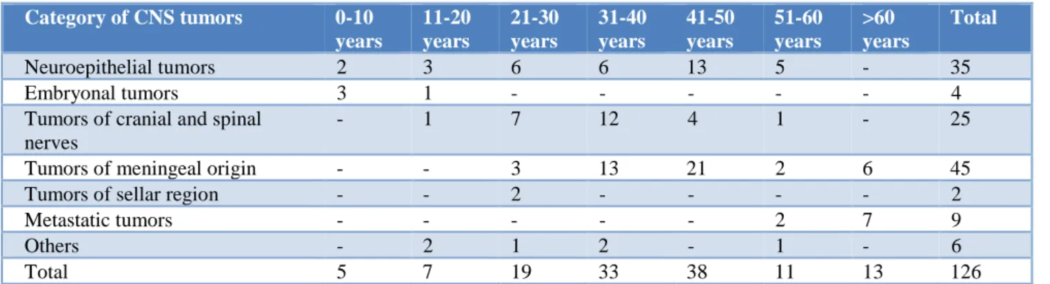
A total of 126 cases of CNS neoplasms were analyzed during this period. Male: Female ratio was 1.25:1.The age groups with tumors included a wide range from 3
years to 69 years with the highest number of cases seen within 41-50 years age group , 38 cases (30.15%) and metastatic tumors are common with age group of 60 and above (9 cases , 7.14%) (Table 1). Tumors were tabulated according to the WHO CNS 2016 classification series. Most common presentation of brain tumors in the present study was headache and vomiting (41 cases 32.54%), seizures (40 cases 31.75%), other symptoms were weakness and focal neuro deficits (25 cases 19.84%), visual disturbances (6 cases, 4.76%), hearing loss and tinnitus (12 cases, 9.52%) and gait disturbances ( 2 cases 1.59%).
Table 1: Age distribution in CNS tumors (n=126).
Category of CNS tumors 0-10
years
11-20 years
21-30 years
31-40 years
41-50 years
51-60 years
>60 years
Total
Neuroepithelial tumors 2 3 6 6 13 5 - 35
Embryonal tumors 3 1 - - - 4
Tumors of cranial and spinal nerves
- 1 7 12 4 1 - 25
Tumors of meningeal origin - - 3 13 21 2 6 45
Tumors of sellar region - - 2 - - - - 2
Metastatic tumors - - - 2 7 9
Others - 2 1 2 - 1 - 6
Total 5 7 19 33 38 11 13 126
In the present study majority of the CNS tumors were meningiothelial tumors (35.71%) followed by neuroepithelial tumors (27.78%) and tumors of cranial and spinal nerves was (19.84%). The tumors were common in the frontal lobe and cerebropontine angle (15.87% each).
Figure 1: Photomicrograph of Pilocytic astrocytoma (H and E, 400 X).
Astrocytic tumors constituted 26 cases (74.28%) of the neuroepithelial tumors. The common histopathological variants of neuroepithelial tumors were diffuse astrocytomas (28.58%), followed by Pilocytic astrocytomas (17.14%) (Figure 1) and glioblastoma
multiforme (17.14%) Meningothelial meningioma (Figure 2,3).
Figure 2: Gross photograph of Meningioma with Duramater.
constituted 55.6% of meningeal tumors, followed by transitional meningioma (20%) and 5 cases of psammomatous meningioma, 3 cases of atypical meningioma (figure 4) and a case of angiomatous meningioma.
Figure 3: Photomicrograph of Meningiotheliomatous
meningioma (H and E, 100 X).
Figure 4: Photomicrograph of Atypical meningioma invading the glial tissue (H and E, 100 X).
Figure 5: Photomicrograph of Metastatic carcinoma in the brain (H and E, 100 X).
Figure 6: Photomicrograph of Schwannoma with Verocay bodies (H and E, 400 X).
Table 2: Distribution of histopathological variants of CNS tumors in the present study (n=35).
Neuroepithelial tumors Grade of tumor Number Percentage
Pilocytic astrocytoma (nos type) Grade-i 6 17.14
Diffuse astrocytoma (nos type) Grade-ii 10 28.58
Anaplastic astrocytoma (nos type) Grade-iii 4 11.43
Glioblastoma multiforme (nos type) Grade-iv 6 17.14
Ependymoma Grade-ii 2 5.71
Oligodendroglioma (nos type) Grade-ii 2 5.71
Neuronal tumor Grade-i 5 14.29
Total - 35 100
Meningeal tumors Grade of Tumor Number Percentage
Meningothelial Grade-1 25 55.56%
Transitional Grade-1 9 20.00%
Psammomatous Grade-1 5 11.11%
Atypical Grade-ii 3 6.67%
Fibrous Grade-1 2 4.44%
Angiomatous Grade-1 1 2.22%
Total - 45 100 %
Cranial and spinal nerves tumors Grade of Tumor Number Percentage
Schwannoma Grade-i 21 84%
Neurofibroma Grade-i 4 16%
Tumors from cranial and spinal nerves, tumors of sellar
region, metastatic tumors (figure 5) and pituitary adenomas (figure 7) showed hundred percent (100%) correlation between radiology and histopathology, while 88.57% of neuroepithelial tumors and 84.44% of meningeal tumors showed correlation between radiology and histopathology (Table 3). Radiology as a diagnostic test for central nervous tumors: shows sensitivity: 66.67 %, specificity: 97.78 %, positive predictive value: 87.5%, negative predictive value: 92.63% and overall accuracy: 91.89 % (Table 4). Mean KI 67 proliferative index (Fig 8) increased as the grade of tumor increased in astrocytic tumors with P value of 0.0006. Mean KI 67 proliferative index increased as the grade of tumor increased in astrocytic tumors with P value of 0.00001 (Table 5).
Figure 7: Photomicrograph of Pituitary adenoma (H and E, 400 X).
Table 3: Histo-radiological correlation taking histopathology as gold standard (n=126).
CNS tumors Number
Correlated with radiology
Did not correlate with Radiology
Overall correlation percentage
Neuroepithelial tumors 35 31 4 88.57%
Embryonal tumors 4 0 4 0%
Tumors of cranial and spinal nerves 25 25 0 100%
Tumors of meningeal origin 45 38 7 84.44%
Tumors of sellar region 2 2 - 100
Metastatic tumors 9 9 - 100
Pituitary adenomas 6 6 - 100
Total 126 111 15 88.09%
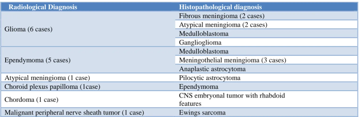
Table 4: Histo-radiological correlation showing deferred cases in the present study (n=15).
Radiological Diagnosis Histopathological diagnosis
Glioma (6 cases)
Fibrous meningioma (2 cases) Atypical meningioma (2 cases) Medulloblastoma
Ganglioglioma
Ependymoma (5 cases)
Medulloblastoma
Meningothelial meningioma (3 cases) Anaplastic astrocytoma
Atypical meningioma (1 case) Pilocytic astrocytoma
Choroid plexus papilloma (1case) Ependymoma
Chordoma (1 case) CNS embryonal tumor with rhabdoid
features
Malignant peripheral nerve sheath tumor (1 case) Ewings sarcoma
DISCUSSION
Tamkeen Masoodi et al1analysed 106 cases of CNS
tumors, the age of the patients ranged from 4 to 82 years with a mean age of 43.29 years. Maximum number of cases (47.1%) were seen in the age group between 31-50 years. Overall males were slightly more frequently
affected than females, the male: female being 1.12:1. Headache was the most common symptom (69.6%) followed by seizures (35.9%). Frontal lobe was the commonest site (20.7%) involved.
3years to 69 years with highest number of cases seen
within 41-50 years age group. Most common presentation of brain tumors was headache and vomiting (32.54%) followed by seizures (31.75%). The tumors were common in the frontal lobe and cerebropontine angle (15.87% each). This is in agreement to the findings of Jalali and Datt 7 and Jamal et al.8
The various patterns of CNS tumors in the study by Tamkeen Masoodi et al were astrocytomas (41.50%), meningiomas (19.81%), pituitary adenomas (11.32%), schwannoma (11.32%), oligodendrogliomas (2.84%), ependymoma (4.72%), medulloblastoma (3.78%).1 In the
present study the various patterns were astrocytomas (20.63%) meningiomas (35.71%), pituitary adenomas (4.76%), nerve sheath tumors (19.84%), oligodendrogliomas (1.59%), ependymoma (1.59%), medulloblastoma (1.59%) and metastatic tumors (7.14%). In the present study meningiomas comprised the major group, which was comparable with studies by Das et al, Suh et al10 and Lee at al.9,11
Lakshmi S analysed 507 CNS tumors, meningiomas comprised 25.25% of cases. The classical meningiothelial variant was the commonest (23.44%).12 In the present
study meningiomas constituted (35.71%) of cases and the commonest variant being meningothelial meningioma constituting 55.56% of cases, followed by transitional meningioma (20.00%) and 5 cases of psammomatous meningioma, 3 cases of atypical meningioma and a case of angiomatous meningioma. Various studies done by AB Shah et al, Khaled R Zalata, Sangamithra et al, S Babu et al showed similar findings.6,13-15
Tumors from cranial and spinal nerves were compared with various studies. In the study by Lilith and Dastur et al, Schellinger et al, Kaye et al, Cheang et al, Herbet H Engelhard et al, Moein P et al, Nitin M. Gadgil et althe percentage of nerve sheath tumors were 39.92%, 24.4%, 32.3%, 52.2%, 21.2%, 33%, 42.36% respectively.16-22 In
the present study all the tumors were nerve sheath tumors constituting 19%, 84% being schwannoma and 16% being neurofibroma.
The diagnostic accuracy of MRI in CNS tumors ,the study by Raj Kumar et al showed sensitivity of 94.5%, specificity of 75%, positive predictive value of 96.29%, and negative predictive value of 66.66%.23 In the present
study the diagnostic accuracy was sensitivity:66.67 %, specificity: 97.78 %, positive predictive value: 87.5%, negative predictive value: 92.63% overall accuracy: 91.89 %.
Thotakura M et al 3 evaluated Ki 67 as proliferative index
in the grading of astrocytomas in order to predict the biological behavior of the tumor and prognosis of patients. His observations were; mean Ki 67 LI in Grade I astrocytomas was 3.36, 7.05 in Grade II astrocytomas, in Grade III astrocytomas and 38.7 in Grade IV astrocytomas.
p values were significant between all grades of astrocytomas except between Grade I and Grade II tumors which was 0.5076. In the present study mean Ki 67 LI in Grade I astrocytomas was 4.66, 8.07 in Grade II astrocytomas, 13.5 in Grade III astrocytomas and 22.93 in Grade IV astrocytomas.
Sashidhar Babu et alin their study Grade-I meningiomas constituted about 90%, Grade-II about 7% and Grade-III about 2% of the meningiomas. Histopathologic grading is one of the important predictors of recurrence. Ki 67 LI correlates with histological grade and recurrence.6
However, all the tumors in each grade do not behave uniformly. The Ki 67 LI increased with the grade of meningiomas and mean Ki 67 LI between Grade I and II and I and III were statistically significant. In the study by Sanghamitra Mukherjee et alMean Ki 67 LI of Grade I meningiomas was 1.4%, Grade II meningiomas was 4.08% and Grade III meningioma showed Ki 67 LI of 15%. In the present study the Ki 67 LI increased with the grade of meningiomas. Mean Ki 67 LI of Grade I meningiomas varied from 3.1 to 7.3% and Grade II meningiomas was 10.8%.15
CONCLUSION
In the present study the commonest tumors consisted of meningiomas, followed by neuroepithelial tumors and tumors from cranial and spinal nerves with a radio histopathological over all correlation of 91.89%. Measurement of proliferative activity are important in determining the grade and recurrence of the tumor. Histopathology grading may sometimes overrate or under rate the actual behavior of the tumor especially in Astrocytomas. Ki 67 is a nuclear marker used to demonstrate proliferative phase of cell cycle. Thus immunohistochemical evaluation with Ki 67 may prove to be a vital supplementary tool for diagnostic and prognostic significance. In the present study the expression of Ki 67 increased as the grade of tumor increased in both astrocytomas and meningiomas. In one case with Ki 67 index greater than 7 in case of meningiomas showed recurrence within a period of 1 year. Determination of Ki 67 should constitute a part of routine investigation in patients with astrocytomas and meningiomas.
Funding: No funding sources Conflict of interest: None declared
Ethical approval: The study was approved by the Institutional Ethics Committee
REFERENCES
1. Masoodi T, Gupta RK, Singh JP, Khajuria A. Pattern of central nervous system neoplasm: A study of 106 cases. JK Pract. 2012;17:42-6.
3. Thotakura M, Tirumalasetti N, Krishna R. Role of
Ki 67 labeling index as an adjunct to the histopathological diagnosis and grading of astrocytomas. J cancer res therapeut. 2014;10(3):641.
4. Quiñones-Hinojosa A, Sanai N, Smith JS, McDermott MW. Techniques to assess the proliferative potential of brain tumors. J neuro-oncol. 2005;74(1):19-30.
5. Louis DN, Perry A, Reifenberger G, Von Deimling A, Figarella-Branger D, Cavenee WK, et al. The 2016 World Health Organization classification of tumors of the central nervous system: a summary. Acta neuropathol. 2016;131(6):803-20.
6. Babu S, Uppin SG, Uppin MS, Panigrahi MK, Saradhi V, Bhattacharjee S, et al. Meningiomas: correlation of Ki 67 with histological grade. Neurol Ind. 2011;59(2):204.
7. Jalali R, Datta D. Prospective analysis of incidence of central nervous tumors presenting in a tertiary cancer hospital from India. J Neuro-oncol. 2008;87(1):111.
8. Jamal S, Mamoon N, Mushtaq S, et al. Pattern of Central Nervous System (CNS) Tumours: A study of 430 cases. Pak J Pathol 2005;16(4):106-109. 9. Das A, Chapman CA, Yap WM. Histological
subtypes of symptomatic central nervous system tumours in Singapore. J Neurol, Neurosurg Psychiat. 2000;68(3):372-4.
10. Suh YL, Koo H, Kim TS, Chi JG, Park SH, Khang SK, et al. Tumors of the Central Nervous System in Korea A Multicenter Study of 3221 Cases. J neuro-oncol. 2002;56(3):251-9.
11. Lee CH, Jung KW, Yoo H, Park S, Lee SH. Epidemiology of primary brain and central nervous system tumors in Korea. J Korean Neurosurg Soc. 2010;48(2):145.
12. Lakshmi S. Meningiomas: a clinico pathological study Int J Med Res Health Sci. 2015;4(4):827-831. 13. Shah AB, Muzumdar GA, Chitale AR.
Meningiomas: report of a hospital-based registry. Ind J pathol microbiol. 2005;48(4):468-71.
14. Khaled R Zalata, Dina A El Tantawy, Azaa Abdel Aziz, Abdel Wahab M Ibraheim, Ahmed H Halaka, Hasan H Gawish, et al; Frequency of CNS tumors in the delta region, Egypt; Ind J pathol Microbiol 2011: 54(2)299-06
15. Mukherjee S, Ghosh SN, Chatterjee U, Chatterjee S. Detection of progesterone receptor and the correlation with Ki 67 labeling index in meningiomas. Neurol Ind. 2011;59(6):817.
16. Lalitha VS, Dastur DK. Neoplasms of the central nervous system-histological types in 2237 cases. Ind J cancer. 1980;17(2):102-6.
17. Schellinger KA, Propp JM, Villano JL, McCarthy BJ. Descriptive epidemiology of primary spinal cord tumors. J neuro-oncol. 2008;87(2):173-9.
18. Kaye AH, Giles GG, Gonzales M. Primary central nervous system tumours in Australia: a profile of clinical practice from the Australian Brain Tumour Register. Aus New Zeal J Surg. 1993;63(1):33-8. 19. Cheang CM, Hwang SL, Hwong SL. An analysis of
intraspinal tumors in south Taiwan. The Kaohsiung J med sci. 1997;13(4):229-36.
20. Engelhard HH, Villano JL, Porter KR, Stewart AK, Barua M, Barker FG, et al. Clinical presentation, histology, and treatment in 430 patients with primary tumors of the spinal cord, spinal meninges, or cauda equina. J Neurosurg: Spine. 2010;13(1):67-77.
21. Moein P, Behnamfar O, Khalighinejad N, Farajzadegan Z, Fard SA, Razavi M, et al.12-year epidemiologic study on primary spinal cord tumors in Isfahan, Iran. J res in med sci: offic J Isfahan Univ Med Sci. 2013;18(1):17.
22. Nitin M Gadgil, Chetan Sudhakar Chaudhari, Sangeeta R Margam, Mohd. UnzerMohd. Umar Khan, Prashant Vijay Kumavat, et al. A clinicopathological study of lesions of spinal cord and its coverings: A tertiary care hospital experience. Ann Pathol Lab Med. 2016;03(03). 23. Raj Kumar, Alex Daniel Prabhu Arul Pitchai,
Srinivasa Mudali. Diagnostic Accuracy of Magnetic Resonance Imaging in Characterizing Intracranial Space Occupying Lesions: A Cross-sectional Study International J Scient Stud.2016;4(3).