Verma Rahul et al JMSCR Volume 06 Issue 04 April 2018 Page 627
Original Research Article
Study of prevalence of primary bone tumors at a tertiary care centre in
Central India
Authors
Verma Rahul
1, Gupta Upendra
2, Uikey Suresh
31
Associate Professor, Department of Orthopedics, Gandhi Medical College, Bhopal 2
Senior Resident, Department of Orthopedics, Gandhi Medical College, Bhopal 3
Assistant Professor, Department of Orthopedics, Gandhi Medical College, Bhopal
Abstract
Introduction: Bone sarcomas account for 0.2% of all malignancies. Many relevant demographic, clinical and epidemiological characteristics have been reported.
Objectives: To analyze the spectrum of primary bone tumors and their relative frequency in Gandhi Medical College and Hamidia Hospital, Bhopal.
Methods: This is a three and a half year study conducted from July 2012 to December 2015. All primary bone tumors confirmed by histopathology have been included.
Results: A total of 124 cases of primary bone tumours were studied. Age range varied from 7 to 92 years with a mean age of 24.4 years. Among these, 55.65% were males and 44.35% were females. On histological examination, 51.6% were benign and 48.4% cases were malignant. Giant cell tumor was the commonest benign tumor (39.7%) and osteosarcoma was the commonest malignant tumor (56.7%). Maximum number
of cases belonged to the 2nd decade followed by 3rd and 4th decade. Clinically, all patients presented with
swelling, hence the most common symptom. 42% patients presented with both pain and swelling.
Conclusion: The pattern and distribution of bone tumors seen at our centre are similar to those reported from other studies. Males were more commonly affected than females with a peak in the second decade of life. Long bones of lower extremity were the most commonly affected sites. Giant cell tumour and osteosarcoma were the commonest benign and malignant varieties respectively.
Keywords: Bone tumors, sarcoma, prevalence.
Introduction
Among the wide array of human neoplasms, primary tumors of bone are relatively uncommon [1]
. This has led to the scarcity of data about their relative frequency. Overall, bone sarcomas account for just 0.2% of all malignancies and the five year overall survival rate is 67.9%[2]. Also, the clinical presentation of these patients is quite
non specific[1]. Majority of the patients report pain and swelling[3]. This non specific presentation poses a diagnostic problem. The challenge is greater in developing countries due to inadequate diagnostic and therapeutic amenities and unawareness. There are worldwide variations in patterns of cancer[4]. Age of the patient, site affected, radiographic and microscopic
www.jmscr.igmpublication.org Impact Factor (SJIF): 6.379
Index Copernicus Value: 71.58 ISSN (e)-2347-176x ISSN (p) 2455-0450
Verma Rahul et al JMSCR Volume 06 Issue 04 April 2018 Page 628 appearances contribute in making the final
diagnosis, hence a multidisciplinary approach is required. Early diagnosis and identification of benign lesions help in performing limb salvage surgeries[5]. This study has been conducted at a tertiary care centre in Central India in order to analyze the prevalence of bone tumors in this geographical area with respect to age, sex, site and clinical symptoms.
Material and Methods
This is a three and a half year study conducted in Gandhi Medical College and Hamidia Hospital, Bhopal from July 2012 to December 2015. The study was retrospective for cases from July 2012 to July 2014, and prospective for cases from then onwards. All primary bone tumors confirmed by histopathology have been included in the study. All non neoplastic lesions and metastatic tumors have been excluded. All patients were registered according to the proforma. Patients were questioned for the symptoms of bone tumors like pain, swelling, weakness, range of motion of the involved joint, any sensory or motor deficit, rapid growth, erythema or pathological fractures.
A detailed clinical examination was done followed by investigations and comprehensive examination of other systems. Finally, patient’s consent was taken, biopsy of tumor was done and sample sent for histopathological examination for confirmation.
Results
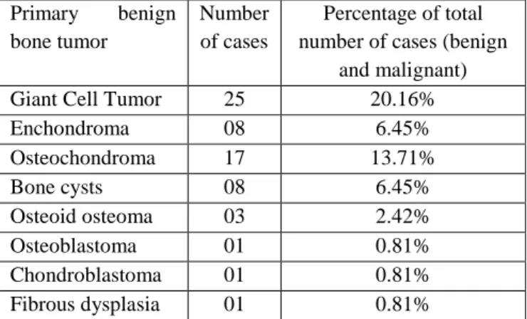
A total of 124 cases of primary bone tumours were studied over a span of three and a half years. The cases were studied with respect to age of patient, sex and site affected and histopathologic diagnosis. Age range varied from 7 to 92 years with a mean age of 24.4 years. Among these, 69 patients (55.65%) were males and 55 patients (44.35%) were females. On histological examination, 64 cases (51.6%) were benign. The distribution of primary bone tumors is represented in Table 1and Table 2. Giant cell tumor was found to be the commonest benign tumor accounting for
Verma Rahul et al JMSCR Volume 06 Issue 04 April 2018 Page 629 commonly affected bone. Other less common
affected bones were foot, hand, fibula, ulna and scapula. 59% of osteosarcomas occurred in femur followed by tibia, humerus and radius. Among Ewing’s sarcoma, tibia was the commonest site (42%) followed by femur, pelvis and humerus. Giant cell tumors and osteochondromas were commonest in femur and tibia. For enchondromas, commonest site was hand (50%) followed by foot (37%). Clinically, all patients presented with swelling, hence the most common symptom. 42% patients presented with both pain and swelling. Occasional patients had limitation of movement and pathological fracture.
Table 1- Distribution of primary benign bone tumors
Primary benign bone tumor
Number of cases
Percentage of total number of cases (benign
and malignant)
Giant Cell Tumor 25 20.16%
Enchondroma 08 6.45%
Osteochondroma 17 13.71%
Bone cysts 08 6.45%
Osteoid osteoma 03 2.42%
Osteoblastoma 01 0.81%
Chondroblastoma 01 0.81%
Fibrous dysplasia 01 0.81%
Table 2- Distribution of primary malignant bone tumors
Primary malignant bone tumors
Number of cases
Percentage of total number of cases (benign and malignant)
Osteosarcoma 34 27.4%
Ewings’ Sarcoma 19 15.32%
Chondrosarcoma 05 4.03%
Multiple myeloma 02 1.61%
Table 3- Site distribution of primary bone tumors
Site affected Number of cases Percentage
Femur 53 42.7%
Tibia 26 21%
Humerus 14 11.3%
Pelvis 08 6.5%
Radius 08 6.5%
Discussion
This study was done to analyze the spectrum of primary bone tumors among patients in Gandhi
Medical College and Hamidia Hospital. The results of our study were obtained after statistical analysis of 124 cases of primary bone tumors (confirmed by histopathology) over a period of three and a half years The data was recorded with respect to age, sex and site affected.
In our study, benign tumors (51.6%) were marginally more common than malignant tumors (48.4%). This is in concordance with other studies [6-8]
.
In our study, primary bone tumors were commonest in 2nd decade followed by 3rd decade. The findings were similar when compared to studies in other regions of India[8-9]. For osteosarcomas and Ewing sarcomas specifically, peak incidence was in 2nd decade as was seen in other studies[10-13]. For Giant cell tumors, incidence peaked in 2nd decade followed by 3rd decade in our study, slightly lower than in other studies, wherein the peak was in 3rd and 4th decade. There were 5 cases of chondrosarcomas with a wide age range and a mean of 37.6 years. Peak incidence for chondrosarcomas in study by Oyemade et al was 3rd and 4th decade. Multiple myeloma was found to be a tumor of older individuals with a mean of 48.5 years, concordant with other studies[7].
Verma Rahul et al JMSCR Volume 06 Issue 04 April 2018 Page 630 The most common sites of primary bone tumors in
our study were femur (42.7%), followed by tibia (21%) and humerus (11.3%). These findings are concordant with other studies[3,8,15]. On analysis of individual types, 59% osteosarcomas, 40% giant cell tumors and 53% osteochondromas involved the femur bone in our study. Findings are similar to other studies [5,16,17].
The most common tumor in our study was osteosarcoma (27.4%), the finding is concordant with a large study conducted in China with a sample size of 9200 patients[4]. However, some other studies found the most common tumor to be osteochondroma [5]. Giant cell tumor was second most common tumor in our study, a finding similar to other studies [4, 5]. Also, in our study the 3rd most common tumor was Ewing’s sarcoma accounting for 15.32% cases, this finding however did not show concordance with other studies. Petca et al also reported Ewing’s sarcoma as the 2nd most common malignant primary bone tumor, most other studies found chondrosarcomas to be more common [15]. It can be considered as a geographical variation. Among the benign tumors, as mentioned earlier Giant cell tumors were the most common in our study (39.06% cases). The finding coincides with the study conducted in Ahmedabad [18]. Although studies conducted in Manipur, Jammu and Pune have reported osteochondromas as the most common benign tumor [5, 8, 9]. Overall, our study showed a higher prevalence of malignant primary bone tumors than benign tumors and a greater number of cases of Ewing’s sarcoma when compared to other studies. The most common clinical presentation of these tumors was swelling. 55% patients presented with only swelling and 42% patients presented with both pain and swelling. Minority of patients presented with other symptoms like pathologic fracture and limitation of movement.
Our study describes the pattern and frequencies of primary bone tumors clinically evaluated at our institute over a three and a half year period. The pattern and distribution of bone tumors seen at our centre are similar to those reported from other
studies. Males were more commonly affected by bone tumours than females with a peak in the second decade of life. Long bones of lower extremity were the most commonly affected sites. Giant cell tumour and osteosarcoma were the commonest benign and malignant varieties respectively. A greater number of cases of Ewing’s sarcoma were noted as compared to other studies. Since bone tumors present with non specific symptoms of pain and swelling, to achieve a high rate of accurate diagnosis of bone tumours, joint clinical, radiological and pathological team work is required. A high index of suspicion is recommended especially in patients in their first two decades of life.
References
1. Dorfman HD, Czerniak B, R. K. WHO classification of tumours of bone. 2002. 225-367 p.
2. Franchi A. Epidemiology and classification of bone tumors. Clin Cases Miner Bone Metab. 2012;9(2):92–5. 3. Sakala D, Munthali JC, Mulla Y. Primary
Malignant Bone Tumours at the University Teaching Hospital in Lusaka Zambia. Med J Zambia. 2016;43(1):24–30.
4. Niu X, Xu H, Inwards CY, Li Y, Ding Y, Letson GD, et al. Primary bone tumors: Epidemiologic comparison of 9200 patients treated at Beijing Ji Shui Tan Hospital, Beijing, China, with 10 165 patients at Mayo Clinic, Rochester, Minnesota. Arch Pathol Lab Med. 2015;139(9):1149–55.
5. Rhutso Y, Laishram R, Sharma LdC, Debnath K. Histopathological evaluation of bone tumors in a tertiary care hospital in Manipur, India. J Med Soc. 2013;27(2):135.
Verma Rahul et al JMSCR Volume 06 Issue 04 April 2018 Page 631 Mruthyunjaya, Rupakumar CS, Gadiyar
HB, et al. Bone tumors in a tertiary care hospital of south India: A review 117 cases. Indian J Med Paediatr Oncol. 2011;32(2):82–5.
8. Gupta D, Gupta RK, Gupta RK. Study of the morphological pattern of non-neoplastic and non-neoplastic bone lesions- a 5 year study. Indian J Pathol Oncol. 2016;3(2):165–73.
9. Bamanikar S, Pagaro P. Histopathological Study of Primary Bone Tumours and Tumour-Like Lesions in a Medical Teaching Hospital. J Krishna Inst Med Sci Univ. 2015;4(2):46–55.
10. Larsson S, Lorentzon R. The incidence of Malignant primary bone tumours in relation to age, sex and site. J bone Jt Surg. 1974;56 B(3).
11. Price CHG, Jeferee GM. Incidence of bone sarcoma in sw england, 1946-74, in relation to age, sex, tumour site and histology. Br J Cancer. 1977;36:511–22. 12. Oyemade GAA, Abioye AA. Primary
Malignant tumors of bone: Incidence in Ibadan, Nigeria. J Natl Med Assoc. 1982;74(1):65–8.
13. Arora RS, Alston RD, Eden TOB, Geraci M, Birch JM. The contrasting age-incidence patterns of bone tumours in teenagers and young adults: Implications for aetiology. Int J Cancer. 2012;131(7):1678–85.
14. Garciadiego-cázares D, Rubio RD. Epidemiological Aspects of Osteosarcoma , Giant Cell Tumor and Chondrosarcoma Musculoskeletal Tumors - Experience of the Epidemiological Aspects of Osteosarcoma , Giant Cell Tumor and Chondrosarcoma Musculoskeletal Tumors - Experience of the National Reha. Asian Pac J Cancer Prev. 2015;15(August
2016):6451–5.
15. Rc Petca, Gavriliu S, Burnei G. Retrospective clinicopathological study of malignant bone tumors in children and adolescents in Romania – single center experience. J Med Life. 2016;9(2):205–10. 16. Patel D, Patel P, Gandhi T, Patel N, Patwa J. Clinicopathological study of bone lesions in tertiary care center – a review of 80 cases. Int J Adv Res. 2015;3(7):1267– 72.