Original Research Article
CT scan vs nasal endoscopy findings in the diagnosis of chronic
rhinosinusitis: our experience
Shruti Baruah*, Pratibha Vyas, Arpit Srivastava
INTRODUCTION
Chronic rhinosinusitis has become a disease that affects a significant number of populations worldwide with a 15% prevalence in India alone.1 It has become the second most prevalent chronic health condition in the US.2
Regardless of its prevalence CRS affects the quality of life remarkably in patients, in terms of lost days of office work, lost school and college days, recurrent hospital visits and multiple antibiotics use.2 Thus, its timely diagnosis and treatment is of utmost importance.
Diagnosing CRS was based solely on patients’ symptoms in the past. However, in the recent years increasing emphasis is being laid on the objective documentation of CRS. According to the AAOHNS 2015 update, CRS has been defined as “twelve weeks or longer of two or more of the following signs and symptoms: mucopurulent drainage (anterior, posterior or both), nasal obstruction(congestion), facial pain-pressure-fullness, or decreased sense of smell and inflammation, documented by one or more of the following findings: purulent mucus or oedema in the middle meatus or anterior ethmoid region, polyps in nasal cavity or middle meatus, ABSTRACT
Background: Diagnostic nasal endoscopy and CT imaging are both widely used essential diagnostic tools for chronic rhinosinusitis (CRS). This study analyses their individual roles in the management of CRS as well as the degree of correlation between the two.
Methods: A prospective observational comparative study was conducted in the Department of Otorhinolaryngology, Mahatma Gandhi Hospital, Jaipur from January, 2017 to June, 2018 on a sample size of 201 patients diagnosed with chronic rhinosinusitis, as per AAOHNS guidelines. DNE and CT PNS were done for all patients enrolled in the study, the findings of each were correlated and their individual sensitivity and specificity for each variable was calculated.
Results: On Comparing CT findings with diagnostic nasal endoscopic findings, Polyps were seen in 91 patients’ CT scans as opposed to 124 on DNE. B/L Polyps on CT imaging vs bilateral ethmoidal polyps visualized during DNE revealed a highly significant “P” value; whereas for antrochoanal polyps or unilateral polyps there was no significant difference. Maxillary sinus involvement is the most commonly observed finding in CT scan of PNS in CRS while deviated nasal septum is the most common finding on a diagnostic nasal endoscopy, seen in 60.7%. For anatomical variants like concha bullosa and paradoxical middle turbinate, no significant difference was seen.
Conclusions: CT scans and DNE are both key pre-operative diagnostic tools for patients of CRS and both are complementary to each other in detecting type and extent of pathology.
Keywords: Diagnostic nasal endoscopy vs CT PNS, Chronic rhinosinusitis, Sensitivity of DNE vs CT PNS Department of Otorhinolaryngology, Mahatma Gandhi University, Jaipur, Rajasthan, India
Received: 23 January 2019
Revised: 20 March 2019
Accepted: 23 March 2019
*Correspondence:
Dr. Shruti Baruah,
E-mail: shruti.baruah@gmail.com
Copyright: © the author(s), publisher and licensee Medip Academy. This is an open-access article distributed under the terms of the Creative Commons Attribution Non-Commercial License, which permits unrestricted non-commercial use, distribution, and reproduction in any medium, provided the original work is properly cited.
radiographic imaging showing inflammation of the paranasal sinuses with or without acute exacerbations”. Thus, for the diagnosis of CRS, the presence of disease in the form of Inflammation needs to be documented either by direct visualization in the form of anterior rhinoscopy or nasal endoscopy or by using imaging modalities.3 With the advent of rigid endoscopes, visualization of the Nasal cavity and sinuses has transformed remarkably especially in the office setting. DNE enables direct endocavitary observation, identification and evacuation of possible secretions, as well as simultaneously evaluating the anatomical and functional state of the Sino-nasal mucosa particularly that of the osteo-meatal complex region. Today, nasal endoscopy often acts as the primary diagnostic as well a therapeutic modality in patients of CRS and associated diseases.4
The introduction of endoscopic sinus surgery by Messerklinger dramatically reformed the principles of treatment of CRS. The discourses by Messerklinger and Wigand necessitated an imaging modality that would provide information about mucosal changes in areas deep into the osteo-meatal complex that were not easily assessed with endoscopy such as frontal sinus, infundibulum, anterior and posterior ethmoidal cells and the ostium of maxillary sinus. CT scan of the PNS soon became the investigation of choice to visualize such areas, inaccessible to the endoscope.5
Hence, nasal endoscopy and computerized tomography (CT) have both revolutionized the understanding and
management of chronic rhinosinusitis in recent times. The objective of this study was to determine the individual role of these modalities in the diagnosis and management of CRS as well as to compare their findings and assess any disparity noted.
METHODS
The present prospective observational study was conducted in the Dept. of Otorhinolaryngology, Mahatma Gandhi Hospital, Jaipur, India between January, 2017 and June, 2018. All patients with symptoms of chronic rhinosinusistis for twelve weeks or longer presenting to the outpatient department and clinically diagnosed as CRS as per the AAOHNS (2015) definition, were enrolled for the study. A total of 201 patients, between the age-group of 16-50 yrs and refractory to medical management of CRS, were evaluated. Patients with suspected malignancy of nose and paranasal sinuses, chronic granulomatous disease, those who have undergone major nasal surgery including FESS in the past, or with any history of facial trauma or with clinical evidence of sinusitis of dental origin were not included in the study.
A detailed history of the patient was taken including history of presenting complaints, past history of same illness, family history and any other relevant history. A detailed general physical examination, systemic examination, anterior rhinoscopy, examination of ear, throat and oral cavity using Bull’s Eye lamp and head mirror were conducted.
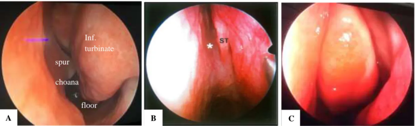
Figure 1: Structures visualized on diagnostic nasal endoscopy. (A) 1st pass structures; (B) 2nd pass structures; (C) 3rd pass structures showing concha bullosa obstructing the region of OMC and middle meatus.
All the study subjects were subjected to diagnostic nasal endoscopy (DNE) using 4 mm “0”degree nasal endoscope and findings were noted in a pre-structured proforma (Figure 1 A-C).
Non-contrast CT scans of nose and paranasal sinuses using Multidetector CT with mainly coronal cuts and axial cuts for sphenoethmoidal recess and onodi cell, and sagittal cuts for frontal sinus, with a gross slice thickness of 3 mm were obtained to aid the diagnosis of CRS
(Figure 2). As all the cases were planned for FESS, routine biochemical and hematological evaluation was also done.
The collected data were analysed with IBM.SPSS statistics software 23.0 version. To find the significance in categorical data Chi-Square test was used similarly if the expected cell frequency is less than 5 in 2×2 tables then the Fisher's exact was used. In all the above statistical tools the probability value.05 was considered as significant level.
A B C
spur Inf. turbinate
choana
Figure 2: CT PNS (2.4 mm cuts) and structures visualized on coronal cuts.
RESULTS
Demographic results
Of the 201 patients, in the age-group of 16 to 50 years, 51 patients (25%) were female and the rest 150 (75%) were males. The maximum burden of the disease was seen in the most productive age group of 21-30 years (43.8%), followed by the age group of 41-50 years (20.4%). The most common symptom of CRS presented in our study was nasal discharge (87%) which was most commonly accompanied by nasal obstruction (69.6%), followed by headache (52.7%).
On DNE
Deviation of nasal septum was the most common finding seen in patients (60.7%). During the first pass inferior turbinate hypertrophy was identified in 23.3% patients. Unilateral antro-choanal polyps were noted in 45 patients, while bilateral ethmoidal polyps were seen in 79 out of 201 patients. During the third pass, pneumatized middle turbinate or concha bullosa was seen in 63 patients (31.3%) and a paradoxically curved middle turbinate was seen in 20% of the patients. An accessory ostium was seen in 65 patients (32.3%) (Figure 3).
Figure 4: Various findings and their frequency of appearance on CT scans of patients.
Table 1: CT vs DNE for DNS.
Value df Asymp. Sig. (2-sided) Exact Sig. (2-sided) Exact Sig. (1-sided)
Pearson Chi-Square 7.845b 1 0.005
Continuity correctionc 7.294 1 0.007
Likelihood ratio 7.871 1 0.005
Fisher's exact test 0.007 0.003
N of valid cases 402
Table 2: CT vs DNE for B/L ethmoidal polyp.
Ethmoidal polyp CT DNE Chi-Square testsa
Absent 78.1% 60.7% Value df
Asymp. Sig. (2-sided)
Exact Sig. (2-sided)
Exact Sig. (1-sided)
Present
21.9% 39.3% Pearson Chi-Square 14.350b 1 0 Continuity correctionc 13.542 1 0 Likelihood ratio 14.501 1 0
Fisher's exact test 0 0
N of valid cases 402
CT PNS findings
All the patients (n=201) underwent CT imaging of the nose and paranasal sinuses pre-operatively using multiplanar device. Images were reconstructed in coronal, axial as well as sagittal planes. The osteomeatal complex was found to be blocked on CT scans of 123 patients (61.2%), among which 59 patients showed OMC blockage bilaterally (29.3%) and 64 unilaterally (31.8%).
while sphenoid sinus involvement, complete or partial, unilateral or bilateral was seen in 16.4% of the patients. Involvement of the frontal recess or frontal sinuses was seen in 23% patients. Deviated nasal septum was seen in 94 patients on CT scan. Presence of anatomical variants like haller cells was seen in about 10%, agger nasi in 16% and onodi cells were seen in 9% of the study population (Figure 4).
Comparison between DNE and CT PNS
Deviated nasal septum was seen in 94 patients on CT scan while a Deviated Nasal Septum was encountered during DNE in 122 patients (Table 1). A bony spur was seen on CT PNS in 50 patients (~25%) and during DNE a bony spur was seen in 65 patients (32.3%). Thus, on comparison of DNS in CT and DNE, a “P” value of 0.005 was obtained, which shows significant difference. On the contrary, no significant difference was seen when comparing Spur in CT with Spur in DNE.
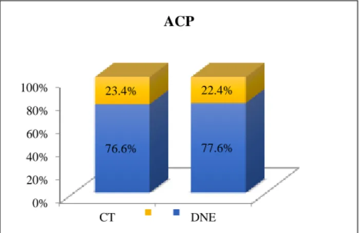
Polyps were seen in CT scans of 91 patients as opposed to 124 on DNE. Of these, DNE revealed 79 patients with bilateral ethmoidal polyps, 44 patients with antrochoanal polyp and 3 patients with isolated maxillary polyp. Thus, on comparing bilateral polyps on CT imaging vs bilateral ethmoidal polyps visualized during DNE revealed a highly significant “P” value; whereas for antrochoanal polyps or unilateral polyps there was good correlation seen as CT scans showed unilateral polyps in 23% and DNE also showed AC polyp in 22% (Table 2, Figure 5).
Figure 5: Comparison of antrochoanal polyps on CT and DNE.
Anatomical variants like concha bullosa were seen in 51 patients on CT while the same was identified in 63 patients during DNE. Paradoxically curved middle turbinates were encountered in 42 patients (20.9%) during DNE while only 34 of these were detected on CT images (16.9%). Both these values showed p>0.05 thus confirming no significant association in their values (Figure 6). Anatomical variants like agger nasi cells, haller cells and onodi cells are all well appreciated on CT scan while polyps, prominent uncinate process and
prominent bulla ethmoidalis were visualized only on diagnostic nasal endoscopy.
Figure 6: Comparison of concha bullosa on CT and DNE.
Sinus haziness could only be seen with CT scan, thus with regards to extent and spread of the disease CT scan gives a better understanding than DNE. Knowing the extent of the disease also helps to plan for better management of the disease.
Presence of polyps or polypoidal disease, on the other hand is better understood on DNE than on CT.
DISCUSSION
Symptomatology
The most common symptom of CRS presented by patients in our study was Nasal Discharge (87%) which was most commonly accompanied by Nasal obstruction (69.6%). This was consistent with a number of studies. A study of 250 patients by Levine et al showed the most common symptom as nasal discharge seen in 51% cases.6 Another study conducted by Kirtane et al also observed that the commonest complaint was nasal discharge occurring in 25 patients (78.1%).7 Other symptoms in our patients included headache (52.7%), post-nasal drip (36.3%), sneezing, nasal bleed and rarely facial pain (2%). There were 34% of patients who developed dry cough because of the phlegm throat or PND.
Diagnostic nasal endoscopy
DNE revealed deviation of nasal septum as the most common finding, seen in 60.7% of the patients, which was in line with the studies conducted by Gautam et al and Venkatchalam et al.8 Septal deviation is thought to laterally compress the middle concha and uncinate process into the infundibulum and thereby cause obstruction of the osteomeatal unit (Davis et al).9 During the first pass inferior turbinate hypertrophy was identified in 23.3% patients. Unilateral antro-choanal polyp was noted in 22.4% patients, while bilateral ethmoidal polyps were seen in 39.3% patients. Levine et al also observed nasal polyposis in 66.1% cases in their study.6 In the
0% 20% 40% 60% 80% 100%
CT DNE
76.6% 77.6%
23.4% 22.4%
ACP
0% 50% 100%
CT DNE
25.4% 31.3%
74.6% 68.7%
Pe
rc
en
ta
g
e
Technique
Concha Bullosa
study by Stammberger et al, polyps were seen in 20% of patients while mucopus was seen in 40%. During the third pass, pneumatized middle turbinate or concha bullosa was seen in 31.3% of patients while, paradoxically curved middle turbinate was seen in 20% of the patients. The studies of Arslan et al and Wani et al demonstrated concha bullosa in 23%.10,11 Concha bullosa is implicated in the pathogenesis of rhinosinusitis because of its tendency to narrow the middle meatus and the infundibulum. An accessory ostium was seen in 32.3% patients. The nasal fontanelles are sites for accessory ostia of maxillary sinus. Mamatha et al in their study reported accessory ostia in 22.5% cases.12 The significance of a particular anatomic variation is determined by its effect on sinus drainage. These anatomic variations should be kept in mind during FESS to prevent complications.
CT PNS findings
CT of the paranasal sinuses should positively be obtained when endoscopic sinus surgery is being considered or planned in patients with CRS or recurrent ARS, refractory to medical management. Kennedy, Zinreich emphasized on the fact that C.T. Scanning of PNS significantly improves ability to diagnose disease in the anterior ethmoid region.13
In our study, the osteomeatal complex was found to be blocked on CT scans of 123 patients (61.2%), which is comparable to a study by Neto et al, who reported 65% cases of block in the osteomeatal complex.14 Furthermore, in our study, 29.3% patients showed OMC blockage bilaterally while 31.8% showed blockage unilaterally. Bolger et al have reported variations of OMC with a frequency of 64.9%.15
Maxillary Sinus involvement being the most commonly observed finding, was seen in the CT scan of 80% patients. This correlated well with the study of Lloyd et al wherein the most common site of involvement was found to be maxillary sinus (83%) followed by anterior ethmoid (63%).17 Unilateral maxillary sinus involvement seen in 42.7% was a more common finding than bilateral involvement which was mostly partial opacification seen in 28.3% of cases. Bilateral complete opacification was least visualized (9%).
The next group of sinuses commonly involved were the anterior group of ethmoid sinuses; seen in 37.3% patients, unilateral or bilateral followed by the posterior ethmoids seen in 28.8% patients. In the studies of Bolger et aland Calhoun et al, the most common site of infection/ inflammation was the anterior ethmoid sinuses (78.2%), and (84.3%) respectively.15,16 Involvement of the Frontal recess or frontal sinuses was seen in 23% patients in our study. The least affected sinus was the Sphenoid Sinus, seen in 16.4% of the patients. These findings were similar to those seen in the study by Zojaji et al, in which the least affected sinuses were the frontal and sphenoid
sinuses, with 10 (20%) and 13 (25%) patients, respectively.18
Presence of anatomical variants like concha bullosa, haller cells was seen in about 25% and 10% respectively, agger nasi in 16% and onodi cells were seen in 9% of our study population. A study by Arslan et al looked into anatomical variants of the paranasal sinus on two mm CT cuts where he found that 30% had concha bullosa while onodi cells at 12% and haller cells were found at 6%.10
Comparison of CT findings with diagnostic nasal endoscopy findings
Among the parameters that were correlated, diagnostic nasal endoscopy was found to be most sensitive investigation for deviated nasal septum followed by polyps. On endoscopy, apart from gross findings like DNS and polyposis, subtle evidence of disease in the osteomeatal area are also identified. Diagnostic endoscopy is a very sensitive and specific tool to diagnose sinonasal disease and to note the pathology in the areas that are inaccessible for visualization by routine anterior rhinoscopy. However, there were a significant percentage of various parameters that could not be visualized at diagnostic endoscopy, such as the sinuses involved, spheno-ethmoid recess, frontal recess, anatomical variants like agger nasi cell, onodi cell and haller cell. This was because it was impossible to pass the endoscope beyond a certain point on DNE mostly due to anatomical abnormalities like a gross deviation of the nasal septum, paradoxical middle turbinate, or a concha bullosa. CT scan of the PNS definitely proved to be very helpful in these cases. CT scans showed a very high sensitivity for anatomical variants as well as a sensitivity of 100% in detecting maxillary sinus disease. The association between diagnostic nasal endoscopy and CT scan PNS was calculated using Chi square test. Since p<0.05 indicates there is association between diagnostic nasal endoscopy and CT scan PNS findings, our study showed significant correlation between the two modalities except for anatomical variants like concha bullosa and paradoxically bent middle turbinate. The diagnostic endoscopic findings correlated well with the computed tomographic findings. Stankiewicy et al on the contrary reported that nasal endoscopy had a sensitivity of 46%, specificity 86%, positive predictive value 74%, and negative predictive value of 64% and showed that there was poor correlation between nasal endoscopy and sinus CT.19 In the study by Pokharel et al, it has been recently recommended that either a CT scan or endoscopic evaluation of nose (preferably with photo or video documentation) should be a part of any prospective clinical trial, as it provides the majority of objective data used to diagnose CRS.20
CONCLUSION
diagnose CRS. CT scan of the PNS help in assessing the extent of sinus disease and to know the vital relations of the paranasal sinuses while DNE helps in understanding the type of pathology. Understanding the advantages and disadvantages of each modality helps us realize that they complement each other to not only provide an objective diagnosis of chronic rhinosinusitis but also give a precise blueprint of the sinonasal passage required to optimally treat it endoscopically.
Funding: No funding sources Conflict of interest: None declared
Ethical approval: The study was approved by the Institutional Ethics Committee
REFERENCES
1. Sood VP. Chronic Rhinosinusitis, ECAB, Elsevier India, 2012.
2. Tan BK, Kern RC, Schleimer RP, Schwartz BS. Chronic Rhinosinusitis: The Unrecognized Epidemic. Am J Respir Crit Care Med. 2013;188(11):1275–7.
3. Rosenfeld RM, Piccirillo JF, Chandrasekhar SS, Brook I, Kaparaboyna AK, Kramper M, et al. Corrigan; Clinical Practice Guideline (Update): Adult Sinusitis Executive Summary. Otolaryngol Head Neck Surg. 2015;152(2S):S1 –S39.
4. Sancheti P, Velankar HK, Setty YK, Mathew MS, Dabholkar YG. Diagnostic Nasal Endoscopy or CT Scan of Paranasal Sinuses: Which One First? Res J Ear Nose Throat. 2017;1:7.
5. Ramakrishnan Y, Zammit-Maempel I, Jones NS, Carrie S. Paranasal sinus computed tomography anatomy: a surgeon's perspective. J Laryngol Otol. 2011;125(11):1141-7.
6. Levine HL. Functional endoscopic sinus surgery: evaluation, surgery, and follow-up of 250 patients. Laryngoscope. 1990;100(1):79–84.
7. Kirtane MV. Functional endoscopic sinus surgery (A preliminary study). Indian J Otolaryngol. 1991;43:126-9.
8. Venkatachalam VP, Bhat A. Functional Endoscopic Sinnus Surgery – A new surgical concept in the management of Chronic sinusitis. Indian Journal of otolaryngology and Head and Neck Surgery. 2000;52:3-16.
9. Davis WE, Templer J, Parsons DS. Anatomy of the paranasal sinuses. Otolaryngol Clin North Am. 1996;29(1):57-74.
10. Arslan H, Aydinlioglu A, Bozkurt M, Anatomical variations of the paranasal sinuses: CT examination
for endoscopic sinus surgery. Auris Nasus Larynx. 1999;26:39–48.
11. Wani AA, Kanotra S, Lateef M, Ahmad R, Qazi SM, Ahmad S. CT scan evaluation of the anatomical variations of the ostiomeatal complex. Indian J Otolaryngol Head Neck Surg. 2009:61(3):163-8. 12. Mamatha H, Shamasundar NM, Bharathi MB,
Prasanna LC. Variations of ostiomeatal complex and its applied anatomy: a CT scan study. Indian J Sci Technol. 2010:3(8):904-7.
13. Kennedy DW, Zinreich SJ, Rosenbaum AE, Johns ME. Functional endoscopic sinus surgery. Theory and diagnostic evaluation. Arch Otolaryngol. 1985;111(9):576-82.
14. Neto SAA, Martins PSL, Souza AS, Baracat ECE, Nanni L. The role of osteomeatal complex anatomical variants in chronic rhinosinusitis. Radiol Bras. 2004;39:227–32.
15. Bolger WE, Butzin CA, Parsons DS. Paranasal sinus bony anatomic variations and mucosal abnormalities: CT analysis for endoscopic sinus surgery. Laryngoscope. 1991;101:56-64.
16. Calhoun KH, Waggenspack GA, Simpson CB Hokanson JA, Bailey BJ. CT evaluation of the paranasal sinuses in symptomatic and asymptomatic populations. Otolaryngol Head Neck Surg 1991;104:480-3.
17. Lloyd GA. CT of the paranasal sinuses: study of a control series in relation to endoscopic sinus surgery. J Laryngol Otol 1990;104(6):477-81. 18. R. Zojaji, M. Mirzadeh, S. Naghibi; Comparative
Evaluation of Preoperative CT Scan and Intraoperative Endoscopic Sinus Surgery Findings in Patients with Chronic Rhinosinusitis. Iranian J Radiol. 2008;5(2):77-82.
19. Stankiewicy JA, Chow JA. Nasal endoscopy and thedefinition and diagnosis of chronic rhino sinusitis. Otolaryngol Head Neck Surg. 2002;126:623.
20. Pokharel M, Karki S, Shrestha BL, Shrestha I, Amatya RCM. Correlations Between Symptoms, Nasal Endoscopy, Computed Tomography and Surgical Findings in Patients with Chronic Rhinosinusitis. Kathmandu Univ Med J. 2013;43(3):201-5.