TECHNICAL UNIVERSITY OF CLUJ-NAPOCA
ACTA TECHNICA NAPOCENSIS
Series: Applied Mathematics, Mechanics, and Engineering Vol. 61, Issue I, March, 2018
ADJUSTMENT OF THE LINEAR CORRELATION BETWEEN THE
SURFACE AREA AND THE VOLUME OF THE LEFT VENTRICLE
José Sérgio DOMINGUES, Marcos DE PAULA VALE, Carlos BARREIRA MARTINEZ
Abstract: This work aims to study the linear correlation between the surface area and the left ventricle volume, considering that this cardiac chamber has the truncated prolate spheroid geometry, and also aims to compare it with a correlation between these two parameters, already described in literature. We used mathematical models to determine the surface area and the left ventricle volume. The volumes that were considered for its real terms and for patients with no cardiac abnormalities, obtained in medical literature. The volumes of this chamber were obtained in the same bibliographic reference. They were determined by MRI and were used to find more realistic correlations.
Key words: Ventricular volume, ventricular surface area, truncated prolate spheroid, left ventricle, mathematical models.
1. INTRODUCTION
Some of the main heart parameters for the left ventricle (LV), which allow us to verify the functioning of the heart, are: ventricular diastolic volume (𝑉𝑉𝑑𝑑) and systolic volume (𝑉𝑉𝑠𝑠), ejection fraction, shortening fraction, and ventricular mass. Usually, they are based on the fact that this chamber has a similar geometry to the one of a revolution ellipsoid, or still, of a truncated prolate spheroid (TPS) [1-5].
However, the surface area of the LV (𝐴𝐴𝑠𝑠) is a little discussed parameter on literature. A possible application of this parameter is on determining the dimensions of the heart portion to be removed of the LV’s free wall when performing a Partial Left Ventriculectomy on patients with dilated myocardiopathy, a surgical procedure which execution was drastically reduced all over the world, but has been successfully conducted, especially on Japan [6-9].
It is also possible that the 𝐴𝐴𝑠𝑠 can be used as a new cardiac normality verification criterion, besides being a determining point of the interruption of growth of the extracellular matrix on the development of artificial organs, in a way that the developed artificial heart has each patient’s specific features [10-13].
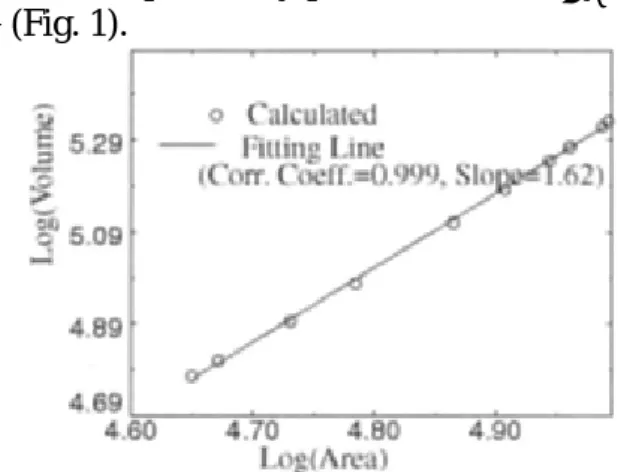
Feng et al. [14] presented a project where they use a mathematical model on ellipsoidal coordinates, which allowed them to obtain an equation for the collection of the 𝐴𝐴𝑠𝑠 values, and another equation to obtain the 𝑉𝑉𝑑𝑑 values. For that, they used the measures obtained on the LV of ten patients, without informing any other feature of the patients, except the 𝐴𝐴𝑠𝑠 and 𝑉𝑉𝑑𝑑 results.
The results obtained present a strong correlation (practically perfect) between 𝐴𝐴𝑠𝑠and
𝑉𝑉𝑑𝑑 (Fig. 1).
Fig. 1. Log-log correlation between 𝐴𝐴𝑠𝑠 and 𝑉𝑉𝑑𝑑, from Feng, Sitek, and Gullberg [14].
The main goal of this project is to verify if this strong correlation remains when 𝐴𝐴𝑠𝑠 is correlated with the 𝑉𝑉𝑑𝑑 values, previously presented on literature, and calculated through traditional medical methods. For that, we present a model equivalent to the one used in [14], but built from Cartesian coordinates, to facilitate the acquiring of the necessary measures for simulations, along with the 𝑉𝑉𝑑𝑑 values, described on literature. Then, the results are presented and compared, where we verify that, when considering the same geometry and the same model for the attainment of 𝐴𝐴𝑠𝑠 and 𝑉𝑉𝑑𝑑 the correlation between them is going to be practically perfect, which won’t happen when the 𝐴𝐴𝑠𝑠 parameter is correlated to the 𝑉𝑉𝑑𝑑 obtained by medical methods.
2. MATHEMATICAL MODELS
2.1 Mathematical models for the LV’s area and volume
The mathematical model we used to calculate the average 𝐴𝐴𝑠𝑠 values is based on Cartesian coordinates, to facilitate the use and attainment of the measures, since it depends only on the LV’s linear measures.
Fig. 2. Representation of the truncated ellipsoid and the TPS that is generated, considered as the LV’s geometry.
We considered that the LV has the geometry of a TPS [1–5] with semi-axis a, b and c, where 𝑐𝑐 >
𝑎𝑎= 𝑏𝑏> 0 and generated by the rotation of the truncated ellipsoid, of axes in the intervals [𝜎𝜎,𝑐𝑐] and [−𝑎𝑎,𝑎𝑎] (Fig. 2).
The equation of the curve 𝑥𝑥(𝑧𝑧) ≥0 of the truncated ellipsoid is given on Eq. (1).
𝑥𝑥(𝑧𝑧) =𝑎𝑎 ∙ �1−𝑧𝑧𝑐𝑐22 (1)
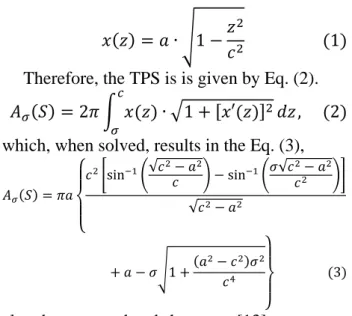
Therefore, the TPS is is given by Eq. (2).
𝐴𝐴𝜎𝜎(𝑆𝑆) = 2𝜋𝜋 � 𝑥𝑥(𝑧𝑧)∙ �1 + [𝑥𝑥′(𝑧𝑧)]2𝑑𝑑𝑧𝑧 𝑐𝑐
𝜎𝜎 , (2)
which, when solved, results in the Eq. (3),
𝐴𝐴𝜎𝜎(𝑆𝑆) =𝜋𝜋𝑎𝑎
⎩ ⎪ ⎨ ⎪
⎧𝑐𝑐2�sin−1�√𝑐𝑐2− 𝑎𝑎2
𝑐𝑐 � −sin−1�𝜎𝜎√𝑐𝑐 2− 𝑎𝑎2 𝑐𝑐2 �� √𝑐𝑐2− 𝑎𝑎2
+𝑎𝑎 − 𝜎𝜎�1 +(𝑎𝑎2− 𝑐𝑐𝑐𝑐42)𝜎𝜎2
⎭ ⎪ ⎬ ⎪ ⎫
(3)
already presented and shown on [13].
2.2 Association of the TPS and LV measures
On Eq. (3), the 𝑎𝑎,𝑐𝑐 and 𝜎𝜎 parameters of the TPS correspond, respectively, to the final diastolic diameter of the LV (D), to the coordinate of the LV apex and to the coordinates of the truncating point of the TPS (which represents the coordinate of the average point of the LV base) (Fig. 3). The ways of determining the measures of these parameters are given by Eqs. (4), (5), and (6) [13].
Fig. 3. Representation of the LV’s short and long axis, adapted from Lang et al. [15].
𝑎𝑎= 𝐷𝐷2 (4)
𝑐𝑐= 23𝐿𝐿 (5)
|𝜎𝜎| =13𝐿𝐿, (6)
where 𝐿𝐿=𝑐𝑐+ |𝜎𝜎| is the dimension of the LV’s long axis, on average hearts, respectively taken. In normal patients, the relation 𝐷𝐷/𝐿𝐿 (short axis/long axis) on the LV varies from 0.45 to 0.62
[16]. Considering this relation as a 𝛿𝛿 ∈
𝐿𝐿= 𝐷𝐷𝛿𝛿 (7)
So, taking Eq. (7) to the Eqs. (5) and (6), it is possible to obtain the |𝜎𝜎| and 𝑐𝑐 values according to D, obtaining:
|𝜎𝜎| =3𝐷𝐷𝛿𝛿 𝑎𝑎𝑎𝑎𝑑𝑑 𝑐𝑐 =23𝐷𝐷𝛿𝛿 (8)
Based on these measures and using the Eq. (3), it is possible to estimate the 𝐴𝐴𝜎𝜎(𝑆𝑆) parameter, relative to the LV’s surface area on average patients.
2.3 TPS volume and the perfect correlation between 𝐀𝐀𝛔𝛔(𝐒𝐒) and 𝐕𝐕𝐝𝐝
The model presented on [14] calculates the 𝑉𝑉𝑑𝑑 and the 𝐴𝐴𝜎𝜎(𝑆𝑆) based on the same geometrical model on ellipsoidal coordinates. With that, there is a self-dependence between the equations that were used, which mathematically assures that there will be a strong correlation between those two parameters (𝑟𝑟= 0,999). In order to illustrate that this dependence will always generate a practically perfect correlation between the parameters, we obtained an equation for the TPS volume, Eq. (9), based on the revolution of the curve on Eq. (1) around the Z axis and we obtain the correlation between
𝐴𝐴𝜎𝜎(𝑆𝑆) and 𝑉𝑉𝑑𝑑.
𝑉𝑉𝑑𝑑 = 𝜋𝜋 � [𝑥𝑥(𝑧𝑧)]2𝑑𝑑𝑧𝑧 𝑐𝑐
𝜎𝜎
∴ 𝑉𝑉𝑑𝑑 = 23𝜋𝜋𝑎𝑎2(𝑐𝑐 − 𝜎𝜎) (9)
3. REAL MEDICAL DATA AND
STATISTICAL ANALYSIS
On a study that aimed to obtain reference measures regarding the left and right ventricles, through magnetic resonance, on average
Brazilian patients, Macedo et al. [17]
particularly presented the average 𝐷𝐷 and 𝑉𝑉𝑑𝑑 values for 107 individuals (asymptomatic and with no heart diseases), in which 54 of them were men, and 53 were women. Images in orthogonal planes, long vertical and horizontal axes, and heart short axes were performed in the four-chamber plane (4ch) to describe the short axes, covering all the LV, from the basis up to the end, using the Steady State Free Precession (SSFP) technique. The 𝐷𝐷 and 𝑉𝑉𝑑𝑑 measures, by
age group and sex, and the calculated values for
a, by Eq. (4), are described on Table 1.
Table 1 Average 𝐷𝐷, 𝑉𝑉𝑑𝑑 and 𝑎𝑎 values, by sex and age group.
Sex Age (years)
D (cm) 𝑉𝑉𝑑𝑑 (mL) 𝑎𝑎 (cm)
Men
20 – 29 5.00 165.3 2.50 30 – 39 5.10 149.3 2.55 40 – 49 5.10 141.3 2.55 50 – 59 4.80 131.9 2.40
≥ 60 4.60 121.6 2.30
Women
20 – 29 4.70 127.0 2.30 30 – 39 4.90 117.6 2.45 40 – 49 4.40 110.0 2.20 50 – 59 4.20 101.5 2.10
≥ 60 4.50 110.2 2.25
The 𝐷𝐷 values are used by Eqs. (4) and (8), and when substituted on Eqs. (3) and (9), determined the average 𝐴𝐴𝜎𝜎(𝑆𝑆) and 𝑉𝑉𝑑𝑑 values.
The calculated values of 𝐴𝐴𝜎𝜎(𝑆𝑆) were correlated to the calculated values of 𝑉𝑉𝑑𝑑 through Equation (9), and also, with the 𝑉𝑉𝑑𝑑 values, obtained on [17]. These correlations were analyzed by Pearson’s coefficient, r, by coefficient of determination, R², and the statistical significance was determined by the p-value, calculated based on Student’s t test, with three degrees of freedom.
From the real medical 𝐷𝐷 values obtained on [17], and considering that 𝛿𝛿 ∈[0.45, 0.62], we performed simulations using three values for 𝛿𝛿: 0.45, 0.62, and also 0.50, which is one of the most used relations on methods that calculate the ventricular mass [5]. So, the correlations between 𝐴𝐴𝜎𝜎(𝑆𝑆) and 𝑉𝑉𝑑𝑑 were obtained.
4. RESULTS AND DISCUSSION
For simulations with self-dependent equations, we obtained 𝑟𝑟= 0.9999 with 𝑝𝑝 =
0.000002 for men, with 𝛿𝛿= 0.45. Still for men, but when 𝛿𝛿 = 0.50 or 𝛿𝛿= 0.62, we obtained
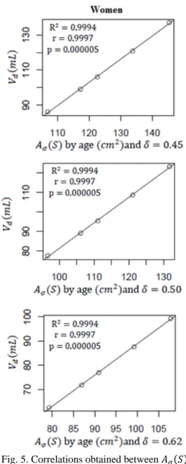
𝑟𝑟= 0.9999 with 𝑝𝑝= 0.000009 (Fig. 4). For women, we obtained 𝑟𝑟= 0.9997 with
𝑝𝑝= 0.000005 for the three values used for parameter 𝛿𝛿 (Fig. 5).
indicates that the models with ellipsoidal and Cartesian coordinates are equivalent, and that, consequently, using Cartesian coordinates in clinical practice is more viable, because it only depends on linear measures which are easily obtained on echocardiography or magnetic resonance exams.
Fig. 4. Correlations obtained between 𝐴𝐴𝜎𝜎(𝑆𝑆) and 𝑉𝑉𝑑𝑑, for men, through dependent models.
The simulations with the equations without self-dependence, and that, therefore, are more realistic, since they use real values of the LV’s diastolic volume, already discussed in scientific literature, were also performed. We obtained
𝑟𝑟= 0.7413 with 𝑝𝑝= 0.1517 on all three values of parameter 𝛿𝛿 for men (Fig. 6).
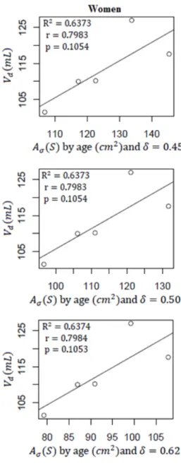
For women, the values obtained were 𝑟𝑟=
0.7983 with 𝑝𝑝= 0.1054, also for all three values of 𝛿𝛿 (Fig. 7).
Fig. 5. Correlations obtained between 𝐴𝐴𝜎𝜎(𝑆𝑆) and 𝑉𝑉𝑑𝑑, for women, through dependent models.
The correlations for the equations model without self-dependence, although strong, displayed values that were inferior (and less significant) to the ones obtained by Feng, Sitek, and Gullberg [14].
5. CONCLUSION
In conclusion, the mathematical models to determine the LV’s surface area and its diastolic volume used by Feng, Sitek, and Gullbert [14] are self-dependent and, therefore, practically
perfect correlations are mathematically
already described in literature. These values were determined by Simpson’s rule, which is the most efficient method to calculate the ventricular volume [5, 18–20].
These correlations, although also strong, have little statistical significance and are very inferior to the ones described by Feng, Sitek, and Gullbert [14]. That way, as opposed to what is presented in that study [14], it is not possible to precisely determine the diastolic volume from the left ventricle’s surface area indirectly.
Fig. 6. Correlations obtained between 𝐴𝐴𝜎𝜎(𝑆𝑆) and 𝑉𝑉𝑑𝑑 calculated by Equation (9), for men.
6. REFERENCES
1. Yeh G.C.K., Martinek J., Comparison of surface potentials due to several singularity representations of the human heart, Bulletin of Mathematical Biophysics. 1957, 19: 293–308.
2. Baccani B., Domenichini F., Pedrizzetti G., Model and influence of mitral valve opening during the left ventricular filling, Journal of Biomechanics. 2003, 36:355–361.
3. Nielsen P.M.F., Le Grice I.J., Smaill B.H., Hunter P.J., Mathematical model of geometry and fibrous structure of the heart, American Journal of Physiology. 1991, 260(4-Pt 2), H1365–78.
5. Ferreira Filho P.R.P., Padrões de Hipertrofia e Geometria do Ventrículo Esquerdo pela Ecocardiografia Transtorácica, Revista Brasileira de ecocardiografia e Imagem Cardiovascular. 2012, 25(2):103–115.
6. Domingues J.S., Barbosa, M.P., Vale M.P., Modelo Matemático para Fatia Cardíaca Removida na Ventriculectomia Parcial, In: 4th National Meeting of Biomechanical Enginering (ENEBI), 2013, Vitória-ES, Brasil. Anais do IV ENEBI, 2013. 7. Domingues J.S., Barbosa, M.P., Vale M.P.,
Mathematical and Computational Aspects in Partial Ventriculectomy, In: 8th International Conference on
Advanced Computational Engineering and Experimenting (ACEX 2014), 2014, Paris, France. 8. Domingues J.S., Barbosa, M.P., Vale M.P.,
Mathematical and Computational Aspects in Partial Ventriculectomy, In: Applications of Computational Tools in Biosciences and Medical Engineering, Advanced Structured Materials. Öchsner A, Altenbach H. Eds. Springer International, 2015, cap. 3, 37–41.
9. Domingues J.S., Vale M.P., Barbosa M.P., Partial left ventriculectomy: have well-succeeded cases and innovations in the procedure been observed in the last 10 years?, Brazilian Journal of Cardiovascular Surgery. 2015, 30(5):579 – 585.
10. Angelo B., Santanu R., Michael S. et al., Arbitrary Self-Assembly of Peptide Extracellular Microscopic Matrices, Angewandte Chemie International. 2012, 51:428 – 431.
11. Paul S.M., Are Artificial Organs Still Needed?, Artificial Organs, 38(10):827 – 828.
12. Michael E., Honey, I shrunk the lungs: Miniature versions of hearts, lungs and other organs are
heralding a bright future for drug research and discovery, Nature. 2015, 519: S16 – S18.
13. Domingues J.S., Vale M.P., Martinez C.B., New mathematical model for the surface area of the left ventricle by the truncated prolate spheroid, The Scientific World Journal. 2017, 2017:1 – 9.
14. Feng B., Sitek A., Gullberg T., The prolate Spheroidal Transform for Gated SPECT, IEEE Transactions on Nuclear Science. 2001, 48(3):872 – 875.
15. Lang R.M., Bierig M., Devereux R.B. et al., Recommendations for chamber quantification, European Journal of Echocardiography. 2006, 4:79 – 108.
16. Graziosi P., Análise ecocardiográfica da estrutura e da função sistólica ventricular esquerda na hipertensão arterial, HiperAtivo, 1998, 5(3):161 – 174.
17. Macedo R., Fernandes J.L., Andrade S.S. et al., Morphological and Functional Measurements of the Heart Obtained by Magnetic Resonance Imaging in Brazilians, Arquivos Brasileiros de Cardiologia, 2013, 101(1):68 – 77.
18. Burkhoff D., Weisfeldt M.L., Cardiac function and curculatory control. In: L. Goldman and I. Andrew (Eds). Goldman-Cecil Medicine, vol. 2, 21th Edition, Elsevier, 2000.
19. Silva A.A., Silva A.F., Ferreira C.B.N.D. et al., Ecocardiograma Transesofágico Intraoperatório, In: A. Bagatini, O. C. Pires, M. F. S. Filho et al. (Eds). ETI Ecocardiografia Transesofágica no Intraoperatório. Sociedade Brasileira de Anestesiologia, 2013.
20. Otto, C.M., Textbook of Clinical Echocardiography, 5th ed., Saunders: Elsevier, 2013.
Ajustarea corelării liniare între suprafața și volumul ventriculului stâng
Rezumat: Această lucrare îşi propune să studieze corelaţie liniară între suprafaţa şi volumul ventriculului
stâng, având în vedere că această cameră cardiace are trunchiate izabella prolate geometrie, şi, de asemenea, isi propune sa comparati-l cu o corelaţie între aceşti doi parametri, deja descrise în literatura de specialitate. Am folosit modele matematice pentru a determina suprafaţa şi volumul ventriculului stâng. Volumele care au fost luate în considerare pentru statutul său real şi pentru pacienţii cu nu anomalii cardiace, obţinute în literatura medicală. Volumele din această cameră au fost obţinute în aceeaşi referinţă bibliografică. Acestea au fost determinate de MRI şi au fost folosite pentru a găsi corelaţiile mai realist.
José Sérgio DOMINGUES, Prof. PhD, Federal Institute of Minas Gerais - Campus Formiga, Mathematics department, Address: Rua São Luiz Gonzaga, s/nº, São Luiz, Formiga - MG, Brazil, 35570-000, Email: [email protected]
Marcos DE PAULA VALE, MD, MSc, Nossa Senhora das Dores Hospital, Chief of Cardiac Surgery, Address: Av. João Soares Silva, 135 - Penha, Itabira - MG, Brazil, 35900-062, Email: [email protected]