Original Research Article
Corneal ulcer: an epidemiological, microbiological and clinical study of
cases attending Assam medical college and hospital, Dibrugarh, India
Mamoni Baruah, Rajiv Kumar Das, Vijaya Agarwalla*, Pranami Basyach
INTRODUCTION
Corneal ulcer is one of the important ophthalmic conditions causing significant morbidity especially in the developing countries.1 Scarring of the cornea developed
as a result of suppurative corneal ulcer is the second commonest cause of preventable blindness after unoperated cataract among people in Asia, Africa and in the Middle East.2 Suppurative corneal ulcers may be
caused by bacteria, fungi, and protozoa. However, within the tropics, as many as two thirds of ulcers may be due to filamentous fungi. This type of ulceration is commonly associated with ocular trauma.3
The epidemiological pattern of corneal ulceration varies significantly from country to country and even from region to region. It is essential to determine the local aetiology within a given region when planning a corneal ulcer management strategy. Several studies have investigated the epidemiology of corneal ulceration, causative micro-organisms, and effective treatments, particularly in the Indian subcontinent but there is lack of data with regard to the north eastern area. The purpose of this study was to evaluate the common etiological agents, predisposing factors, age, gender and occupational distribution and to study the clinical features and management of all corneal ulcers seen at Assam Medical Department of Ophthalmology, Assam Medical College and Hospital, Dibrugarh, Assam, India
Received: 07 January 2020 Revised: 11 January 2020 Accepted: 29 January 2020
*Correspondence: Dr. Vijaya Agarwalla,
E-mail: agarwallavijaya@gmail.com
Copyright: © the author(s), publisher and licensee Medip Academy. This is an open-access article distributed under the terms of the Creative Commons Attribution Non-Commercial License, which permits unrestricted non-commercial use, distribution, and reproduction in any medium, provided the original work is properly cited.
ABSTRACT
Background: Corneal ulcer is one of the important ophthalmic conditions causing significant morbidity especially in the developing countries. This study was carried out to evaluate the common etiological agents, predisposing factors, age, gender and occupational distribution and to study the clinical features and management of all corneal ulcers. Methods: A total of 50 cases of corneal ulcers who attended the Ophthalmology outpatient department (OPD) of Assam medical college and hospital, Assam, India, over a 6 months period were included in the study. A detailed history was taken and examination done as per the proforma. Microscopy and culture were performed on all corneal specimens obtained.
Results: Corneal ulcers were common in 3rd to 5th decades of life with Male to Female ratio of 1.3:1. Majority of patients were farmers or hired agricultural workers. Ocular trauma was the major predisposing factor in majority of cases (32%). Out of 50 cases, 31 (62%) were culture positive. 19 were bacterial isolates and 12 were fungal isolates. All patients were treated according to standard treatment protocol and majority (95.5%) patients responded well to treatment.
Conclusions: This study has revealed that suppurative corneal ulcers are caused by both bacterial and fungal agents with bacterial preponderance in this geographical area. Early and accurate diagnosis and intensive treatment is the need of hour for saving the eye and preventing the catastrophe of lifelong blindness.
Keywords: Corneal ulcer, Corneal scraping, Culture, Ocular trauma
College and Hospital, Assam, India, over a 6 month
period from 1 November 2018 to 30 April 2019.
METHODS
All cases of corneal ulcers who attended the Ophthalmology outpatient department (OPD) of Assam Medical College and Hospital, Assam, India, over a 6 months period from 1 November 2018 to 30 April 2019 were included in the study. Corneal ulcer was diagnosed as loss of corneal epithelium with underlying stromal infiltration with signs of inflammation with or without hypopyon.3
For each patient, a standardised proforma was filled out documenting sociodemographic details including history of trauma, duration of symptoms, predisposing risk factors, time taken to present to hospital. All clinical data including treatment and follow up details were collected.
Ophthalmic examination
Visual aquity was recorded and all patients underwent a detailed slit lamp examination along with fluorescent staining. Corneal ulcer was examined and all the details along with presence of foreign body, hypopyon and associated ocular conditions were looked for. Syringing of the nasolacrimal passage was done.
Laboratory investigations
RBS, R/E blood were done. Corneal scraping material were collected before starting any specific therapy. The ulcer was cleaned with sterile normal saline, and 0.5% proparacaine was applied as a local anesthetic agent. With the help of slit lamp microscope corneal scraping was performed using a sterile Bard-Parker surgical blade number 15. The clinical material obtained was directly inoculated onto blood agar, and sabouraud’s dextrose agar (SDA) making multiple “C” shape marks and stained for Gram stain and KOH wet mount. If, by microscopy, hyphae were observed in corneal tissue, but failed to grow in culture, the causative organism was reported as fungal.
In diagnosed cases of bacterial corneal ulcer, patients were treated with 4th generation fluoroquinolone (0.3% gatifloxacin) or fortified antibiotic drops (vancomycin, amikacin, gentamicin) either as monotherapy or combination.
Fungal corneal ulcers were treated with topical natamycin 5%. Few cases were also treated with voriconazole.
In culture negative cases, patients were treated on the basis of clinical findings and response to treatment.
RESULTS
From 1 November 2018 to 30 April 2019 there were 50 patients with the clinical diagnosis of corneal ulceration
that were examined at the Ophthalmology OPD of Assam Medical College and Hospital, Assam. Of the total 50 patients, 29(58%) were males and 21 (42%) were females with ulceration occurring in both groups most frequently in the middle decades of life (Table 1), (Table 2).
Table 1: Age distribution.
Age Number of cases Percentage(%)
11-20 1 2
21-30 10 20
31-40 6 12
41-50 13 26
51-60 10 20
61-70 8 16
>70 2 4
Table 2: Sex distribution.
Sex Number of cases Percentage(%)
Males 29 58
Females 21 42
The predominance of corneal ulceration in males was most pronounced in the middle years with an overall ratio of male to female patients of 1.3:1.
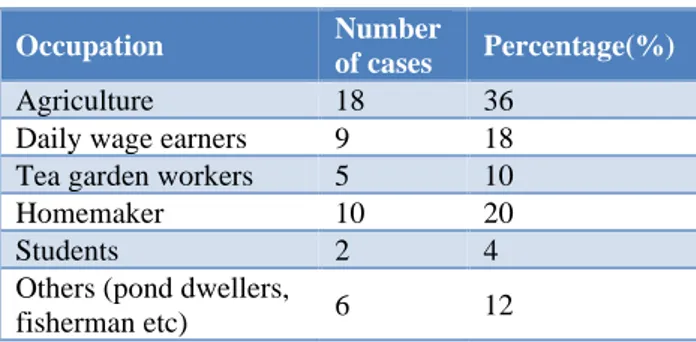
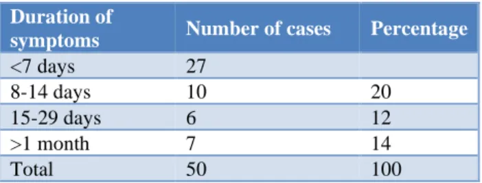
The majority were farmers or hired agricultural workers (Table 3). Incidence was more among rural population (66%) as they are more involved in works prone to corneal ulcer (Table 4). The duration of the patients’ symptoms before their evaluation was also determined (Table 5). Out of 50 cases, 27 cases sought treatment between 1-7 days but 7 patients (14%) waited for more than a month before coming to the hospital for evaluation. The delayed presentation was due to financial limitation, lack of awareness of the problem and topographical constrains to attend health care.
Table 3. Occupational distribution.
Occupation Number
of cases Percentage(%)
Agriculture 18 36
Daily wage earners 9 18 Tea garden workers 5 10
Homemaker 10 20
Students 2 4
Others (pond dwellers,
fisherman etc) 6 12
Table 4: Rural-urban distribution.
Number of cases Percentage
Rural 33 66
Table 5: Duration of symptoms before evaluation.
Duration of
symptoms Number of cases Percentage
<7 days 27
8-14 days 10 20
15-29 days 6 12
>1 month 7 14
Total 50 100
Of the 50 patients 13 (26%) sought medical help before their initial examination. Of the 13 who received medical attention only 2 (15.3%) were seen by ophthalmologists, 5 (38.4%) received advice from pharmacists, and 6 (46.1) went to village healers. It is of interest that 7 (14%) of the total of 50 patients were using some kind of herbal topical medication before examination.
Table 6: Predisposing factors.
Predisposing factor Number of
cases Percentage
Ocular trauma 16 32
Dacryocystitis 5 10
Entropion 2 4
Systemic factors
Diabetes mellitus 1 2
Anemia 2 4
Table 7: Culture results in study.
Culture Number
of patients Percentage
Culture positive 31 62
Culture negative 19 38
Culture results
Fungal 12 24
Bacterial 19 38
Sterile (negative) 19 38 Bacterial results
Streptococcus
pneumoniae 9 47.36
Gram positive cocci 6 31.5 Gram negative diplococci 2 10.5
Diphtheroids 1 5.2
Unidentified 1 5.2
Fungal results
Fusarium 7 58.3
Aspergillus 1 8.3
Unidentified 4 33.3
A history of recent ocular trauma was obtained in 16 (32%) of the 50 patients (Table 6). Agents responsible for the trauma were mainly agricultural and animal products. It is to be noted that these trauma cases were exclusively related to fungal ulcers. These patients were either culture positive for fungus or were started on antifungal treatment based on clinical findings and showed good
response to treatment. Co-existing ocular diseases like dacrocystitis was present in 5 (10%) cases and entropion in 2 (4%) cases. Systemic factors like diabetes mellitus and anemia were present in 2% and 4% of cases respectively.
Out of 50 patients, 21 patients (42%) had hypopyon at presentation. Out of 50 cases, 31 (62%) were culture positive and out of 31 culture positive, 19 were bacterial isolates and 12 were fungal isolates. Among bacterial isolates, Streptococcus pneumoniae was the most common cause and among 12 fungal cases, 7 cases (58.3%) were Fusarium spp., 1 (8.3%) case was Aspergillus spp and 4 (33.3%) were unidentified hyaline fungal species (Table 7).
DISCUSSION
Corneal ulcer is a major health problem in developing world causing prolonged ocular morbidity and loss of vision. Even with appropriate treatment, there is a high incidence of visual loss due to the development of dense corneal scar.
In the study, 29 (58%) patients were male while 21 (42%) patients were females with a male:female ratio of 1.3:1. This ratio is near to that reported by Srinivasan M. et al, (1.6:1).3 Males form the majority of working class, hence
exposure to risk factors is more. Majority of the patients (58%) were in 3rd to 5th decade that is middle age group, as they are more involved in outdoor and physical activities and are exposed to risk factors more frequently. In a study by Panda A. et al performed on thousand eyes of thousand patients, 50% of the patients with corneal ulcer were aged between 36 and 65 years.4 The present
study showed similar age distribution.
The 33 (66%) patients in present study belonged to the rural area. This is chiefly because our hospital caters to a large extent to the villages in and around Dibrugarh district. A similar propensity of rural patients was found in a study conducted in Jammu by Gupta et al. where 65% patients came from a rural background.5
Out of 50 cases, 27 (54%) cases sought treatment between 1-7 days similar to a study conducted in south India where 60% of the patients reported within a week time3 but 7 patients (14%) waited for more than a month before coming to the hospital for evaluation. The delayed presentation was due to financial limitation, lack of awareness of the problem and topographical constrains to attend health care.
In present study, it was found that 18 (36%) out of 50 patients were involved in farming activities. Similar results were also found in the studies by Gopinathan J et al, thus suggesting that corneal ulcers are more prevalent in farmers and other outdoor workers.6 Others were involved in
In the study, out of 50 cases, 16 (32%) cases had ocular
trauma which accounted for most common cause of corneal ulcer followed by dacryocystitis (10%). By far the most common predisposing risk factor for corneal ulcer in south India was a history of corneal injury.7 The
other predisposing factors were entropion (4%) and systemic factors like diabetes mellitus and anemia were present in 2% and 4% of cases respectively.
In present study culture was positive in 31 (62%) cases similar to Srinivasan et al (68.4%) and study conducted in Ghana (57.3%).3,8 Bacterial isolates (38%) outnumbered
fungal isolates (24%) in contrast to previous study conducted in Assam by Nath et al where fungal etiology was established in 60.6% cases.9 Such phenomena may
be the result of difference in climate and environment across time and space. But this is similar to a study conducted in Nepal by Suwal et al. which found bacterial isolates (56%) more than the fungal isolates (44%).10
The prevalence of fungal pathogens in a study conducted by Sharma S et al in South India was 44% and by Dunlop AA et al in Bangladesh was 36%.11,12 In the present study
the incidence of fungal ulcer is lower (24%) compared to the above studies. Among the fungal positive cultures, 58.3% were Fusarium species, 8.3% were Aspergillus species, unidentified Hyaline fungi were 33.3%. The similar results were reported by Srinivasan et al where Fusarium was the commonest identified fungus followed by Aspergillus.3
Of total 50 cases, bacterial isolates were observed in 19 cases (38%) similar in study by Srinivasan M et al in Madurai south India.3 There is a paucity of information in
the literature regarding the aetiology of bacterial corneal ulcers in Assam and other north eastern states and, therefore, comparisons are only possible with similar geographical and climatic regions outside the state. Streptococcus pneumonia (47.36%) was the most common isolate in present study similar in study by Upadhyaya et al while Staphylococcus aureus was reported to be the most common isolate in Basak et al in West Bengal and Pseudomonas species were identified as the commonest bacterial isolate in a study by Dunlop et al in Bangladesh.12-14
Standard treatment protocols were followed in treating the cases. These cases were either admitted to the hospital or were in regular follow up for monitoring. Some patients were lost to follow up. Cases of bacterial corneal ulcer were treated with 4th generation fluoroquinolone (0.3% gatifloxacin) or fortified antibiotic drops (vancomycin, amikacin, gentamicin) either as monotherapy or combination. Most of fungal corneal ulcer cases were treated with natamycin 5% and 1 case was treated with voriconazole. Culture negative cases were treated based on clinical suspicion and response to treatment.2 Cases prone to perforation were applied
Bandage contact lens. Out of 45 follow up cases in this study, majority (95.5%) patients responded well to
treatment. But on follow up it was observed that all patients presented with varying degrees of corneal opacities after healing.
CONCLUSION
Corneal ulcer is a major cause leading to prolonged ocular morbidity and loss of vision. It has been seen that the microbiological etiology of infective keratitis shows a wide regional variation. In present study, it was found that that in study region bacterial corneal ulcers predominate. Common etiological factors are mainly gram positive organisms and Fusarium species. Early and accurate diagnosis and intensive treatment with appropriate antimicrobial therapy is the need of the hour for saving the eye of the patient and preventing the catastrophe of lifelong blindness. Awareness of changes in aetiology and antimicrobial resistance, when this information is available are critical to managing keratitis cases.
Funding: No funding sources Conflict of interest: None declared
Ethical approval: The study was approved by the Institutional Ethics Committee
REFERENCES
1. Bharathi MJ, Ramakrishnan R, Vasu S. Aetiological diagnosis of microbial keratitis in South India a study of 1618 cases. Indian J Med Microbiol. 2002;20(1):19.
2. Thylefor B. Epidemiological patterns ocular trauma. Aust Naj Ophthalmol. 1992;20:95-8.
3. Srinivasan M, Gonzales CA, George C, Cevallos V, Mascarenhas JM, Asokan B, et al. Epidemiology and aetiological diagnosis of corneal ulceration in Madurai, south India. British J Ophthalmol. 1997;81(11):965-71.
4. Panda A, Ahuja R, Sastry SS. Comparison of topical 0.3% ofloxacin with fortified tobramycin plus cefazolin in the treatment of bacterial keratitis. Eye. 1999;13(6):744.
5. Gupta R, Singh C, Mahajan B, Khurana AK. Microbiological regional profile of infective keratitis. Indian J Clinic Experimen Ophthalmol. 2015;1(4):264-72.
6. Gopinathan U, Sharma S, Garg P, Rao GN. Review of epidemiological features, microbiological diagnosis and treatment outcome of microbial keratitis: experience of over a decade. Indian J Ophthalmol. 2009;57(4):273.
7. Dandona R, Dandona L. Review of findings of the Andhra Pradesh eye disease study: policy implications for eye-care services. Indian J Ophthalmology. 2001;49(4):215.
9. Nath R, Baruah S, Saikia L, Devi B, Borthakur A,
Mahanta J. Mycotic corneal ulcers in upper Assam. Indian J Ophthalmol. 2012;60(4):336.
10. Suwal S, Bhandari D, Thapa P, Shrestha MK, Amatya J. Microbiological profile of corneal ulcer cases diagnosed in a tertiary care ophthalmological institute in Nepal. BMC Ophthalmol. 2016;16(1):209.
11. Sharma S, Srinivasan M, George C. The current status of Fusarium species in mycotic keratitis in South India. Indian J Medical Microbiol. 1993;11(2):140.
12. Dunlop AA, Wright ED, Howlader SA, Nazrul I, Husain R, McClellan K, et al. Suppurative corneal ulceration in Bangladesh: a study of 142 cases examining the microbiological diagnosis, clinical and epidemiological features of bacterial and fungal keratitis. Australian New Zealand J Ophthalmology. 1994;22(2):105-10.
13. Upadhyay MP, Karmacharya PC, Koirala S, Tuladhar NR, Bryan LE, Smolin G, et al. Epidemiologic characteristics, predisposing factors, and etiologic diagnosis of corneal ulceration in Nepal. Am J Ophthalmol. 1991;111(1):92-9. 14. Basak SK, Basak S, Mohanta A, Bhowmick A.
Epidemiological and microbiological diagnosis of suppurative keratitis in Gangetic West Bengal, eastern India. Indian J Ophthalmol. 2005;53(1):17.