GE Healthcare
Life Sciences
Instructions 11-0026-01 AD Affinity Chromatography
MabSelect SuRe™
MabSelect SuRe is an alkali-tolerant protein A-derived medium for capturing monoclonal antibodies from large volumes of feed by packed bed chromatography.
• Alkali-tolerant protein A-derived ligand allows the use of 0.1–0.5 M sodium hydroxide for cleaning-in-place (CIP). • Improved product quality and reduction in overall costs. • Eliminates the need for expensive and corrosive CIP reagents. • High dynamic binding capacity (DBC) reduces process time
and amount of medium used.
• High-flow agarose matrix allows processing of large volumes of feed.
• Simple scale-up to production-sized BPG™, AxiChrom™ and Chromaflow™ columns.
Table of contents
1 Description... 2
2 Process development ... 5
3 Recommended screening conditions ... 7
4 Removal of leached ligand from final product ... 9
5 Packing columns... 10
6 Evaluation of column packing ... 16
7 Cleaning-In-Place (CIP) ... 19
8 Sanitization ... 21
9 Storage ... 22
10 Scaling up... 22
11 Troubleshooting ... 23
12 Ordering information ... 24
1 Description
The protein A-derived MabSelect SuRe ligand is produced in
Escherichia coli. Fermentation and subsequent purification are performed in the absence of animal products. The ligand has been specially engineered to create an affinity medium with enhanced alkali stability and high binding capacity for IgG. The specificity of binding to the Fc region of IgG is similar to that of conventional Protein A and provides excellent purification in one step. Alkali tolerance, high capacity and low ligand leakage plus the rigid base matrix, make MabSelect SuRe ideal for the purification of monoclonal antibodies for clinical applications.
Figure 1 shows stability in alkaline conditions of MabSelect SuRe in terms of dynamic binding capacity. A pressure/flow curve is shown in Figure 2.
.
Fig 1. Dynamic binding capacity for MabSelect and MabSelect SuRe after CIP with 0.1 or 0.5 M NaOH, 15 min contact time, for 0–120 cycles.
Each cycle in Figure 1 consisted of:
• 10 column volumes binding buffer, pH 7.4, • 6 column volumes elution buffer, pH 3.0, • 3 column volumes of binding buffer, pH 7.4, • 0.1 or 0.5 M NaOH, 15 min contact time, and • 6 column volumes binding buffer, pH 7.4.
The dynamic binding capacity (DBC, 10% breakthrough) for MabSelect SuRe was measured every 20 cycles. The capacity of conventional Protein A media (MabSelect) is also included in the figure for comparison.
100.0
80.0
60.0
40.0
20.0
0.0
MabSelect 0.1 M NaOH MabSelect SuRe 0.5 M NaOH MabSelect SuRe 0.1 M NaOH
0 20 40 60 80 100 120
CIP cycles
Capacit
y
Fig 2. Pressure/flow profile for MabSelect SuRe in a packed bed (bed height 20 cm) in BPG 300 column (i.d. 300 mm).
Table 1. Characteristics of MabSelect SuRe.
Composition Rigid, highly cross-linked agarose Average particle size (d50v)1 85 μm
Ligand Alkali-tolerant, protein A-derived (E. coli) Coupling chemistry Epoxy
Dynamic binding capacity2 Approx. 35 mg human IgG/ml medium Chemical stability Stable in all aqueous buffers commonly
used in Protein A chromatography. pH working range 3 to 12
Cleaning-in-place stability3 0.1–0.5 M NaOH Maximum flow velocity4 500 cm/h Temperature stability5 2°C to 40°C Delivery conditions 20% ethanol
Regulatory support Regulatory support file is available. No material of animal origin is used in the manufacturing process
800
600
400
200
0
0.1
0.00 0.2 0.3
Pressure (MPa)
V
e
locit
y
1d
50v is the medium particle size of the cumulative volume distribution. 2Determined at 10% breakthrough by frontal analysis at a flow velocity of
500 cm/h in a column with a bed height of 20 cm, i.e. residence time is 2.4 min. Residence time is equal to bed height (cm) divided by nominal flow velocity (cm/h) during sample loading. Nominal flow velocity is equal to volumetric flow rate (ml/h) divided by column cross-sectional area (cm2).
3See Figure 1 and section 7 Cleaning-In-Place (CIP).
4In BPG 300 column, bed height 20 cm, operating pressure < 2 bar, 25°C. 5Recommended long-term storage conditions: 2°C to 8°C, 20% ethanol.
2 Process development
For initial studies of MabSelect SuRe in small-scale columns, we recommend prepacked HiScreen™ columns or HiScale™ 16 columns with 10 cm bed height. Choose a residence time (see Table 1, footnote 2) that fulfills your demand on dynamic binding capacity and nominal flow velocity according to Figure 3. Make sure the chosen bed height and velocity do not conflict with the large-scale pressure/flow limitations.
1000 900 800 700 600 500 400 300 200 100 1 1 2 2 2 3 3 3 4 4 5 5 5 7.5 7.5 7.5 10 10 15 15 V elocit y (cm/h)
Figure 3 shows the possible combinations of bed height and operational nominal flow velocity for MabSelect SuRe. The figure also displays the residence time in the interval 1–15 minutes for any bed height and velocity. Included are also pressure drop limitations and packing limitations at large scale. The solid curved line shows the calculated large-scale column pressure restriction which is 2 bar according to specification (500 cm/h at 2 bar and 20 cm bed height). The dashed vertical line indicates that operating at below 10 cm bed height is not favorable. The reason for this is that large diameter columns have a very different aspect ratio, and that packing short wide beds is a greater challenge.
Figure 3 can be used as a guide when determining suitable bed height and operating velocity in terms of residence time and thus capacity and pressure drop.
Figure 4 shows the relation between dynamic binding capacity and residence time for MabSelect SuRe.
Fig 4. Relation between dynamic binding capacity and residence time for MabSelect SuRe for a purified monoclonal antibody.
50.0 45.0 40.0 35.0 30.0 25.0 20.0 15.0 5.0 0
0 1 2 3 4 5 6 7
Residence time (min)
DBC at 10% br
eakr o u g h (m g /ml) 10.0
3 Recommended screening
conditions
Note: To save material, screening can also be performed using PreDictor™ plates.
Examples of suitable buffers:
• Buffer A: 20 mM sodium phosphate, 150 mM NaCl, pH 7.2 • Buffer B: 0.1 M sodium citrate, pH 3.0–3.6.
Experimental conditions:
• Equilibrate the column with 5 column volumes of buffer A. • Apply a small sample of antibody.
• Wash the column with 5 column volumes of buffer A. • Elute the column with a linear gradient of 10 column volumes
to 100% buffer B.
• Collect fractions into titrating diluent (e.g. 1.0 M Tris-HCl, pH 8.0 so that the diluent volume equals 5% of the programmed fraction volume).
• Regenerate the column with 5–10 column volumes of 100% buffer B.
• Wash the column with 3 column volumes of buffer A. • Perform CIP with 5 column volumes of 0.1–0.5 M NaOH. • Re-equilibrate the column with buffer A.
Washing
To minimize the use of buffer, however, we recommend optimizing the washing procedure with respect to residence time, volumes, pH and conductivity.
Elution
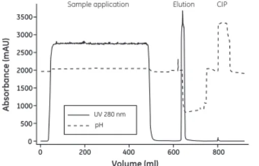
When optimizing elution conditions, determine the highest pH that allows efficient desorption of antibody from the column. This will prevent denaturing sensitive antibodies due to exposure to low pH. Step-wise elution (Figure 5) is often preferred in large-scale applications since it allows the target monoclonal antibody to be eluted in a more concentrated form, thus decreasing buffer consumption and shortening cycle times. It might be necessary to decrease the flow rate due to the high concentrations of protein in the eluate.
Example
Figure 5 shows an example of purification of a monoclonal antibody from a clarified mammalian cell culture on MabSelect SuRe. The load was 21 mg antibody/ml column volume (CV), and the yield was 94% of highly purified antibody. An XK 16/20 column with a CV of 20 ml and a bed height of 10 cm was used.
Fig 5. Purification of a monoclonal antibody from a mammalian cell culture on MabSelect SuRe.
Dynamic binding capacity
The dynamic binding capacity for the target antibody should be determined by frontal analysis using real process feedstock. The dynamic binding capacity is a function of the sample residence time and should therefore be defined over a range of different sample residence times.
Elution CIP Sample application 3500 3000 2500 2000 1500 1000 500 0
0 200 400 600 800
pH UV 280 nm
Volume (ml) Ab s orbance (mA U )
4 Removal of leached ligand from
final product
The ligand leakage from MabSelect SuRe is generally low. For example, the eluate from the purification run shown in Figure 5 contained 3 ppm (ng ligand/mg antibody) of leached ligand. However, in many monoclonal antibody applications it is a requirement to eliminate leached ligand from the final product. There are a number of chromatographic solutions, such as cation and anion exchange chromatography, or multimodal anion exchange chromatography, which can be used to remove leached ligand. For more details about removal of leached ligand and antibody aggregates, see the application note Two step purification of monoclonal IgG1 from CHO cell culture supernatant
5 Packing columns
R
e
comm
e
nd
e
d columns
Table 2. Recommended columns for MabSelect SuRe
1Bed volume range calculated from 10 cm bed height to maximum bed height. 2 Intelligent Packing method according to MabSelect SuRe can be used. 3The pressure rating of BPG 450 is too low to use with MabSelect media. 4See Application note: Methods for packing MabSelect media in production scale
columns (11-0007-52).
5Larger pack stations might be required at larger diameters.
All large-scale columns can be supplied as variable bed height columns. Do not choose large diameter columns if the bed height is low.
For practical instructions in good packing techniques, see the CD-ROM Column Packing - The Movie (18-1165-33). For more details about packing HiScale columns, see instructions HiScale™ columns (16, 26, 50) and accessories (28-9674-70). For information on packing of process scale columns, please contact your local GE Healthcare representative.
Column Inner diameter
(mm) Bed volume
1 Bed height (cm) Lab scale
HiScale 16/20 16 20–40 ml max 20
HiScale 16/40 16 20–70 ml max 35
HiScale 26/20 26 53–106 ml max 20
HiScale 26/40 26 53–186 ml max 35
HiScale 50/20 50 196–393 ml max 20
HiScale 50/40 50 196–687 ml max 35
Production scale
AxiChrom2 50–200 0.2–12.5 l max 30
AxiChrom2 300–1000 7–314 l max 30
BPG 3 100–300 1–28 l max 40
Pac
k
ing HiScal
e
columns
Packing preparations
Materials needed
MabSelect SuRe HiScale column
HiScale packing tube (depending on bed height) Plastic spoon or spatula
Glass filter G3
Vacuum suction equipment Filter flask
Measuring cylinder 20% ethanol with 0.4 M NaCl
Equipment
ÄKTA™ system, or a stand-alone pump such as Pump P-900, depending on the flow rate required, can be used for packing. Equilibrate all materials to room temperature.
Definitions
The bed height of a gravity settled bed differs from the bed height of a bed settled at a given flow (consolidated). Therefore, the compression factor (CF) has to be separated from the packing factor (PF).
Lsettled Bed height measured after settling by gravity.
Lcons Consolidated bed height
Bed height measured after settling the medium at a given flow velocity.
Lpacked Packed bed height
CF Compression factor CF = Lsettled/Lpacked PF Packing factor PF = L /L
Preparation of the slurry
To measure the slurry concentration, let the media settle in 20% ethanol at least overnight in a measuring cylinder or use the method for slurry concentration measurement described in application note 28-9259-32. This method can also be used with HiScale columns.
Washing the medium
Mount a glass filter funnel onto a filtering flask. Suspend the medium by shaking and pour into the funnel and wash according to the following instructions:
• 5 times with 5 ml 20% ethanol with 0.4 M NaCl/ml medium • Gently stir with a spatula between additions.
• Move the washed medium from the funnel into a beaker and add 20% ethanol with 0.4 M NaCl to obtain a 50% slurry concentration.
Packing the column
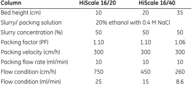
Table 3. Main features of the packing method for HiScale 16/20 and HiScale 16/40
Column HiScale 16/20 HiScale 16/40
Bed height (cm) 10 20 35
Slurry/ packing solution 20% ethanol with 0.4 M NaCl
Slurry concentration (%) 50 50 50
Packing factor (PF) 1.10 1.10 1.06
Packing velocity (cm/h) 300 300 300
Packing flow rate (ml/min) 10 10 10
Flow condition (cm/h) 750 450 260
Table 4. Main features of the packing method for HiScale 26/20 and HiScale 26/40
Table 5. Main features of the packing method for HiScale 50/20 and HiScale 50/40
Column HiScale 26/20 HiScale 26/40
Bed height (cm) 10 20 35
Slurry/ packing solution 20% ethanol with 0.4 M NaCl
Slurry concentration (%) 50 50 50
Packing factor (PF) 1.15 1.13 1.10
Packing velocity (cm/h) 300 300 300
Packing flow rate (ml/min) 27 27 27
Flow condition (cm/h) 750 450 260
Flow condition (ml/min) 66 40 23
Column HiScale 50/20 HiScale 50/40
Bed height (cm) 10 20 35
Slurry/ packing solution 20% ethanol with 0.4 M NaCl
Slurry concentration (%) 50 50 50
Packing factor (PF) 1.15 1.10 1.06
Packing velocity (cm/h) 300 300 300
Packing flow rate (ml/min) 100 100 100
Flow condition (cm/h) 750 450 260
Packing procedure
1 Assemble the column according to the column instructions (HiScale columns (16, 26, 50) and accessories, code no 28-9674-70).
2 Mount the column tube in a stand.
3 Connect the bottom adapter unit to the pump or a syringe and prime the bottom net with a slow flow of packing solution. This is easiest done if the nets are dry but if air is trapped under the net it can be removed by a light suction with a syringe.
4 Mount the bottom adapter unit in the bottom of the column tube and tighten the o-ring.
5 Fill the column with approximately 1 cm packing liquid using the pump/syringe. Disconnect the pump/syringe and put a stop plug on the outlet.
6 Mount the packing tube on top of the column tube.
7 Connect the top adapter to the pump and prime it with a slow downward flow. The net needs to be facing the roof as this is done. If air is trapped under the net it can be removed by a light suction with a syringe.
8 Fill the column with slurry suspended in packing solution. If needed, top up the slurry with extra packing solution so the top adapter dips into the slurry to avoid air under the net.
9 Mount the top adapter unit on top of the packing tube. Tighten the o-ring firmly and remove the bottom stop plug.
10Start a downward flow with packing velocity according to Table 3, 4 and 5.
11Let the flow run until the bed has consolidated.
12Use the scale on the column to measure the bed height. There might be a build up of media at the column wall after the bed is consolidated and to easier see where the top of the bed is, a light source can be used.
13Calculate the final bed height by dividing the consolidated bed height with the desired packing factor.
Lpacked = Lcons/PF
15Dismount the top adapter from the packing tube.
16Over a beaker or a sink, detach the packing tube from the column.
17Remount the top adapter in the column tube. Make sure no air is trapped under the net and lower the adapter down to 1 to 2 cm above the bed, making sure the surface is not disturbed.
18Tighten the O-ring on the adapter. Remove the bottom stop plug and carefully start turning the end cap down. While spilling out liquid through the bottom, proceed turning until the calculated final bed height is reached.
19Make sure that the pressure peaks that occur during turning the end knob down do not exceed the pressure specifications of the media.
20Start a downward flow to flow condition the bed. The flow rate is shown in Table 3, 4 and 5.
21Let the flow run for about 10 column volumes. The column is ready to be tested.
6 Evaluation of column packing
Test the column efficiency to check the quality of packing. Testing should be done after packing, at regular intervals during the working life of the column or when separation performance is seen to deteriorate. The best method of expressing the efficiency of a packed column is in terms of the height equivalent to a theoretical plate (HETP) and the asymmetry factor (As). These values are easily
determined by applying a sample such as 1% acetone solution to the column. Sodium chloride can also be used as a test substance. Use a concentration of 0.8 M NaCl in water with 0.4 M NaCl in water as eluent. For more information about column efficiency testing, consult the application note Column efficiency testing
(28-9372-07).
Note: The calculated plate number will vary according to the test conditions and it should only be used as a reference value. It is important that test conditions and equipment are kept constant so that results are comparable. Changes of solute, solvent, eluent, sample volume, flow velocity, liquid pathway, temperature, etc. will influence the results.
For optimal results, the sample volume should be at maximum 2.5% of the column volume and the flow velocity 30 cm/h. If an acceptance limit is defined in relation to column performance, the column plate number can be used as one of the acceptance criteria for column use.
Method for measuring HETP and As
Calculate HETP and AS from the UV curve (or conductivity curve) as
follows:
The concept of reduced plate height is often used for comparing column performance.
The reduced plate height, h, is calculated as follows:
As a guideline, a value of < 3 is very good.
The peak should be symmetrical, and the asymmetry factor as close to 1 as possible
(A typical acceptable range could be 0.8 < AS< 1.8).
A change in the shape of the peak is usually the first indication of bed deterioration due to excessive use.
Peak asymmetry factor calculation: L = bed height (cm)
N = number of theoretical plates
VR = volume eluted from the start of sample application to the peak maximum
Wh = peak width measured as the width of the recorded peak at half of the peak height VR and Wh are in the same units
d50v = mean diameter of the beads (cm)
a = ascending part of the peak width at 10% of peak height
b = descending part of the peak width at 10% of peak height
L N HETP =
VR
Wh
N = 5.54 × 2
HETP
d50v
h =
b a As =
Figure 6 shows a UV trace for acetone in a typical test
chromatogram from which the HETP and As values are calculated.
Fig 6. A typical test chromatogram showing the parameters used for HETP and As calculations.
VR
Wh
50%
10% Volume
a b
7 Cleaning-In-Place (CIP)
Cleaning-in-place (CIP) is the removal of very tightly bound, precipitated or denatured substances from the purification system. If such contaminants are allowed to accumulate, they may affect the chromatographic properties of the column, reduce the capacity of the column and, potentially, come off in subsequent runs. If the fouling is severe, it may block the column, increase back pressure and reduce flow rate.
Regular CIP prevents the build up of contaminants in the packed bed, and helps to maintain the capacity, flow properties and general performance of MabSelect SuRe. We recommend performing a blank run, including CIP, before the first run with antibody feed.
MabSelect SuRe is an alkali-tolerant medium allowing the use of NaOH as CIP agent. NaOH is widely accepted for cleaning due to the low cost and the ability to dissolve proteins and saponify fats. For difficult cases were CIP with NaOH is not sufficient to restore the column performance an extended protocol including wash with 100 mM thioglycerol pH 8.5 followed by CIP with 0.1 - 0.5 M NaOH is recommended. For more details, see the application note
High-throughput process development for design of cleaning-in-place protocols (28-9845-64).
CIP protocol
1 Wash the column with 3 column volumes of binding buffer.
2 Wash with at least 2 column volumes of 0.1–0.5 M NaOH. Contact time 10–15 minutes.
3 Wash immediately with at least 5 column volumes of sterile and filtered binding buffer at pH 7–8.
CIP is usually performed immediately after the elution. Before applying the alkaline NaOH CIP solution, we recommend equilibrating the column with a solution of neutral pH in order to avoid the direct contact between low-pH elution buffer and high-pH NaOH solution on the column. Mixing acid and alkaline solutions might cause a rise in temperature in the column. NaOH concentration, contact time and frequency are typically the main parameters to vary during the optimization of the CIP. The nature of the feed material will ultimately determine the final CIP. However, the general recommendation is to clean the column at least every 5 cycles during normal use. Depending on the nature of the contaminants, different protocols may have to be combined, for example 0.1 M NaOH every cycle and 0.5 M NaOH every 10 cycles.
8 Sanitization
Sanitization reduces microbial contamination of the chromatographic bed to a minimum. MabSelect SuRe is alkali-tolerant allowing the use of NaOH as sanitizing agent. NaOH is very effective for inactivating viruses, bacteria, yeasts, and endotoxins. In addition, NaOH is inexpensive compared with other sanitizing agents.
Sanitization protocol
1 Wash the column with 3 column volumes of binding buffer.
2 Equilibrate the column with 0.1–0.5 M NaOH.
3 Use a contact time of at least 15 minutes for 0.5 M NaOH or 30 minutes for 0.1 M NaOH (see also the note below).
4 Wash immediately with at least 5 column volumes of sterile and filtered binding buffer at pH 7–8.
For more challenging microbial contamination, a mixture of 30% to 40% 1- or 2-propanol in 0.5 M NaOH could be used for sanitization. Note: Higher concentrations of NaOH and/or longer contact time
inactivates microorganisms more effectively. However, these conditions might also lead to a decrease in the dynamic binding capacity. The conditions for sanitization should therefore be evaluated to maximize microbial killing and to minimize loss of capacity.
9 Storage
Store unused media in its container at a temperature of 2°C to 8°C. Ensure that the screw top is fully tightened.
Equilibrate packed columns in buffer containing 20% ethanol or 2% benzyl alcohol to prevent microbial growth.
After storage, equilibrate with starting buffer and perform a blank run, including CIP, before use.
10 Scaling up
After optimizing the antibody fractionation at laboratory scale, the process can be scaled up to pilot and process scales.
• Keep the residence time constant in order to maintain the dynamic binding capacity.
• Select bed volume according to required binding capacity. Verify the purification step with the new bed height, if it is changed.
• Select column diameter according to your volume throughput requirements. Then determine the bed height to give the desired residence time. Bed heights of 10–25 cm are generally considered appropriate. Note that the backpressure increases proportionally with increasing bed height at constant nominal velocity.
• Keep sample concentration and elution conditions constant. See also Figure 3 for appropriate windows of operation for MabSelect SuRe.
11 Troubleshooting
The list describes faults observed from the monitor curves. Fault Possible cause/corrective action High backpressure during
the run
• Change the in-line filter. • The column is clogged. Perform CIP. • The adapter net/filter is clogged. Clean
or replace the net/filter. Unstable pressure curve
during
sample application
• Remove air bubbles that might have been trapped in the sample pump. • Degas the sample using a vacuum
degasser or an air trap. Gradual broadening of the
eluate peak
• Might be due to insufficient elution and CIP caused by contaminants accumulating in the column. Optimize the elution conditions, the CIP protocol and/or perform CIP more frequently. • Perform wash with thioglycerol followed
by CIP with NaOH, according to Section 7.
Gradual decrease in yield • Too high sample load. Decrease the sample load.
• Precipitation during elution. Optimize the elution conditions.
• Might be due to insufficient elution and CIP. Optimize the elution conditions, the CIP protocol and/or perform CIP more frequently.
Gradual increase in CIP peaks
• Might be due to insufficient elution or CIP. Optimize the elution conditions, the CIP protocol and/or perform CIP more frequently.
12 Ordering information
Product Quantity Code No
MabSelect SuRe 25 ml 17-5438-01
200 ml 17-5438-02
1 l 17-5438-03
5 l 17-5438-04
10 l 17-5438-05
Related product Quantity Code No HiTrap™ MabSelect SuRe 5 × 1 ml 11-0034-93
1 × 5 ml 11-0034-94
5 × 5 ml 11-0034-95
HiScreen MabSelect SuRe 1 × 4.7 ml 28-9269-77 PreDictor MabSelect Sure, 6 μl 4 × 96-well filter plates 28-9258-23 PreDictor MabSelect Sure, 20 μl 4 × 96-well filter plates 28-9258-24 PreDictor MabSelect Sure, 50 μl 4 × 96-well filter plates 28-9258-25
HiScale 16/20 1 28-9644-41
HiScale 16/40 1 28-9644-24
HiScale 26/20 1 28-9645-14
HiScale 26/40 1 28-9645-13
HiScale 50/20 1 28-9644-45
Related literature Code No.
Data Files MabSelect SuRe 11-0011-65
AxiChrom Columns 28-9290-41
BPG columns 18-1115-23
Chromaflow columns 18-1138-92
Application notes MabSelect SuRe – Leakage and Toxicity 11-0011-64 MabSelect – Column packing 11-0007-52 Two step purification of monoclonal IgG1
from CHO cell culture supernatant
28-9078-92 High-throughput process development for
design of cleaning-in-place protocols
For local office contact information, visit www.gelifesciences.com/contact GE Healthcare Bio-Sciences AB Björkgatan 30
751 84 Uppsala Sweden
www.gelifesciences.com/protein-purification
GE Healthcare Europe GmbH Munzinger Strasse 5, D-79111 Freiburg, Germany GE Healthcare UK Ltd Amersham Place Little Chalfont
Buckinghamshire, HP7 9NA UK
GE Healthcare Bio-Sciences Corp 800 Centennial Avenue P.O. Box 1327 Piscataway, NJ 08855-1327 USA
GE Healthcare Japan Corporation Sanken Bldg.
3-25-1, Hyakunincho Shinjuku-ku, Tokyo 169-0073 Japan
GE, imagination at work and GE monogram are trademarks of General Electric Company. ÄKTA, AxiChrom, BPG, BioProcess, Chromaflow, HiScale, HiScreen, HiTrap, MabSelect, MabSelect SuRe, PreDictor and Sepharose are trademarks of GE Healthcare companies. All third party trademarks are the property of their respective owners.
© 2004-2011 General Electric Company – All rights reserved. First published Jun. 2004
All goods and services are sold subject to the terms and conditions of sale of the company within GE Healthcare which supplies them. A copy of these terms and conditions is available on request. Contact your local GE Healthcare representative for the most current information.