CliniCal Case
agnieszka socha
a, B, e, F, iwona niedzielska
a, eExophytic Tumours of Skin of the Head – Case Study
and Review of the Literature
Guzy egzofityczne skóry głowy – opis przypadków
i przegląd piśmiennictwa
Department of Maxillofacial and Oral surgery, silesian Medical University in Katowice, Poland
A – concept; B – data collection; C – statistics; D – data interpretation; E – writing/editing the text;
F – compiling the bibliography
Abstract
Basal Cell Carcinoma (BCC) and squamous Cell Carcinoma (sCC) – the most common of nonmalanotic skin cancers are at the same time the most common cancers in general, rarely resulting in metastasis, but of great risk of local recurrence. The authors present factors which are responsible for developing nonmalanotic skin tumors, such as environmental factors (ultraviolet radiation, arsenic exposure, phototeraphy, radioteraphy) and endogenic (immunosuprecy, patient age, virus infection). They present review of proposed and used method of treatment BCC and sCC. in the following paper three cases of patients operated in their clinic are presented, which proves that there is a lack of social awareness of the necessity of treatment of such cancers and oncological vigilance among general practitioners. The authors want to stress the necessity for establishing algorithms of after surgery treatment and long term observation of patient treating for nonmalanotic skin cancers (Dent. Med. Probl. 2013, 50, 2, 229–237).
Key words: basal cell carcinoma, squamous cell carcinoma, dynamic phototeraphy, Mohs microsurgery, immu-nosupression.
Streszczenie
amelanotyczne nowotwory skóry, do których należą raki podstawnokomórkowe (BCC) i kolczystokomórkowe (sCC), są najczęściej spotykanymi nowotworami skóry, rzadko dającymi przerzuty, ale o dużym ryzyku wznowy miejscowej. W pracy przedstawiono czynniki wpływające na rozwój amelanotycznych guzów skóry, takie jak czyn-niki środowiskowe (promieniowanie ultrafioletowe, ekspozycja na arszenik, fototerapia, radioterapia) i wewnątrz-pochodne (immunosupresja, wiek pacjenta, zakażenia wirusami). Dokonano przeglądu rekomendowanych i sto-sowanych metod leczenia zmian typu BCC i sCC skóry głowy. Przedstawiono także opis trzech przypadków ope-rowanych w Klinice autorów, które przekonują, iż brak jest świadomości społecznej dotyczącej potrzeby leczenia takich zmian i czujności onkologicznej lekarzy pierwszego kontaktu. autorzy zwracają uwagę na konieczność opracowania algorytmów postępowania pozabiegowego i długotrwałej obserwacji pacjentów leczonych z powodu amelanotycznych nowotworów skóry (Dent. Med. Probl. 2013, 50, 2, 229–237).
Słowa kluczowe: rak podstawnokomórkowy, rak kolczystokomórkowy, terapia fotodynamiczna, mikrochirurgia Mhosa, immunosupresja.
Dent. Med. Probl. 2013, 50, 2, 229–237
issn 1644-387X © Copyright by Wroclaw Medical University and Polish Dental society
Basal Cell Carcinoma – BCC and squamous Cell Carcinoma – sCC are the most common skin cancers. They are classified into the category of the so-called non-melanoma tumours, do not have precursors among melanocytes cells and despite the growing social awareness of the detrimental
effect of sun exposure, their frequency has been steadily growing by 3–8% annually since 1960 in the whole world [1, 2]. in the last decade of the 20th
century, the number of sCC and BCC cases have increased profoundly [3]. Globally, the frequency of BCC cases has been increasing by 10%. it is
es-sure, immunosuppression, light complexion (type i and ii by Fitzpatric), and in the cases of sCC, al-so HPV and HiV infection [1, 6, 7]. sun exposure, mainly UVB of wavelength between 280 and 320 nm, is the prime risk factor causing the develop-ment of BCC. The appearance of sCC on the skin is connected with cumulated sun exposure of the most exposed area of the skin. appearance of BCC have low correlation with cumulated dose and is most popular on skin areas moderately exposed to sun light, like torso among men and lower limbs among women [3]. The greater risk of BCC can be found among patients of Caucasian race with light hair and eyes, who admitted to recreational sun-bathing with sunburns [3].
The chosen treatment of nonmelanotic skin cancers is surgery; however, pharmacological treatment is also used on surface changes, photo-dynamic therapy and radiotherapy [5, 8, 9].
The increase in the number of skin cancers lately has been correlated with the increase in skin cancers of head and neck areas. The size of can-cers, with which patients contacted Department of Maxillofacial surgery of silesian Medical school in Katowice proves that there is a lack of social awareness of cancers.
Case Reports
Case 1
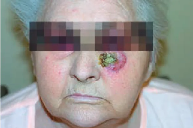
Female patient K.K. age 83, no. of medical his-tory: 11/437c, had been admitted to the Clinic in June 2011, for the surgical removal of a skin tu-mour located on the left suborbital area persisting for several years, with a growth tendency (Figs 1, 2). skin lesions were not associated with pain, but with periodical bleeding. The patient did not con-nected the lesion with injury. The patient had mul-tiple morbid conditions (pancytopenia, le, hiper-tonia, ischemia, atherosclerosis, vertebral arthrop-athy). The patient had been treated with renal
cortex hormones for several years because of le. The patient had not been able to move on her own. after extraoral examination, an exophytic tumour was discovered sized 5 × 4 × 2 cm, located on the left suborbital area with direct contact with lower eyelid of the left eye and soft tissues of lateral sur-face of the nose. in the laboratory examination of blood and urine, nothing varying from norm had not been discovered. in imaging study stated:
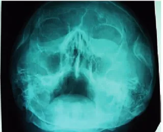
sinus X-ray examination by Waters view “On the section of facial skeleton on the left side oval shadow visible sized 30 × 40 mm, located on sec-tion of lower part of orbitae, projecting on the left zygomatic bone and partly on the left sinus max-ille. Visible bone parts without lesions in X-ray” (Fig. 3).
CT scan of bony face showed “Tumorous in-filtration of tissue density located on the left cheek area, with direct contact with eyelid of the left eye and soft tissues of side area of the nose on the same side. This infiltration is unevenly increased after intravenously addend contrast medium, in
podoczodołowej lewej (aP)
Fig. 2. Patient K.K. age 83 – left suborbital area tumor (left side)
Ryc. 2. Pacjentka K.K. lat 83 – guz okolicy podoczodołowej lewej (strona lewa)
the long axis measured on skin level it has 36 mm, in sagittal diameter it has 22 mm and in CC axis measured from lower eyelid 3 cm, which is typi-cal for cancer growth. The tumour is not infiltrat-ed into bones; however, it infiltrates soft tissues, merging with muscles of levator labii superioris and alae nasi. The tumour is partly vasculatured from arteria facialis and arteria angularis and is not connected with musculi oculomotorica, al-though encapsulates in great extent the lower eye-lid, practically from one to another eye anguli.
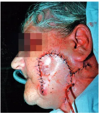
after consultations and preparations of the patient to the surgery dated July 6, 2011 under gen-eral anaesthesia (through the trachea with intuba-tion through the nose) the lesion of the left un-dereye was excised with 5 mm margin of healthy tissues the tumour of left undereye area sized 5 × 4.5 × 2.5 cm (Figs. 4, 5). next with excision along the rim of left chick bone, preauricular and in neck
fold on the left side the flap of the skin was mobil-ised, to close postexcised defect (Figs. 6, 7).
The following result has been provided after Fig. 3. Patient K.K. age 83 – sinus X-Ray Waters
pro-jection
Ryc. 3. RTG zatok wg Watersa pacjentki K.K. lat 83
Fig. 4. Patient K.K. age 83 – intrasurgery picture, tumor excised with margin of tissues
Ryc. 4. Pacjentka K.K. lat 83 – zdjęcie śródoperacyjne – guz wycięty z marginesem tkanek
Fig. 5. Patient K.K. age 83 – tumor after excision
Ryc. 5. Pacjentka K.K. lat 83 – guz po wycięciu
Fig. 6. Patient K.K. age 83 – intrasurgery picture, tis-sues mobilization, with an aim of closing defect area
Ryc. 6. Pacjentka K.K. lat 83 – zdjęcie śródoperacyjne – mobilizacja tkanek w celu zamknięcia powstałego ubytku skóry twarzy
Fig. 7. Patient K.K. age 83 – patient after tumor exci-sion and plastic of skin
Ryc. 7. Pacjentka K.K. lat 83 po zabiegu wycięcia guza i plastyce płatowej
mour with 4-year-long history located on the left area of the forehead. in the last year, the skin le-sions have been steadily growing. accompanying morbid conditions: hipertonia arteriaris, diabetes, stabilised hypothyroidism, hypoacustic. in labora-tory examination of blood and urine, high level of glucose had been diagnosed, apart from that noth-ing extraordinary. Physical examination revealed a hard, pedicled tumour located on the left area of the forehead sized 2 × 2 cm, brown-blue colour, covered with calloused epitelium.
On 19th July, 2011, under local anaesthesia of the left forehead area, the tumour was excised with a 2 cm margin of health tissues. next, the H-Plastic was performed, the wound had been tightly sewn up to a satisfactory aesthetic effect.
The following result was provided after the histopathological examination: “Carcinoma Ba-socellulare.” [no of examination 1369219/H- -1369223/H, examiner Piotr Paleń, pathomorphol-ogy and cytolpathomorphol-ogy specialist]. Healing took place without any complications. Recently, there have not been any symptoms of cancer recurrence.
Case 3
The female patient B.a. age 89, no. of medical history: 2000/306, was admitted to the Clinic for the purpose of surgically removing a skin tumour lo-cated on the left cheek. The main reason the patient contacted the clinic was because of a recurring, dif-ficult to stem bleeding from the whole surface of the tumour. The patient received treatment for diabetes and hypertension. in the laboratory examination of blood and urine, high level of glucose was diagnosed. a physical examination revealed a pediculed, pain-less tumour sized 10 × 10 × 8 cm, dark brown co-lour, calloused surface, bleeding after touch (Figs. 8, 9). From anamnesis: the lesions appeared 8 years be-fore admission to hospital in the form of crust/ulter-atio sized 1.5 cm. The crust peeled and was detached by daily hygienic routine. One year before admission to hospital the lesion grew.
after preparing the patient under general an-aesthesia through the trachea, on 24th May, 2000,
the operation was done by excising the tumour with margin of health tissues. next, the plastic surgery was performed by taking the piece of skin from the neck area (Fig. 10). The following result was given Fig. 8. Patient B.a. age 89 – face with exophytic tumor in left cheek area (aP)
Ryc. 8. Pacjentka B.a. lat 89 – twarz z egzofitycznym guzem w obrębie policzka lewego (aP)
Fig. 9. Patient B.a. age 89 – face with exophytic tumor in left cheek area (side picture)
Ryc. 9. Pacjentka B.a. lat 89 – twarz z egzofitycznym guzem w obrębie policzka lewego (widok z boku)
following a histopathological examination: “Carci-noma planoepitheliale praecipue keratodes”.
The patient had been periodically consulted in the clinic. There were wound complications because of a salival fistula. in the period of 5 years after the surgery, neither the cancer recurred nor was there any metastasis in thearea of lymphatic noduli (Fig. 11).
Discussion
Basal Cell Carcinoma – BCC and squamous Cell Carcinoma – sCC are the most common skin cancers. Basal Cell Carcinoma rarely results in metastasis to distant organs contrary to squamous Cell Carcinoma [4, 10].
One of the most important oncogenetic fac-tors in the development of Basal Cell Carcinoma is ultraviolet radiation, which apart from its car-cinogenic effect, disturbs skin homeostasis, which leads to immunosuppression, and eases the dereg-ulation of cell cycle [8]. it has been found that 80– 85% of Basal Cell Carcinoma grows on the skin of the head and neck, 10% on the skin of the torso and only a small fraction on different areas of the skin [9]. BCC appears in many clinical forms and histopathological variety. Clinical physicians most often diagnose superficial and nodulus forms, roughly 90% of lesions located on the head and neck are tumour like. 60% of superficial forms of tumours are located on the thorax [11]. in the ar-ea of the har-ead and neck, superficial forms usually are recognised in females, most often on the face, lip area and neck. in the case of males, they usu-ally appear on ears, the temple and hairless skin on the head [11]. in the cases of BCC described in this paper, the superficial form was found. lesions were single focused.
Usually, Basal Cell Carcinoma can be found isolated. nevertheless, it is important to mention genetic, autosomatic illnesses: Gorlina-Goltz syn-drome, also known as nevoid Basal Cell Carcino-ma syndrome nBCCs. it is caused by a mutation in the area of suppressor genes PTCH. in patients with Gorlina-Goltz syndrome, apart from multi-ple lesions of BCC type, epidermal cysts can also be found, along with fissures of the palm or plan-tal, calcify of falx cerebri, odontogenic cysts, ker-atocystic tumor of maxillary bones and skeleton abnormalities [12, 13].
Contrary to BCC with metastasis frequency at the level 0.03–0.1% [4, 14], sCC result in metas-tasis in 2.5% of cases [14]. similarly to BCC, the growth of sCC is influenced by environmental factors, such as ultraviolet radiation, mainly UVB with wave length 280–320 nm. sCC grow on skin chronically damaged by the sun [15]. sCC is often preceded by precancerous conditions of the skin, which include: skin photodamage, actinic kera-toses, xeroderma pigmentosum, cornu cutaneum, other dermatosis and morbus Boweni, which is considered to be an intraepidermal form of sCC, where basal layer of epidermis is not changed in histopathological examination [15].
it has also been found that sCC and BCC are types of complication of skin chronic le, mainly in Fig. 10. Patient B.a. age 89 – after surgery picture,
plastic of tissues from rotated skin
Ryc. 10. Pacjentka B.a. lat 89 – zdjęcie pooperacyjne – plastyka tkanek zrotowanym płatem skóry
Fig. 11. Patient B.a age 89 – face (aP) 5 year after sur-gery, lack of cancer metastasis
Ryc. 11. Pacjentka B.a. lat 89 – twarz (aP) 5 lat po lecze-niu operacyjnym, brak wznowy procesu nowotworowego
patients and purposefully treated patients after organ transplantation. ngujen et al. have not proved an in-crease in sCC and BCC among HiV infected patients; however, among seropositive patients they noted in-creased mortality because of sCC [7]. among pa-tients in all stages of aiDs, local recurrence, metas-tasis and mortality did not correlate with the number of opportunistic infections and of CD4 cells. There-fore, according to ngujen et al. [7], HiV positive pa-tients should be subjected to aggressive therapy, re-gardless of the immunosuppression stage and prog-nosis because of general condition.
immunosuppression, which is essential among organs recipients, may cause carcinogenesis. 40% of cancers among patients using immunosuppression are skin cancers, out of which 90% are of BCC and sCC types. 82% of patients after a kidney trans-plant will get skin cancer within 20 years. Rang-wala and Tsai [19] claim that skin cancers corre-late with a decreased level of CD4, and a reduction in immunosuppression lowers the risk of getting skin cancer. Patients with lymphoplactic leukemia have eight times greater risk of getting nonmala-notic skin cancer; recurrence is 7–14 times great-er, even after Mohs surgery, and mortality, due to sCC metastasis, is greater [19].
The influence of azatiopryn on increasing the risk of getting sCC has been described, which may be caused by an increase in photosensitivity and mutagenesis triggered by UVa, by means of di-rect metabolic incorporation 6 – thioguanin into Dna. an increase in the number of mutagenic fo-cus TP53 in skin treated with azatiopryn and de-crease of reparative activity (Dna in keratinocyte) has been proven.
Research of the correlation between Papilloma Virus (HPV) and increased risk of skin cancer has been made. an increase in the risk of getting cer-vical cancer, nail cancer and genital cancer is con-nected with a serotype HPV e6 and e7 [19], but with no proved effect on getting sCC. They may probably act as cofactors in the genetic predisposi-tions and exposure to environmental factors.
on the rear surface of the chest, next, on face and upper limbs. The size of the tumours can reach 40 cm. They usually infiltrate the adjoining tis-sues, muscles and bones [21]. in the first case, the tumour had been growing for several years and in last time growth accelerated substantially, which corresponds with academic literature. The tumour infiltrated subcutaneous layer and muscles, which has been proven by the CT scan.
The preferred treatment of BCC and sCC is surgery, which is agreed by most of the authors. The required margin of healthy tissues is usual-ly in the range of 3–4 mm [4]. in the case of giant BCC some authors recommend the removal of the tumour with a margin up to 1 cm [21]. in the first described case, the authors used a minimum mar-gin of 5 mm, which according to a histopatholog-ical examination was radhistopatholog-ical excision. in all the presented cases the single skin plastic was used, achieving complete closure of postresection de-fect, which is recommended by above quoted au-thors [3, 4, 8, 17, 22, 23].
according to Berlin and others [23], an in-complete excision of BCC on the first attempt re-sults in metastasis in 12–41% of cases. Hansen and others, in years 2005–2007, carried out in austra-lia research on the frequency of incomplete exci-sion of BCC and sCC. in the case of BCC, the in-complete excision rate was 6.4%; however, in areas of head and neck skin, the rate increased to 9.8% and was considerably higher than in other ana-tomical areas. Most cases of incomplete excision were found in aesthetically important areas such as nose and ear – 19.1%. in the case of sCC, the in-complete excision rate was comparable with BCC and reached 6.3%. The ratio was higher in the ar-eas of the head and neck – 11%, which most prob-ably results from the tendency to narrow the mar-gin of health tissues from functional and aesthet-ical reasons [24]. in described cases the authors achieved margins of tissues after excision of the tumour free from the cancer. The age of patients in the presented cases was a great facilitation that
allowed easier immobilisation of skin for closing the loss and lack of necessity to choose between aesthetic and radicalism of surgery.
To limit the extent of surgery the authors can also consider the use of artificial skin to cover vast postresective defects, when there is no possi-bility of reconstruction with pieces of skin taken from adjusting areas or with patients in bad physi-cal condition. Gohari et al. [25] used human skin substitute with patients after resection of patho-logical lesions and in cases of Mohs surgery after skin cancer. significantly better cosmetic effect has been obtained in comparison to secondary in-tention healing; the scars have been more elastic and less vascularised while the patients were more content with the esthetical effect. artificial skin can also be used on areas difficult to heal, to re-construct skin over banes and cartilages and in areas prone to scaring and fibrosis [25]. However, this method is expensive and it should be consid-ered whether similar effect in post-surgery wound healing can be achieved with less expensive meth-ods.
sobjanek et al. [26] claim that the anatomy of auricle and histological type of nMsC, result in quite high ratio of metastasis. The important fac-tor here is the thickness of the skin and the prox-imity of cartilage, which can act as a barrier for cancer cells; however, on the other hand, it can al-so provide a surface for lesions spreading. The au-thors stress the high ratio of recurrence after ex-cision and histopathological examination of sCC, which amounts to 14–16% [26]. Much better re-sults can be obtained using Mohs surgery. above quoted authors claim that in their own cases, me-tastasis ratio after using Mohs surgery of auricle reached 4–7% [26]. Mohs surgery enables histo-pathological confirmation of oncological radi-calism and stays as standard solution in high risk BCC treatment and treatment of sCC [4]. This op-erational method is recognised for histopatho-logical, intraoperation examination, and further excision of lesion only in places, where cancer is found under microscopic examination. The meth-od allows for precise mapping of the tumour and minimal excision of tissues; however, on the oth-er hand, it is time consuming and requires signif-icant experience of surgeons. still, it is the pre-ferred method in cases of BCC recurrence on face skin areas, in the case of large tumours, over 2 cm, and also, when a tumour is located in places im-portant for aesthetical reasons [4, 27]. similar rec-ommendation of using Mohs surgery are applied in cases of sCC, meaning, in cases of tumours of great metastasis or recurrence risk, fast growing lesions, recurred tumours, indistinct clinical mar-gin tumours or in case of sCC developing in old
scar. it is successfully applied in lips areas, sexual organs and nails [27].
Chiller et al. propose curettage before excision BCC and sCC. They use sharp, round instruments for curettage of lesion of the skin to remove weak-ly attached tissues, which helps surgeons to bet-ter distinguish tumour boundaries. according to the authors, such an approach results in 32% im-provement in radicalism of removal BCC in areas of head and neck skin; on the other hand, there was no improvement in cases of sCC [28].
another possibility in treating BCC, especial-ly in areas important from aesthetical reasons, is photodynamic therapy. Widely used photosensi-tizer is 5-aminolevulinic acid or its ester, which is a biosynthetic precursor of protoporphyria iX. it can be applied externally or orally [29]. Barolet and Boucher [9] used 20% ala injection to recur-rent, nodular BCC of nose, without recurrence in 24 months of observation. application of ala to a lesion resulted in better penetration of photosen-sitizer and a higher level of protoporphyna iX in lesions compared to external application.
a pharmacological method of nonsurgical treatment of skin cancers is imiquimod. it is an immunological response modulator, revealing an-tiviral and anticancer activity. imiquimod is com-bined with Toll-like 7 receptor, which activates dendritic cells, macrophages and nomocytes for synthesis and releases inflammation cytokines, which in turn modulate the correct immunolog-ical response of the cells. The final result is gen-eration of cytotoxic cells T and T helper in immu-nological response type i. imiquimod also induces apoptosis in sCC and BCC cells in vitro [2, 30].
in cases of primary or recurrent tumours of BCC and Css, radiotherapy was applied. it was al-so applied as adjuvant therapy in cases of incom-plete excision of tumour or in cases when patients refused surgery. attention should be paid to the fact that the illuminated area is promoted for can-cer growth and this method of therapy should be avoided when treating young people. in the treat-ment of smaller lesions, cauterization can be ap-plied, cryotherapy [4] and 5 fluorouracil in oint-ment. in isolated cases, superselective, intra-ar-terial infusion of chemotherapeutics (cisplatin) was applied with the result of complete loss of le-sion [31].
as an alternative method of preventing and treating BCC in the future, pharmacological sub-stances influencing PTCH and sMO genes can be used. On the molecular level mutation of gen p53 has been proven in cases of sCC and BCC [2, 22, 32, 33]. lam and others [34] claims that in 30–40% of BCC genes PTCH are inactivated. Two mutations activating genes sMO (Hh signalling pathway)
in-amination of the skin is recommended in a peri-od of 6 months after the first diagnosis, especial-ly among elderespecial-ly people [35]. There still is a lack of unambiguous guidelines for the length of the
lesions in early stages are key to effective treat-ment of skin cancers. Presented cases can serve as an example for lack of social awareness and onco-logical vigilance of general practitioners.
References
[1] Paradisi a., Waterboer T., sampogna F., Tabolli s., simoni s., Pawlita M., abeni D.: seropositivity for hu-man papillomavirus and incidence of subsequent squamos cell and basal cell carcinomas of the skin in patients with a previous nonmelanoma skin cancer. Br. J. Dermatol. 2011, 165, 782–791.
[2] Madan V., lear J.T., szeimies R-M.: non-melanoma skin cancer. lancet 2010, 375, 673–685.
[3] Gailani M.R., Bale a. e.: Developmental genes and cancer: role of patched in basal cell carcinoma of the skin. J. natl. Cancer inst. 1997, 89, 1103–1109.
[4] samarasinghe V., Madan V., lear J.T.: Focus on basal cell carcinoma. J. skin Cancer 2011, doi:10.1155/2011/328615. [5] schroeder M., Kestlmeier R., schlegel J., Trappe a.e.: extensive cerebral invasion of a basal cell carcinoma
of the scalp. eur. J. surg. Oncol. 2001, 27, 510–511.
[6] Masini C., Fuchs P.G., Gabrielli F., stark s., sera F., Ploner M., Melchi C.F., Primavera G., Pirchio G., Picconi O., Petasecca P., Cattaruzza M. s., Pfister H. J., abeni D.: Papillomavirus infection and cutaneous squamos cell carcinoma in immunocompetent individuals. arch. Dermatol. 2003, 139, 890–894.
[7] nguyen P., Vin-Christian K., Ming M.e., Berger T.: aggressive squamos cell carcinomas in person infected with the human imminodeficiency virus. arch. Dermatol. 2002, 138, 758–763.
[8] nakayama M., Tabuchi K., nakamura Y., Hara a.: Basal cell carcinoma of head and neck. J. skin Cancer. 2011, doi:10.1155/2011/496910.
[9] Barolet D., Boucher a.: no-needle jet intradermal aminolevulinic acid photodynamic therapy for recurrent nodular basal cell carcinoma of the nose: a case report. J. skin Cancer 2011, doi:10.1155/2011/790509.
[10] Harwood M., Wu H., Tanabe K., Bercovitch l.: Metastatic basal cell carcinoma diagnosed by sentinel lymph node biopsy. J. am. acad. Dermatol. 2005, 53, 475–478.
[11] lesiak a., słowik-Rylska M., Bogaczewicz J., Jochymski C., Kozłowski W., sysa-Jędrzejowska a., Rogowski-Tylman M., narbutt J.:expressionof selected cell cycle-related suppressor proteins in superficial and nodular sybtypes of basal cell carcinoma. Dermatol. Clin. 2008, 10, 185–189.
[12] Wang s.Q., Goldberg l.H.: Multiple polypoid basal cell carcinomas on the perineum of a patient with basal cell nevus syndrome. J. am. acad. Dermatol. 2007, 57, 36–37.
[13] Kaczmarzyk T., stypułkowska J., Tomaszewska R., Czopek J.: Tumor odontogenes and tumor like laesions of jaws bones. eds.: Quintessence 2009, 83–85.
[14] Ducic Y., Marra D.e.: Metastatic basal cell carcinoma. am. J. Otolaryngol. Head neck Med. surg. 2011, 32, 455–458. [15] Brzeziński P., Poklękowska K.: Morbus Boweni – clinic, dermathoscopy, histopathology. n. Dermatol. Online
2011, 2, 154–155.
[16] Kar B. R., nair V., ebenezer G., Job C.K.: squamous cell carcinoma of the scalp arising from chronic cutaneous lupus erythematosus: report of two indian patients. indian J. Dermatol. Venereol. leprol. 2004, 70, 236–238. [17] Johson H., Bossenbroek n., Rosenman K., Meehan s., Robles M., Pomeranz M.: Chronic cutaneous lupus
er-ythematosus in vitiligo. Dermatol. Online J. 2008 14, 10–12.
[18] Daldon P. e., Macedo de souza e., Cintra M. l.: Hypertrophic lupus erythematosus: a clinicopathological study of 14 cases. J. Cutan. Pathol. 2003, 30, 443–448.
[19] Rangwala s., Tsai K.Y.: Roles of the immune system in skin cancer. Br. J. Dermatol. 2011, 165, 953–965. [20] lorenzini M., Gatti s., Giannitrapani a.: Giant basal cell carcinoma of the thoracic wall: a case report and
[21] archontaki M., stavrianos s., Korkolis D., arnogiannaki n., Vassiliadis V., liapakis i., Christ H., Rapidis a., Kokkalis G.: Giant basal cell carcinoma: clinicopathological analysis of 51 cases and review of the lit-erature. anticancer Res. 2009, 29, 2655–2664.
[22] Padilla s.R., sebastian s., Jiang Z., nindl i., larson R.: Gene expression patterns of normal human skin, ac-tinic keratosis, and squamos cell carcinoma. arch. Dermatol. 2010, 146, 288–293.
[23] Berlin J., Katz K.H., Helm K.F., Maloney M.e.: The significance of tumor persistence after incomplete excision of basal cell carcinoma. J. am. acad. Dermatol. 2002, 46, 549–553.
[24] Hansen C., Wilkinson D., Hansen M., soyer P.: Factor contributing to incomplete excision of nonmelanoma skin cancer by australian general practitioners. arch. Dermatol. 2009, 145, 1253–1260.
[25] Gohari s., Gambla C., Healey M., spaulding G., Gordon K.B., swan J., Cook B., West D.P., lapiere J-CH.: evaluation of tissue-engineered skin (human skin substitute) and secondary intention healing in the treatment of full thicknesswounds after Mohsmicrographic or excisional surgery.Dermatol. surg. 2002, 28, 1107–1114. [26] sobjanek M., Kozicka D., Michajłowski i., Drogoszewska B., Kolano P., Włodarkiewicz a.,
Roszkie-wicz J.:skin cancers of the auricle: clinical, histological and surgical treatment analysis of 100 patients. Dermatol. Clin. 2011, 13, 127–132.
[27] Dudra-Jastrzębska M.: Micrographic surgery by Mohs´ Method. new Med. 2007, 3, 66–71.
[28] Chiller K., Passaro D., McCalmont T., Vin-Christian K.: efficacy of curettage before excision in clearing sur-gical margins of nonmelanoma skin cancer. arch. Dermatol. 2000, 136, 1327–1332.
[29] agostinis P., Berg K., Cengel K. a., Foster T. H., Girotti a.W., Gollnick s. O., Hahn s.M., Hamblin M. D., Juzeniene a., Kessel D., Korbelik M., Moan J., Mroz P., nowis D., Piette J., Wilson B. C., Golab J., Photo-dynamic therapy of cancer: an update. Ca Cancer J. Clin. 2011, 61, 250–281.
[30] Brown V., atkins C., Ghali l., Cerio R., Harwood C., Proby Ch.: safety and efficacy of 5% imiquimod cream for the treatment of skin dysplasia in high-risk renal transplant recipients. arch Dermatol. 2005, 141, 985–993. [31] Takaoka K., noguchi K., Toyohara Y., Hiromoto T., Okui s., sakurai K., Urade M.: Keratotic basal cell
car-cinoma of the tongue: case report. Br. J. Oral Maxillofac. surg. 2009, 47, 230–232.
[32] Zhang W., Hanks a. n., Boucher K., Florell s.R., allen s. M., Brash D. e., Grossman D.: UVB-induced apoptosis drives clonal expansion during skin tumor development. Carcinogenesis 2005, 26, 249–257.
[33] Frey l.M., Houben R., Brocker e-B.: Pigmentation, melanocyte colonization, and p53 status in basal cell carci-noma. J. skin Cancer 2011, doi:10.1155/2011/349726.
[34] lam Ch-W., Xie J., To K-F., ng H-K., lee K-Ch., Yuen n., lim P-l., Chan l., Tong s-F, McCormik F.: a fre-quent activated smoothened mutation in sporadic basal cell carcinomas. Oncogene 1999, 18, 833–836.
[35] Flohil s.C., Koljenović s., de Haas e.R.M., Overbeek l.i.H., de Veris e., nijsten T.: Cumulative risk and rates of subsequent basal cell carcinomas in the netherlands. Br. J. Dermatol. 2011, 165, 874–881.
Address for correspondence:
agnieszka socha
Department of Maxillofacial and Oral surgery silesian Medical University in Katowice Francuska 20-24
40-027 Katowice Poland
e-mail: agadydo@wp.pl Tel.: +48 32 259 13 61 Received: 10.09.2012 Revised: 13.11.2012 accepted: 8.04.2013
Praca wpłynęła do Redakcji: 10.09.2012 r. Po recenzji: 13.11.2012 r.