CLINICAL CASES
Suwarna Dangore-Khasbage
A, D, Rahul Bhowate
B, F, Shirish Degwekar
C, EGingival Enlargement as a Diagnostic Indicator
in Systemic Diseases – Two Case Reports
Przerost dziąseł jako objaw diagnostyczny chorób układowych
– opis dwóch przypadków
Oral Medicine and Radiology, Datta Meghe Institute of Medical Sciences, India
A – research concept and design; B – collection and/or assembly of data; C – data analysis and interpretation;
D – writing the article; E – critical revision of the article; F – final approval of article
Abstract
Gingival enlargement is quite a common oral manifestation and is caused by a variety of etiological factors. Though the most common reason for gingival enlargement is the presence of local irritants, in some cases it could be a find-ing enlightena find-ing us that sometha find-ing is wrong with the general health of the patient. It is an indication to proceed with immediate investigation, as gingival enlargement in such patients could be an early manifestation of a life threatening disorder. In these kinds of patients, dental therapy done without systemic evaluation could be danger-ous or even fatal. Here we describe two cases reported to an oral diagnostician with a complaint of gingival enlarge-ment and bleeding gums of a short duration. A detailed history, increased suspicion of systemic conditions which are known to cause gingival enlargement and appropriate investigations led to diagnosis of their major systemic disease. This article stresses the role of gingival enlargement as a diagnostic indicator in major systemic illness as well as awareness of the oral diagnostician in similar diseases (Dent. Med. Probl. 2015, 52, 1, 112–116).
Key words: gingival enlargement, systemic diseases, oral manifestations.
Słowa kluczowe: przerost dziąsła, choroby układowe, objawy w jamie ustnej.
Dent. Med. Probl. 2015, 52, 1, 112–116
ISSN 1644-387X © Copyright by Wroclaw Medical University and Polish Dental Society
Gingival enlargement occurs due to several causes, one of which is systemic illness [1]. Two mechanisms take part in the pathogenesis of gin-gival enlargement due to systemic causes [2]: 1. Magnification of an existing inflammation initi-ated by local irritants, which includes some hor-monal conditions (pregnancy & puberty), nutri-tional deficiency disorders (scurvy) and drug in-duced enlargement (phenytoin, cyclosporin A, calcium-channel blockers); 2. Gingival enlarge-ment independent of the inflammatory status of the gingiva like neoplastic enlargement.
In some cases, gingival enlargement could be a warning sign to proceed with investigations to diagnose systemic illness. Here we describe two case reports of female patients reported with gin-gival enlargement and bleeding of a short duration and an oral diagnostician was the first examiner
to suspect a systemic disease based on oral find-ings. Dental professionals must be sufficiently fa-miliarized with the clinical manifestations of such systemic diseases.
Case Reports
Case 1
An 11-year-old girl reported with complaints of bleeding, swollen gums for 1 month and facial swelling on the right side as well as the periorbit-al area of the left side for 2-3 days, which was of sudden onset and progressive in nature. Extra-oral swelling was painful. Pain was mild to moderate in severity, throbbing, continuous and non-radiat-ing. On review of systemic illness, she gave
a his-tory of violaceous rashes, joint pain & weakness in the lower limbs with restriction of motion for ap-proximately 1 month duration. She was a known case of anemia diagnosed 2 years before and was treated symptomatically.
On general examination, she was febrile (38°C), had pallor and submandibular lymphade-nopathy. She had pain with motion of her knees along with ecchymoses on both legs over the knee and ankle region, but there was no apparent joint swelling. Facial examination showed diffuse swell-ing on the right side and periorbital edema on the left side (Fig. 1). Intraoral findings were general-ized inflammatory gingival enlargement which was more prominent in upper right and lower right quadrant (Fig. 2). Gingiva was edematous, purple blue, soft, tender and with bleeding noted on provocation. No signs of stippling were present.
Acute gingival enlargement due to leukemia or platelet disorder and space infection on the right side of face were considered as a provision-al diagnosis while nutritiona provision-al anemia with scur-vy and collagen disorder were the possible differ-ential diagnosis.
Blood examination revealed normal findings except low hemoglobin percentage (5.2 gm %) and
reduced RBC count, which ruled out leukemia and platelet disorder. We could not measure serum vi-tamin C levels at our institution but, because of the patient’s dietary history (diet devoid of fruits and vegetables) and clinical features, we suspect-ed scurvy. She therefore was treatsuspect-ed with rehydra-tion, supplementation of ascorbic acid 500 mg dai-ly for 2 weeks as an empirical therapy, as well as antibiotics & analgesics. She was transfused with packed red cells which raised her hemoglobin lev-el to 9.4 gm/dL. Oral supplementation of folic acid, iron, and multivitamins were added and the child was subsequently discharged. She was encouraged to eat a balanced diet with plenty of fruits and veg-etables. There was significant reduction in ecchy-moses and gingival swelling after 2 weeks. Diag-nosis was confirmed by observing the response of the patient to ascorbic acid intake. She had been advised to continue vitamin C, 50 mg/day for 6 months. This patient showed rapid symptomatic improvement with replacement therapy, and was able to stand and walk in about 4 weeks after the start of the treatment.
Case 2
A female, 32 years old, reported with a chief complaint of generalized gingival enlargement and bleeding gums of sudden onset for 1 month. She gave a history of weight loss, fever, fatigue and anorexia for a month. There was no history of any systemic illness and similar gingival enlargement in the past. On general examination, she was fe-brile, had pallor, tachycardia, tachypnea and re-gional lymphadenopathy.
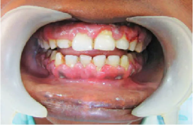
Intraoral examination showed generalized diffuse gingival enlargement, more on the pala-tal aspect of the maxillary anterior teeth and lin-gual aspect of the mandibular anteriors (Fig. 3, 4). Gingiva was swollen, edematous, pale pink, soft, tender and bleeding on probing was present. Even though there was the presence of local irritants, no bone resorption was evident in panoramic radio-graph. On the basis of the clinical findings, chron-ic inflammatory gingival enlargement and acute gingival enlargement due to systemic cause were considered in differential diagnosis.
Taking into consideration the detailed history and presence of clinical features, primary impor-tance was given to investigation before any dental treatment. The results of a hematological exami-nation led to a surprising & confirmatory diagno-sis. Her hematological findings showed a decrease in red blood cells (7 million/mm3) with lowered hematocrit and hemoglobin levels (6.4 gram %); a low platelet count (14,000/mm3) and increased number of white blood cells (62,300/mm3).
Hyper-Fig. 1.
Diffuse swelling on right side and perior-bital edema on left side of face
Fig. 2.
Gingival enlargement on palatal aspect of maxillary teeth
cellular bone marrow with immature myeloid cells was observed in a bone marrow biopsy which was suggestive of acute myelogenous leukemia. She was quickly referred to an oncologists. The patient was treated with chemotherapy and supportive/symp-tomatic drugs by the team of oncologist. Unfortu-nately she succumbed to death after 2 months due to complications of the chemotherapy.
Discussion
Gingival enlargement is quite a common oral manifestation but it should not be taken for grant-ed and neglectgrant-ed. An oral diagnostician should consider systemic illness under differential diag-nosis and there should be a procedure to evaluate the case thoroughly, to rule out underlying serious systemic diseases like leukemia, scurvy, drug his-tory like dilantin sodium, cyclosporin, nifedipine, etc. and certain syndromes associated with it.
Scurvy is a historical, nearly-forgotten disease associated with starving populations deprived of
vitamin C intake [3]. It is rarely seen in developed countries where fortified food products and multi-ple supmulti-plements fill the market. However, poor diets devoid of fresh fruits and vegetables can still cause this disease [4]. Thus, administration of ascorbic acid results in significant improvement [5, 6].
Our first patient’s initial diagnostic work-up considered conditions that are more common than scurvy like leukemia and platelet disorders, which were then excluded by hematological exam-ination. As no common cause for gingival enlarge-ment and bruises/hematoma was found, poor di-etary habits with lack of fruit and vegetables led us to consider scurvy in our differential diagnosis. Of course the dramatic response to vitamin C supple-mentation confirmed the diagnosis.
According to Health Canada’s food and nutri-tion guidelines, the recommended requirement of vitamin C is between 75 and 90 mg per day. Clin-ical manifestations of scurvy can be seen within 8 to 12 weeks of irregular or inadequate intake [7].
Vitamin C deficiency produces an abnormal collagen that affects blood vessel integrity, leading to capillary fragility, perivascular edema, and red cell extravasations. Classic signs of vitamin C defi-ciency include weakness, anemia, tooth loss, gum bleeding, bruises, and petechiae [4]. This deficien-cy does not itself cause gingival inflammation but it causes hemorrhage, collagen degeneration and edema of gingival connective tissue, which mod-ify the usual gingival response to dental plaque. So, local irritation is necessary for the initiation of this type of enlargement [2].
Noordin et al. [6] and Heird [8] have given a thorough description of the radiological chang-es in the long bonchang-es. Noordin et al. [6] also men-tioned that skull changes may produce a “hair-on- -end” or crew-cut appearance secondary to mar-row hyperplasia in response to anemia. However, no sphenoid changes are reported.
Diagnosis of vitamin C deficiency is usual-ly based on the characteristic clinical picture, the radiographic appearance of the long bones, and a history of poor vitamin C intake [6]. Bruising, perifollicular hemorrhages, gingival bleeding and weakness usually improve within 1 to 2 weeks [7]. In the present case, the patient also responded no-ticeably to ascorbic acid therapy.
Acute myelogenous leukemia (AML) is a ma-lignant disease of bone marrow. The various oral manifestations are gingival swelling, gingival bleeding, large, irregular, foul smelling oral ulcers, oral infections, and laryngeal pain [9–11]. Leuke-mic cells may infiltrate the palate, alveolar bone, dental pulp, 5th and 7th cranial nerves and the pa-tient may complain of toothache, tooth mobility and paresthesia. Nevertheless, the treatment-relat-Fig. 3. Gingival enlargement on labial and buccal
aspect of maxillary and mandibular teeth
Fig. 4. Gingival enlargement on palatal aspect of maxil-lary teeth
ed orofacial complications in children are cranio-facial deformities, deficient mandibular develop-ment and dental anomalies [9].
Oral changes are 3 times more common in acute leukemia than chronic leukemia, amongst which leukemic gingival enlargement is seen more in AML than in ALL [2, 10, 12, 13]. In leukemia, gingival enlargement is due to either leukemic in-filtration or reactive hyperplasia, and to differenti-ate between them, biopsy is required. However, in this case, biopsy should be performed with extreme caution or highly contraindicated due to the risk of bleeding and infection [12]. Nevertheless, in the earliest phase of gingival enlargement, the leuke-mic infiltrate may be minimal and obscured by the dominant local inflammatory component, thereby masking the neoplastic element [14]. In our second case, we avoided the gingival biopsy but the hema-tological profile and bone marrow examination ef-ficiently diagnosed acute myelogenous leukemia.
The role of a local irritant as an inducer for leukemic infiltration in gingiva or the possibili-ty of leukemic infiltration in edentulous patients is a controversial point [1, 12, 13, 15].
Carran-za and Newman reported that the alveolar mu-cosa couldn’t be involved in leukemia infiltration whereas the study by Bashar et al. [12] observed leukemic infiltration of alveolar mucosa in an edentulous patient.
During acute phases of leukemia, only emer-gency periodontal care is taken [2]. Demirer et al. [5] stated that the elimination of local factors and maintaining ideal systemic condition might result in full success in periodontal healing. In these kinds of cases, dental therapy performed without thorough investigations could lead to seri-ous complications and could be fatal. Surgical in-tervention may aggravate the situation by exacer-bation of acute symptoms.
Early detection of systemic diseases like leuke-mia and scurvy can lead to appropriate treatment and alleviation of untoward side effects. This is an area where the dentist may well save a life, honor-ing his or her performance as health professional and understanding the patient as a whole. This ar-ticle reinforces the importance of a careful, com-plete history, diagnosis and investigation in cases of gingival enlargement.
Acknowledgment. We wish to thank Datta Meghe Institute of Medical Sciences for approving this study. Special thanks to our patients for their cooperation.
References
[1] Neuman M.G., Takei H.H., Carranza F.A.: Carranza’s Clinical periodontology. Philadelphia: W.B. Saunders Company, New York 2002, 9th ed, 204–228.
[2] Reddy S.: Gingival enlargements. [In:] Essentials of clinical periodontology and periodontics. Ed.: Reddy S. 2nd ed.
Jaypee Brothers pvt Ltd, 2008, 159–163.
[3] Martini E.: How did Vasco de Gama sail for 16 weeks without developing scurvy? Lancet 2003, 361,1480. [4] Velandia B., Centor R.M., McConnell V., Shah M.: Scurvy is still present in developed countries. J. Gen.
Intern. Med. 2008, 23, 1281–1284.
[5] Demirer S., Özdemir H., Şencan M., Marako I.: Gingival hyperplasia as an early diagnostic oral manifestation in acute monocytic leukemia: A case report. Eur. J. Dent. 2007, 2, 111–114.
[6] Noordin S., Baloch N., Sohail M., Memon A.S., Ahmad T.: Skeletal manifestations of scurvy: a case report from Dubai. Case Rep. Orthoped. 2012, 12, 624–628.
[7] Léger D.: Scurvy reemergence of nutritional deficiencies. Can. Fam. Phys. 2008, 54, 1403–1406.
[8] Heird W.C.: Vitamin deficiencies and excesses. [In:] Nelson Textbook of Pediatrics. Eds.: Behrman R.E., Kiegman R.M., Arvin A.M., Nelson W.E. 17th ed. WB. Saunders, Philadelphia 2004, 177–190.
[9] DeRossi S.S., Garfunkel A., Greenberg M.A.: Burket’s Oral Medicine, Diagnosis and Treatment. [In:] Hematologic diseases. Eds.: Greenberg M.S., Glick M. 10th ed. Harcourt private limited, New Delhi 2003, 429–453. [10] Hou G.L., Huang J.S., Tsai C.C.: Analysis of oral manifestations of leukemia: a retrospective study. Oral Dis.
1997, 3, 31–38.
[11] Gleeson P.: Spontaneous gingival haemorrhage: case report. Aust. Dent. J. 2002, 47, 174–175.
[12] Bashar A.H., Hassan Y.I.. Raja K.K., Ferial A.H., Mirza K.B.: Gingival fine needle aspiration cytology in acute leukemia. Oral Med. Pathol. 2002, 31, 55–58.
[13] Jinbu Y., Naito H., Noguchi T., Yoko A., Ozawa K.: Unusual tumor formation in the tongue of an of an acute myelocytic leukemia patient: report of a case. Oral Med. Pathol. 2000, 29, 53–56.
[14] Wu J., Fantasia J.E., Kaplan R.: Oral manifestations of acute myelomonocytic leukemia: a case report and review of the classification of leukemias. J. Periodontol. 2002, 73, 664–668.
[15] Cooper C.L., Loewen R., Shore T.: Gingival hyperplasia complicating acute myelomonocytic leukemia. J. Can. Dent. Assoc. 2000, 66, 78–79.
Address for correspondence
Suwarna Dangore-Khasbage Oral Medicine and Radiology
Datta Meghe Institute of Medical Sciences India
E-mail: [email protected] Conflict of interest: None declared Received: 22.08.2014
Revised: 29.08.2014 Accepted: 3.09.2014