Address for correspondence
Haytham Behery E-mail: behery@live.com
Funding sources
None declared
Conflict of interest
None declared
Received on July 4, 2018 Reviewed on September 16, 2018 Accepted on October 29, 2018 Published online on December 31, 2018
Abstract
Background. Bulk-fill composites were developed to simplify composite placement and minimize po-lymerization shrinkage stresses, which can improve gingival marginal adaptation in deep class II cavities. Objectives. The objective of this study was to compare the gingival microleakage of class II cavities re-stored with bulk-fill composites to that of incrementally rere-stored ones with a conventional composite at 2 storage periods.
Material and methods. Forty freshly extracted intact molars were employed. Two standardized class II slot cavities, 3-millimeter-wide buccolingually, with the gingival floor 0.5 mm below the cementoenamel junction (CEJ) and the axial wall depth of 1.3 mm were prepared in each tooth (80 cavity preparations). The prepared teeth were divided equally into 3 bulk-fill groups (Tetric EvoCeram® Bulk Fill, X-tra Fil® and QuiXX®) and 1 con-trol group (TPH Spectra® HV). Each group was subdivided into 2 equal subgroups (n = 10) according to the storage period in distilled water (24 h and 6 months). The Adper® Single Bond Plus adhesive was used with all the restorative materials. The cavities in the experimental groups were restored with 4-millimeter bulk-fill com-posites in 1 increment, while the cavities in the control group were restored with 2 increments of the thickness of 2 mm. The polymerization light was applied from the occlusal surfaces. The teeth were then immersed in 2% procion red dye solution, sectioned and examined under a stereomicroscope to determine the extent of dye penetration. The data was statistically analyzed using the Kruskal–Wallis test and the Mann–Whitney U test. Results. The Kruskal–Wallis test revealed no significant differences in the mean microleakage scores among all the groups after 24-hour and 6-month storage (p = 0.945 and p = 0.928, respectively). The Mann–Whitney U test revealed an increase in the mean microleakage scores in all the groups after 6-month storage; however, the scores were not significantly different from the means obtained after 24 h (p = 0.259 for Tetric EvoCeram Bulk Fill; p = 0.205 for X-tra Fil; p = 0.166 for QuiXX; p = 0.155 for TPH Spectra HV).
Conclusions. Gingival microleakage of bulk-fill composites in class II cavities was not significantly diffe-rent from that of incrementally restored ones with a conventional composite. The increase in the mean gin-gival microleakage of the specimens stored for 6 months was not statistically significantly different in com-parison to the values obtained after the 24-hour storage period for each composite.
Key words: composite resin, microleakage, bulk-fill, dental restorations
Słowa kluczowe: żywica kompozytowa, mikroprzeciek, bulk-fill, wypełnienia dentystyczne
Cite as
Behery H, El-Mowafy O, El-Badrawy W, Nabih S, Saleh B. Gingival microleakage of class II bulk-fill composite resin restorations. Dent Med Probl. 2018;55(4):383–388. doi:10.17219/dmp/99264
DOI
10.17219/dmp/99264
Copyright
© 2018 by Wroclaw Medical University and Polish Dental Society
This is an article distributed under the terms of the Creative Commons Attribution Non-Commercial License (http://creativecommons.org/licenses/by-nc-nd/4.0/)
Gingival microleakage of class II bulk-fill composite resin restorations
Mikroprzeciek dziąsłowy wypełnień klasy II z żywic kompozytowych
typu bulk-fill
Haytham Behery
1,A–F, Omar El-Mowafy
1,A,C–F, Wafa El-Badrawy
1,A,E,F, Sameh Nabih
2,A,E,F, Belal Saleh
2,A,E,F1 Department of Restorative Dentistry, Faculty of Dentistry, University of Toronto, Canada 2 Department of Operative Dentistry, Faculty of Dental Medicine, Al-Azhar University, Cairo, Egypt
A – research concept and design; B – collection and/or assembly of data; C – data analysis and interpretation; D – writing the article; E – critical revision of the article; F – final approval of the article
Introduction
Polymerization shrinkage of composite resins and the accompanying shrinkage stress build-up represent the major drawbacks in using direct composite resin restor-atives. This is due to the fact that the forces connected with the shrinkage stress may disrupt the bond to the cavity walls, leading to micro-gaps, which can result in the passage of the saliva and oral fluids along the tooth restoration interface (i.e., microleakage). Microleakage is a commonly encountered problem with posterior com-posite restorations, especially at gingival margins placed apically to the cementoenamel junction (CEJ), as in deep class II cavities.1 It represents a matter of concern, as it can lead to staining at the margins of restorations and recurrent caries with subsequent pulp pathology. Other problems related to direct composite restorations are the limited depth of the cure, technique sensitivity and time-consuming placement procedure. Dentists have al-ways sought a fast and reliable restoration technique that would allow for a reduction of the number of composite layers placed, thus reducing the effort and time consumed in such a routine procedure. Bulk-fill composite resins seem to fulfill this desire.
Several developments in the field of composite restora-tion techniques have been made, such as optimizing the polymerization light intensity,2 the application of a flow-able resin as a liner3 and the incremental placement.4 These innovations have been introduced to minimize the shrinkage stress, and improve the marginal integrity and the durability of composite restorations.5
Bulk-fill composites were developed in an attempt to simplify and expedite the composite placement technique.5 According to their manufacturers, they have a higher depth of polymerization, which could eliminate the need for lay-ering. They are also claimed to generate low polymerization shrinkage stresses,6,7 owing to the use of modified resin-filler technologies,8,9 that minimize the volumetric shrink-age and/or modify the visco-elastic behavior of bulk-fill composites by decreasing their elastic moduli and increas-ing their flow capacity. The reduction in shrinkage stresses of bulk-fill composites, if true, can result in improving the marginal integrity and the durability of the bond of com-posite restoration to the tooth structure.10,11
This study was conducted to assess the gingival micro-leakage of class II slot cavity preparations restored with 3 types of bulk-fill composite resins in comparison to those incrementally restored with a conventional composite resin at 2 different storage periods (24 h and 6 months). The research null hypotheses were the following:
– there is no significant difference in the gingival micro-leakage between the tested bulk-fill composites and the conventional composite;
– there is no significant difference in the gingival micro-leakage of each of the 4 tested composites at 2 different storage periods (24 h and 6 months).
Material and methods
Forty freshly extracted intact human molars were em-ployed. The teeth were sterilized with gamma irradiation, thoroughly rinsed and scaled to remove any plaque, cal-culus or attached periodontal tissues. The teeth were then stored in distilled water in a refrigerator. The teeth were mounted vertically in acrylic resin bases 2 mm apical to CEJ. Class II slot cavities were prepared on both proxi-mal sides of each molar, using carbide bur No. 56 (Great White® Series; SS White Burs, Inc., Lakewood, USA) in a water-cooled high-speed handpiece. A total of 80 slot cavities were prepared, each with the dimensions: the width of 3 mm buccolingually, the axial depth of 1.3 mm and the gingival floor located 0.5 mm below CEJ. A new bur was used for every 4 cavity preparations. The dimen-sions of each cavity were verified with a digital caliper (Mastercraft, Toronto, Canada).
The prepared teeth were divided into 4 groups (10 molars each, with 20 class II slot cavities) and assigned to 4 com-posite resins groups – 3 bulk-fill: Tetric EvoCeram® Bulk Fill (Ivoclar Vivadent, Amherst, USA), X-tra Fil® (VOCO America, Inc., Indian Land, USA), and QuiXX® (Dentsply
Caulk, Milford, USA); and 1 control: TPH Spectra® HV
(Dentsply Caulk) (Table 1). Each group was subdivided into 2 equal subgroups (n = 10) according to the storage period in distilled water (24 h and 6 months).
A Tofflemire metal matrix retainer/band was secured around each prepared tooth to establish the proper proxi-mal anatomic contour. The metal matrix band was sup-ported externally with a low-fusing compound to main-tain its adaptation to the cavity margins.1 For all groups, the same etch-and-rinse adhesive system was used (Scotchbond® Etchant and Adper® Single Bond Plus; 3M ESPE, St. Paul, USA). The restorative procedure was per-formed according to the manufacturer’s instructions.
The 3 bulk-fill composites were applied in a single in-crement of 4 mm in thickness, while the conventional composite (TPH Spectra HV) was applied in 2 horizontal increments, each 2-millimeter-thick. An LED light po-lymerization unit (Demi®LED Light Curing System; Kerr Corporation, Orange, USA) was used for the polymeriza-tion of all the composites. Each bulk-fill composite res-toration was subjected to 20 s of irradiation, while in the case of the TPH Spectra HV restoration, each increment was subjected to 20 s of irradiation. The irradiance of the light polymerization unit was periodically checked with checkMARC (BlueLight Analytics, Inc., Halifax, Canada). The irradiance was found to be 1,120 mW/cm² on aver-age. The specimens were stored in distilled water at 37°C for the aforementioned storage periods.
Two coats of nail varnish were applied on the tooth sur-faces, except for 1 mm from the restoration margins. Af-terward, the teeth were immersed in 2% procion red dye solution (Imperial Chemical Industries, London, England) for 24 h at 37°C, and then rinsed under running water
for 5 min. Each tooth was sectioned mesio-distally into 2 halves with a microslicing machine (IsoMet; Buelher, Lake Buff, USA).1,12 The half with the deepest dye pen-etration was used to represent the tooth.1,12 The extent of dye penetration was determined by an examination with a stereomicroscope (Wild M3Z Stereo Microscope; Leica Microsystems (Schweitz) AG, Heerbrugg, Switzer-land) at a magnification ×10, according to a 5-point scale: – 0: no dye penetration;
– 1: dye penetration limited to the outer half of the gin-gival floor;
– 2: dye penetration extended along the entire gingival floor (i.e., beyond the outer half of the gingival floor); – 3: dye penetration extended along the gingival wall and
up to half of the axial wall;
– 4: dye penetration extended along the gingival floor and the entire axial wall.
In addition, a digital camera (Canon PowerShot S 120; Canon, Inc., Tokyo, Japan) was used to capture photo-graphic images for each selected section (Fig. 1,2).
The data was tabulated and statistically analyzed. The means and standard deviations (SDs) were calculated and statistically analyzed using the non-parametric Krus-kal–Wallis test and the Mann–Whitney U test. The sig-nificance level was set at 0.05. The statistical analysis was
performed with SPSS v. 20 for Windows (IBM Corp.,
Armonk, USA).
Results
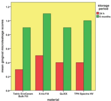
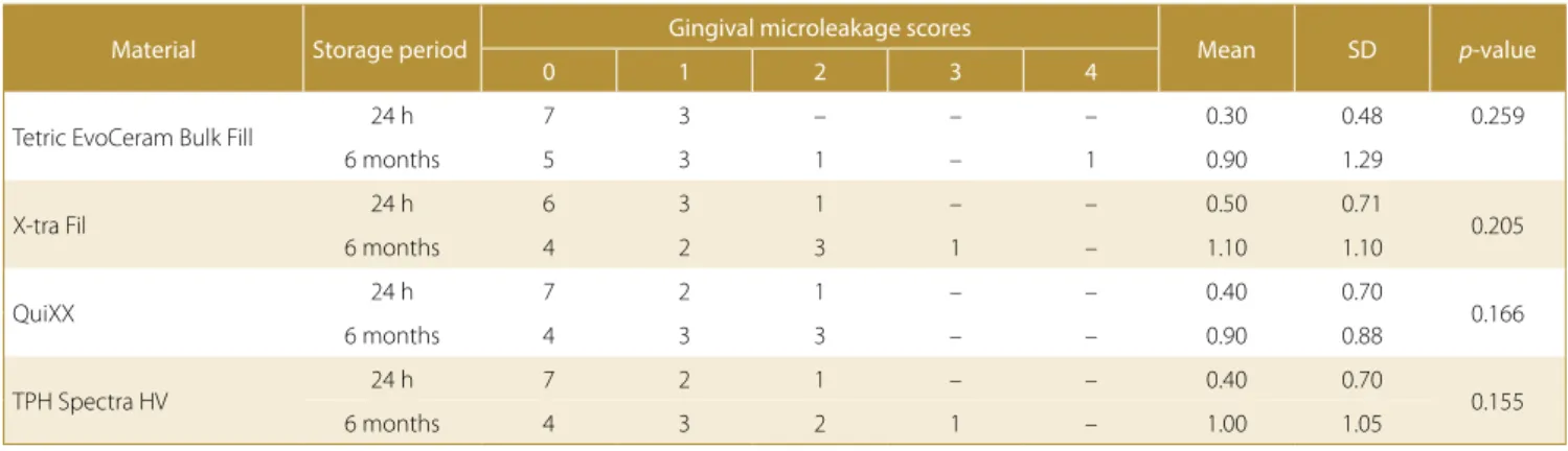
The mean, standard deviation, median, and range val-ues of the gingival microleakage scores for all the 4 groups are recorded in Table 2 and Fig. 3,4.
Table 1. Materials used in the study
Material Product description Main components of the restorative material Manufacturer
Tetric EvoCeram Bulk Fill
light-cured, methacrylate-based bulk-fill composite resin
monomer matrix of dimethacrylates, fillers
(barium glass, ytterbium trifluoride, mixed oxide, prepolymer)
Ivoclar Vivadent, Amherst, USA
X-tra Fil light cured, methacrylate-based bulk-fill composite resin
Bis-GMA, UDMA, TEGDMA, fillers
(barium-boron-alumino-silicate glass)
Voco America, Inc., Indian Land, USA
QuiXX light cured, methacrylate-based bulk-fill composite resin
UDMA,TEGDMA, di- and tri-methacrylate resins, carboxylic acid-modified dimethacrylate, fillers
(strontium-alumino-sodium-fluoro-silicate glass)
Dentsply Caulk, Milford, USA
TPH Spectra HV light cured, methacrylate-based composite resin
urethane-modified Bis-GMA resin, TEGDMA, fillers
(silanated barium, aluminoborosilicate glass, silanated barium-boron-fluoro-alumino-silicate glass, silicon dioxide)
Dentsply Caulk
Scotchbond Etchant 35% phosphoric acid gel 3M ESPE, St. Paul, USA Adper Single Bond Plus two step etch-and-rinse adhesive system 3M ESPE Bis-GMA – bisphenol A glycidyl dimethacrylate; TEGDMA – triethylene glycol dimethacrylate; UDMA – urethane dimethacrylate.
Fig. 1. Photographs of a representative specimen from each group stored for 24 h (from left to right: Tetric EvoCeram Bulk Fill, X-tra Fil, QuiXX, and TPH Spectra HV)
Fig. 2. Photographs of a representative specimen from each group stored for 6 months (from left to right: Tetric EvoCeram Bulk Fill, X-tra Fil, QuiXX, and TPH Spectra HV)
Fig. 3. Bar chart representing the mean microleakage scores of the tested groups at each storage period
After 24-hour storage, the mean gingival microleakage scores for Tetric EvoCeram Bulk Fill, X-tra Fil, QuiXX, and TPH Spectra HV were 0.3, 0.5, 0.4, and 0.4, respec-tively. The Kruskal–Wallis test revealed no statistically significant differences in the mean microleakage scores among the groups (p = 0.945).
After 6-month storage, the mean microleakage scores for Tetric EvoCeram Bulk Fill, X-tra Fill, QuiXX, and TPH Spectra HV were 0.9, 1.1, 0.9, and 1.0, respectively. The Kruskal–Wallis test revealed no significant differ-ences in the mean microleakage scores among the groups (p = 0.928).
In spite of the increase in the mean microleakage scores
after 6 months, the Mann–Whitney U test revealed no
significant differences in the mean microleakage scores for each composite after 6-month storage as compared to the mean microleakage scores obtained after 24-hour storage (p = 0.259 for Tetric EvoCeram Bulk Fill; p = 0.205 for X-tra Fil; p = 0.166 for QuiXX; and p = 0.155 for TPH Spectra HV).
Discussion
One of the key functions of dental restorations is to seal the exposed dentin and to protect the pulp against the oral environment. An insufficient seal at the tooth/restoration interface may lead to microleakage, described as a clini-cally undetectable passage of bacteria, fluids, molecules, or ions between the cavity wall and the restorative ma-terial.13,14 Microleakage tests are widely used to evaluate the marginal sealing of composite restorations.15 Previous studies reported that composite restorations exhibited higher microleakage at gingival margins than at occlusal margins.16–19 Gingival microleakage is more frequently observed in deep class II cavities, where gingival margins are placed apical to CEJ.1 In this study, gingival microle-akage in class II composite restorations was assessed with gingival margins of 0.5 mm apical to CEJ.
Several methods have been used to detect microleak-age, such as the use of dyes, artificial caries, air pressure, bacteria, radioactive isotopes, neutron activation analy-sis, and scanning electron microscopy. The use of dyes as tracers is one of the most common methods of detecting microleakage in vitro. Different types of dyes with differ-ent particle sizes are used for microleakage assessmdiffer-ent, such as procion red dye, basic fuschin and methylene blue.13 This technique is highly feasible and carries no radiation hazards. In addition, the dye has the advantage of its contrasting color to the tooth and the restoration, without reacting chemically with specimens. In this study, 2% procion red dye solution was used as a tracer for mi-croleakage assessment.
Gamma irradiation was used to sterilize the teeth, as it has no adverse effect on the enamel hardness or its resistance to demineralization.20 In addition, gamma ir-radiation is an effective method of sterilizing the teeth, which neither alters the dentin structure and its surface morphology nor affects the strength of the shear bond to the dentin.21,22
A 2-step etch-and-rinse adhesive system (the Adper Single Bond Plus adhesive with Scotchbond Etchant) was used in this study with the 4 tested composite resins, as it is considered to result in optimum bonding. Adper Single
Table 2. Gingival microleakage scores, means and standard deviations (SDs) of the 4 tested groups at each storage period
Material Storage period Gingival microleakage scores Mean SD p-value
0 1 2 3 4
Tetric EvoCeram Bulk Fill 24 h 7 3 – – – 0.30 0.48 0.259
6 months 5 3 1 – 1 0.90 1.29
X-tra Fil 24 h 6 3 1 – – 0.50 0.71 0.205
6 months 4 2 3 1 – 1.10 1.10
QuiXX 24 h 7 2 1 – – 0.40 0.70 0.166
6 months 4 3 3 – – 0.90 0.88
TPH Spectra HV 24 h 7 2 1 – – 0.40 0.70 0.155
6 months 4 3 2 1 – 1.00 1.05
Fig. 4. Box plot representing the median and range values of the microleakage scores of the tested groups at each storage period (the circle represents the outlier)
Bond Plus is an ethanol-based adhesive which contains 2-hydroxyethyl methacrylate (HEMA) and is thought to be able to maintain the collagen fibrils in an expanded form after the evaporation of the solvent, improving the monomer infiltration and the formation of a proper hy-brid layer.23–25
In this study, the null hypotheses were not rejected. The results revealed no statistically significant differences in the means of gingival microleakage scores among the 4 composite resin restoratives tested after 24-hour and 6-month storage periods. This may indicate that polym-erization and/or polympolym-erization shrinkage of the bulk-fill composites (Tetric EvoCeram Bulk Fill, X-tra Fil and QuiXX) was comparable to that of the incrementally placed composite (TPH Spectra HV). These findings are in agreement with the findings reported by Ahmedet al., who found that the type of the composite material used (Filtek® Z250 vs Filtek LS (3M ESPE)) had no significant effect on gingival microleakage, rather the bonding agent type was more critical and had a significant effect.12 Fur-thermore, Rengo et al. found no significant differences in the microleakage of class II cavities restored with bulk-fill composites (G-aenial® Universal Flo bulk-fill, G-aenial Flo bulk-fill and Kalore® bulk-fill (GC Corporation, Tokyo, Japan)) in comparison to that of the cavities incrementally restored with conventional composites (G-aenial Univer-sal Flo, G-aenial Flo and Kalore (GC Corporation)), using the same adhesive system (G-aenial Bond (GC Corpora-tion)) with all the groups.26
The statistical analysis did not reveal significant differ-ences in the mean microleakage scores, when the values obtained for each group after 6 months were compared with the values obtained after 24 h, in spite of an obvi-ous increase in the mean score. It is speculated that with longer storage periods, statistically significant differences may be detected.
The lack of significant differences in the mean microleak-age scores between the specimens stored for 24 h and those stored for 6 months in each restorative group is in agreement with the findings reported in other studies. De Munck et al.14 reported that the effect of artificial aging methods, such as water storage and thermocycling, on mi-croleakage is minimal.27–29 These findings were in accor-dance with those of Mahmoud and Al-Wakeel, who found that the marginal adaptation of ormocer-, silorane- and methacrylate-based composite resin restorative systems bonded to dentin cavities was not affected by aging times (immediately after polymerization, after 1 month, and after 1 year of water storage and thermocycling).30 Khos-ravi et al. found that water storage (24 h, 3 months and 6 months) had no significant effect on gingival microleak-age of class II cavities restored with methacrylate-based and silorane-based composite resins.31 In another study, it was found that water storage for 6 months had no significant effect on the microleakage scores with some composite restorative system products; however, with other
prod-ucts, significant differences were detected.32 Nevertheless, 6-months water storage can only be considered short-term; it is possible that with long-term water storage, sig-nificantly higher microleakage scores may be encountered. However, further research is needed in this respect.
Conclusions
Within the limitations of this in vitro study, it can be concluded that gingival microleakage of class II compos-ite restorations was not significantly affected by the type of composite restoration used (i.e., bulk-fill composite or conventional composite) even after 6 months of water storage. Increasing the storage time from 24 h to 6 months had no significant effect on gingival microleakage for each of the 4 types of class II composite restorations used.
References
1. El-Mowafy O, El-Badrawy W, Eltanty A, Abbasi K, Habib N. Gingi-val microleakage of Class II resin composite restorations with fiber inserts. Oper Dent. 2007;32:298–305.
2. Uno S, Asmussen E. Marginal adaptation of a restorative resin polymerized at reduced rate. Scand J Dent Res. 1991;99:440–444. 3. Alomari QD, Reinhardt JW, Boyer DB. Effect of liners on cusp
deflec-tion and gap formadeflec-tion in composite restoradeflec-tions. Oper Dent. 2001;26:406–411.
4. Lee MR, Cho BH, Son HH, Um CM, Lee IB. Influence of cavity dimen-sion and restoration methods on the cusp deflection of premolars in composite restoration. Dent Mater. 2007;23:288–295.
5. Behery H, El-Mowafy O, El-Badrawy W, Saleh B, Nabih S. Cuspal deflection of premolars restored with bulk-fill composite resins.
J Esthet Restor Dent. 2016;28:122–130.
6. Ilie N, Hickel R. Investigations on a methacrylate-based flowable com-posite based on the SDR technology. Dent Mater. 2011;27:348–355. 7. Rothmund L, Reichl FX, Hickel R, et al. Effect of layer thickness
on the elution of bulk-fill composite components. Dent Mater. 2017;33:54–62.
8. Misilli T, Gonulol N. Water sorption and solubility of bulk-fill com-posites polymerized with a third generation LED LCU. Braz Oral Res. 2017;31:e80.
9. Leprince JG, Palin WM, Vanacker J, Sabbagh J, Devaux J, Leloup G. Physico-mechanical characteristics of commercially available bulk-fill composites. J Dent. 2014;42:993–1000.
10. Kim RJ, Kim YJ, Choi NS, Lee IB. Polymerization shrinkage, modu-lus, and shrinkage stress related to tooth-restoration interfacial debonding in bulk-fill composites. J Dent. 2015;43:430–439. 11. Schneider LF, Cavalcante LM, Silikas N. Shrinkage stresses
generat-ed during resin-composite applications: A review. J Dent Biomech. 2010;2010:131630.
12. Ahmed W, El-Badrawy W, Kulkarni G, Prakki A, El-Mowafy O. Gin-gival microleakage of class V composite restorations with fiber inserts. J Contemp Dent Pract. 2013;14:622–628.
13. Kidd EA. Microleakage: A review. J Dent. 1976;4(5):199–206. 14. De Munck J, Van Landuyt K, Peumans M, et al. A critical review of
the durability of adhesion to tooth tissue: Methods and results.
J Dent Res. 2005;84:118–132.
15. Klein CA, Jr., da Silva D, Reston EG, Borghetti DL, Zimmer R. Effect of at-home and in-office bleaching on marginal microleakage in composite resin restorations using two adhesive systems. J Con-temp Dent Pract. 2018;19:248–252.
16. Eakle WS, Ito RK. Effect of insertion technique on microleakage in mesio-occlusodistal composite resin restorations. Quintessence Int. 1990;21:369–374.
17. Ciucchi B, Bouillaguet S, Holz J. Proximal adaptation and margin-al semargin-al of posterior composite resin restorations placed with direct and indirect techniques. Quintessence Int. 1990;21:663–669.
18. Ferrari M, Davidson CL. Sealing performance of Scotchbond Multi-Purpose-Z100 in class II restorations. Am J Dent. 1996;9:145–149. 19. Hilton TJ, Schwartz RS, Ferracane JL. Microleakage of four class II
resin composite insertion techniques at intraoral temperature.
Quintessence Int. 1997;28:135–144.
20. Rodrigues LK, Cury JA, Nobre dos Santos M. The effect of gamma radiation on enamel hardness and its resistance to demineraliza-tion in vitro. J Oral Sci. 2004;46:215–220.
21. White JM, Goodis HE, Marshall SJ, Marshall GW. Sterilization of teeth by gamma radiation. J Dent Res. 1994;73:1560–1567.
22. Sperandio M, Souza JB, Oliveira DT. Effect of gamma radiation on dentin bond strength and morphology. Braz Dent J. 2001;12:205–208. 23. Tay FR, Gwinnett AJ, Pang KM, Wei SH. Resin permeation into acid-conditioned, moist, and dry dentin: A paradigm using water-free adhesive primers. J Dent Res. 1996;75:1034–1044.
24. Perdigao J, Van Meerbeek B, Lopes MM, Ambrose WW. The effect of a re-wetting agent on dentin bonding. Dent Mater. 1999;15:282–295. 25. El-Bouhi YMM. Bond performance and nanoleakage of resin/den-tin interface under different intrapulpal pressures. PhD thesis. Cairo University, Egypt, 2009.
26. Rengo C, Spagnuolo G, Ametrano G, et al. Marginal leakage of bulk fill composites in Class II restorations: A microCT and digital micro-scope analysis. Int J Adhes Adhes. 2015;60:123–129.
27. Gwinnett AJ, Yu S. Effect of long-term water storage on dentin bonding. Am J Dent. 1995;8:109–111.
28. Gale MS, Darvell BW. Thermal cycling procedures for laboratory testing of dental restorations. J Dent. 1999;27:89–99.
29. Wahab FK, Shaini FJ, Morgano SM. The effect of thermocycling on microleakage of several commercially available composite Class V restorations in vitro. J Prosthet Dent. 2003;90:168–174.
30. Mahmoud SH, Al-Wakeel Eel S. Marginal adaptation of ormocer-, silorane-, and methacrylate-based composite restorative systems bonded to dentin cavities after water storage. Quintessence Int. 2011;42:e131–e139.
31. Khosravi K, Mousavinasab SM, Samani MS. Comparison of micro-leakage in Class II cavities restored with silorane-based and meth-acrylate-based composite resins using different restorative tech-niques over time. Dent Res J (Isfahan). 2015;12:150–156.
32. Crim GA. Effect of aging on microleakage of restorative systems.