Apexification is a way of root canal treatment in the case of irreversible pulpitis or pulp necrosis in immature teeth. During this procedure, there might be some problems caused by absence of api− cal constriction, thin root walls and root canal with the shape of diverted cone. It makes huge difficul−
ties while preparation and final obturation of the root canal. Apexification conduces to stimulating further root formation or creating hard tissue at the apex. Until today, in this method, the best known and the most widely used materials are those based on the calcium hydroxide. Despite many benefi−
L
IDIAP
OSTEK−S
TEFAŃSKA, A
NNAJ
ODŁOWSKA, I
WONAW
YSOCZAŃSKA−J
ANKOWICZApplication of the Mineral Trioxide Aggregate (MTA)
in Apexification – Case Reports
Zastosowanie Mineral Trioxide Aggregate (MTA)
w procedurze apeksyfikacji – opis przypadków
Department of Pediatric Dentistry Medical University of Silesia
Dent. Med. Probl. 2009, 46, 2, 247–251 ISSN 1644−387X
PRACE KAZUISTYCZNE
© Copyright by Wroclaw Medical University and Polish Stomatological Association
Abstract
Background. The purpose of apexification is to finish root end formation or stimulatation of mixed periodontal− pulpal tissue to form mineralized barrier closing root canal.
Objectives. The aim of the work was to assess application of MTA in apexification in 2 patients in developmental age.
Material and methods.Apexification with use of MTA was carried out in 2 patients with immature central max− illary incisors. Root canals were instrumented with step−down technique. The layer of MTA (3–4 mm of thickness) contacting with radiological apex were applied under control of endodontic microscope and with the aim of spe− cial applicator. Root canals were obturated using vertical condensation with AH plus and gutta−percha points and crowns were restored with composite material.
Results. Control x−rays after 3, 6 and 12 months showed correct condition of root end and no pathological lesions in periapical tissues.
Conclusion.Until now results of researches show that MTA might be used in apexification with very good result (Dent. Med. Probl. 2009, 2, 00–00).
Key words: apexification, MTA.
Streszczenie
Wprowadzenie. Apeksyfikacja to procedura mająca na celu zakończenie procesu formowania wierzchołka korzenia lub pobudzenia mieszanej tkanki miazgowo−ozębnowej do wytworzenia zmineralizowanej bariery zamykającej światło kanału korzeniowego. Cel pracy. Ocena zastosowania preparatu MTA w procesie apeksy− fikacji u dwóch pacjentów w wieku rozwojowym.
Materiał i metody. Apeksyfikację z zastosowaniem preparatu MTA wykonano u 2 pacjentów w niedojrzałych zębach siecznych centralnych szczęki. Kanały korzeniowe opracowano techniką step−down. Pod kontrolą mikroskopu endodontycznego i za pomocą specjalnego podajnika nakładano warstwę 3–4 mm preparatu MTA stykającą się z wierzchołkiem radiologicznym. Kanały wypełniono metodą kondensacji pionowej z wykorzys− taniem preparatu AH plus i ćwieków gutaperkowych, a korony zębów odbudowano materiałem kompozytowym.
Wyniki.Kontrolne zdjęcia RTG po 3, 6 i 12 miesiącach wykazały prawidłowy stan wierzchołka korzenia i brak zmian patologicznych w tkankach okołowierzchołkowych. Wnioski.Dotychczasowe wyniki badań wskazują, iż materiał MTA może być wykorzystywany z bardzo dobrym rezultatem w procedurze apeksyfikacji (Dent. Med. Probl. 2009, 2, 00–00).
cial features, such as: antiseptic, bacteriostatic and odontotropic action, these materials are increas− ingly criticized because of disintegration in tissue liquids and poor adhesion to tooth tissues. Although these materials are very effective in apexification they have fundamental defect. The treatment is long−lasting what is conducive to acci− dental fracture of weakened root, and sometimes immediate restoration of damaged tooth hard tis− sues can not be done [1].
In many research centers, clinicians try to short− en apexification time by the use of agents that might be “artificial apical stop” and enable immediate final root canal obturation. Until now, (were used) dentin chips, calcium hydroxide, hydroxyapatite, TCP and other [2, 3]. Authors deal with this topic dishonor them. MTA (Mineral Tioxide Aggregate) is a modern material recommended for apexifica− tion. Material is built of hydrophile particles of cal− cium, magnesium, silicone, ferrum oxides that in wet conditions create colloidal gel. The aim of the work was to assess application of MTA in apexifi− cation in 2 patients in developmental age.
Review of Cases
Apexification with the use of MTA was car− ried out in 2 patients in central maxillary incisors. Instrumentation of root canal with step−down tech− nique were made after fixation of rubber dam and determination of working length with Ingle`s method. Root canal was rinsed with 0.1% sodium hypochlorite and 3% hydrogen peroxide. Final rinse was carried out with physiological salt solu− tion in order to clean root canal from other liquids. Calibrated paper points to the working length were used to dry root canal. About 3–4 mm layer of MTA was applied at the apex under control of endodontic microscope and with the aid of special
applicator. Then, sterile, wet cotton pellet was inserted into root canal for 24 hours and crown was restored with glass−ionomer cement. After this period remaining part of root canal was obtu− rated with lateral condensation with use of AH plus and gutta−percha points, glass−ionomer based material was applied and crowns restored with composite material.
Case 1.
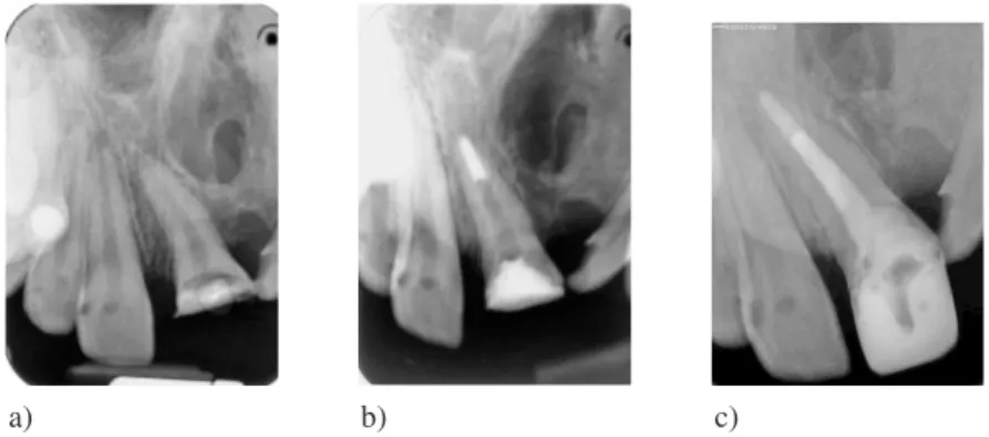
Patient K.W., aged 8, called for Pediatric Dentistry Department of Silesian Medical University (SUM) because of injury of tooth 11 and 21. The injury was on the playground. As a result of injury crowns were fractured in the area of enamel and dentine. At the day of injury, teeth were in the initial phase of eruption. Additionally, tooth 11 was in the II degree of wobbling, and the tooth 21 in the III degree of wobbling. After tak− ing first control x−ray the root fracture was sus− pected (fig.1A). Restoration of crowns was post− poned for 3 months in order to wait for further eruption of crowns and ability testing pulp vitality. The dentine surface was secured with glass− ionomer cement Interface®. After one week, tooth 21 changed color for dark red and was sensitive to percussion. The decision for root canal treatment and apexification was made. After obtaining an access cavity and determining working length root canal was instrumented and cleaned by rinsing with 1% NaOCl, 3% H2O2 and 0.9% NaCl. The
apical part of the root canal was filled with MTA (3–4 mm) under control of endodontic microscope (fig.1B). Additionally, the quality of the procedure was controlled with taking x−ray. Next for 24 hours wet, sterile cotton pellet was inserted into root canal and crown was filled temporarily with glass−ionomer cement Inofill Molar®. Root canal was obturated with vertical condensation tech−
a) b) c)
Fig. 1. A) Patient K.W. tooth 21 – first, B) Tooth 21 – control after control x−ray before endodontic treatment appli− cation of MTA; C) Tooth 21 –after 12 months of observation
Ryc. 1A) Pacjent K.W. Ząb 21 – pierwsze zdjęcie RTG przed leczenim endodontycznym; B) Ząb 21 – po zastosowa− niu MTA; C. Ząb 21 – 12 miesięcy po leczeniu, widoczna bariera zmineralizowana w okolicy wierzchołkowej
nique. Restoration of fractured crown was made with composite material Charisma® (Kulzer). After 3, 6 and 12 months from procedure, control x−ray was taken and the condition of periapical region was estimated (fig.1C). The clinical condi− tion of the tooth was also controlled on every visit.
Case 2.
Patient Ś.A., aged 13, called for Pediatric Dentistry Department of SUM because of tooth 21 crown fracture. This girl was treated in SUM Orthodontic Department because of cleft palate. An x−ray showed immature roots of teeth 11 and 21 and distal inclination tooth 21 (fig. 2A). On the first visit, after local anesthesia with Septanest 1: 200, the pulp was extirpated, root canal was rinsed and instrumentated with step−down method. Root canal was filled with Biopulp – material based on the calcium hydroxide, for 3 weeks. Under control of endodontic microscope 3 mm of apical part was filled with MTA (fig. 2B). The cotton pellet infil− trated with 0.9% NaCl was inserted into root canal for 24 hours. After this period root canal was filled using vertical condensation technique with AH plus as sealer. On the next, visit gingival cauteri− zation was made – subgingival palatal part of root was exposed, anatomical impressions were made and the occlusion was noticed. Crown was restored with the use of fiber glass post Glassix® (Nordin) and composite material in the compro− mise situation between position of tooth and esthetic location in the dental arch. After 3, 6 and 12 months, control x−rays were made (fig. 2C).
Discussion
The purpose of apexification is to finish root end formation or stimulation of mixed periodon−
tal−pulpal tissue to form mineralized barrier clos− ing root canal. Until recently, in this procedure, mostly materials based on calcium hydroxide were used. Their anti−inflammatory feature, antiseptic action, neutralizing acid by−products, activating alkalized phosphate are used in the apexification. However, the necessity of replacing this material every 2–3 months is conducive to reinfection and aborting treatment by patients. In addition long time of waiting for hard barrier formation (3–21 months) causes that final obturation of root canal can not be done, what increases susceptibility to fracture [4, 5]. Long−lasting treatment is also con− ducive to delaying restoration of missed hard tis− sue of the tooth crown. For prophylaxis of maloc− clusion and ensuring patient`s comfort in esthetics of front region of dentition, the clinician has to prepare temporary prosthetic restoration. In the researches of various authors we observed numer− ous trials of shortening treatment time by one−time application of material in the apical area of the root to isolate root canal from periapical region. It might enable final obturation of the canal and restoration of the crown. However, materials men− tioned above used for this procedure did not ful− filled every conditions in achieving the best results in apexification.
MTA (Mineral Trioxide Aggregate) is a mod− ern material used in biological treatment of the pulp. Clinical use of this material is: direct pulp capping, apexification, fixing root perforations, ret− rograde filling of the root end, root canal obtura− tion, treatment of resorptions. It contains hydrophile particles of calcium, magnesium, sili− cone, ferrum oxides that in wet conditions create colloidal gel. MTA is biocompatible, resistant to compression and is not cytotoxic [6]. Setting time is about 3–4 hours, while working time – about 5 minutes [7]. After connection with water, (arises) highly alkaline (pH 12.5) colloidal gel, which
a) b) c)
Fig. 2A) Patient Ś.A. Tooth 21 – first noticeable mineralised barrier in apical region control x−ray before treatment;
B)Tooth 21 – control after; C)Tooth 21 – after 12 month of application of MTA observation noticeable mineralised barrier in apical region
Ryc. 2A)Pacjent S.A. Ząb 21 – pierwsze zdjęcie RTG przed leczeniem; B) Ząb 21 – kontrola po zastosowaniu MTA;
when hardening, gives insoluble barrier. High pH is important at inhibition of many bacteria, also of resistant Enterococcus faecalis [5]. Strength to compression after 21 days from setting is 70 MPa. This value is comparable with results of IRM and SuperEBA and concurrently clearly less from amalgam – 311 MPa [8]. Powder should be mixed with distilled water directly before use in 3:1 pro− portion till wet sand consistency. Specially designed applicator for MTA facilitate applying. Working time for MTA is about 4 minutes, after that it is drained. Another addition of water extends working time and improve consistency [9].
Many authors [10–18] confirm effectiveness of MTA used in various dental procedures, also in apexification. According to Shabahang et al. [19] MTA, stimulates creating of bone and cementum, additionally these authors observed less inflamma− tion after its use than other tested materials.
Koch et al. [20, 21], in researches on human osteoblasts, showed that MTA stimulates cytokines release. Cytokines coordinate bone metabolism by stimulating proliferation bone cells. These results suggest possibility of stimulat− ing hard tissue formation by MTA. While examin− ing MTA seal (microleakage), some authors con− cluded that bacterial leakage is less in the case of MTA in the compare with SuperEBA, IRM and amalgam [22–24]. On the basis of our research and those conclusions, the undoubted advantage of MTA is the possibility of use in one−time method in opposition to materials based on Ca(OH)2. It is
also confirmed by researches included in this work. MTA placed in the apical part of the root makes hard barrier that effectively seal root canal and the final obturation of the root is possible
without moving obturating material into the peri− apical tissues. Radiological control made in 3, 6 and 12 months after procedure also confirmed excellent biocompatibility of MTA with periapical tissues, that structure on the x−ray has not changed. Additionally, it ensures increased resistance of the root to potential injuries, because of good connec− tion with root tissues.
Torabinejad and Chivian [8] recommend before application of MTA initial use of calcium hydroxide for a week in order to bring under con− trol bleeding from periapical region. Authors of this work also applied disinfection insert with cal− cium hydroxide.
Endodontic microscope significantly facili− tates apexification due to visualization of root end, and also the MTA applicator that helped in pre− cisely sealing periapical area. In the second case in accordance with recommendation of Taito et al. [25] thin root walls were reinforced with fiber glass post.
It should be remembered that final effect and time of closing apex not only depends on kind of used material but also on factors connected with treatment process, well right diagnosis, instrumen− tation method and root canal disinfection. General health condition of patient is also very important because regeneration abilities of periodontal struc− tures depend on him.
The authorst concluded, until now results of researches show that MTA might be used in apex− ification with very good result. It is confirmed by control x−rays after 3, 6 and 12 months that show correct condition of apex and absence of inflam− mation in periapical tissues.
References
[1] JODłOWSKAA.: Ocena skuteczności materiałów na bazie wodorotlenku wapnia i hydroksyapatytu zastosowanych w celu apeksyfikacji korzeni niedojrzałych u dzieci. Rozprawa doktorska, SUM 2007.
[2] HAMK.A., WITHERSPOOND.E., GUTMANNJ.L., RAVINDRANATHS., GAITT.C., OPPERMANL.A.: Preliminary eval− uation of BMP−2 expression and histological characteristics during apexification with calcium hydroxide and min− eral trioxide aggregate. JOE 2005, 31, 275–278.
[3] PRADHAND.P., CHAWLAH.S., GAUBAK., GOYALA.: Comparative evaluation of endodontic management of teeth
with unformed apices with mineral trioxide aggregate and calcium hydroxide. J. Dent. Child. 2006, 73, 79–84. [4] CIEPłYJ.: Zastosowanie MTA w leczeniu zębów stałych niedojrzałych niedojrzałych martwą miazgą – opis przy−
padków. Poradnik Stomat. 2006, 6, 11–12, 6–10.
[5] FRIDLANDM., ROSADOR., ENGCH.: MTA solubility: A long term study. JOE 2005, 31, 376–379.
[6] RIBEIROD.A., DUARTEM.A., MATSUMOM.A., MARQUESM.E., SALVADORID.M.: Biocompatibilityin vitrotests of mineral trioxide aggregate and regular and white portland cements. J. Endod. 2005, 31, 605–607.
[7] DRABARCZYK−NASIŃSKAM., KACPRZAKM.: Nowoczesne leczenie endodontyczne – materiały i metody wypełnia− nia kanału korzeniowego. Nowa Stomat. 2001, 6, 3, 11–16.
[8] TORABINEJADM., CHIVIANN.: Clinical applications of mineral trioxide aggregate. J. Endod. 1999, 25, 197–205. [9] ŁASZKIEWICZ J., CIESIELSKIP.: Zastosowanie materiału ProRoot MTA w leczeniu endodontycznym – przegląd
piśmiennictwa. Poradnik Stomat. 2004, 4, 11, 32–36.
[10] SCHWARTZR.S., MAUGER M., CLEMENTD.J., WALKERW.A.: Mineral Trioxide Aggregate: A new material for
[11] AEINEHCHIM., ESLAMIB., GHANBARIHAM., SAFFARA.S.: Mineral trioxide aggregate (MTA) and calcium hydrox− ide as pulp−capping agents in human teeth: preliminary report. Int. Endod. J. 2003, 36, 225–231.
[12] FARACOI.M., HOLANDR.: Response of the pulp of dogs to capping with mineral trioxide aggregate or a calcium hydroxide cement. Dent. Traumatol. 2001, 17, 163–166.
[13] PARIROKHM., ASGARYS., EGHBALM.J., STOWES., ESLAMIB., ESKANDARIZADEA., SHABAHANGS.: A comparative study of white and grey mineral trioxide aggregate as pulp capping agents in dog‘s teeth. Dent. Traumatol. 2005, 21, 150–154.
[14] KARABUCAKB., LID., LIMJ., IQBALM.: Vital pulp therapy with mineral trioxide aggregate. Dent. Traumatol. 2005, 21, 240–243.
[15] BODEMO., BLUMENSHINES., ZEHD., KOCHM.J.: Direct pulp capping with mineral trioxide aggregate in a prima− ry molar: a case report. Int. J. Paediatr. Dent. 2004, 14, 376–379.
[16] EIDELMANE., HOLANG., FUKSA.B.: Mineral trioxide aggregate vs. formocresol in pulpotomized primary molars: a preliminary report. Pediatr. Dent. 2001, 23, 15–18.
[17] HOLLANDR., SOUZAV., MURATAS.S., NERYM.J., BERNABEP.F., OTOBONI−FILHOJ.A., DEZAN−JUNIORE.: Healing process of dog dental pulp after pulpotomy and pulp covering with mineral trioxide aggregate or Portland cement. Braz. Dent. J. 2001, 12, 109–113.
[18] ZARZECKAJ., GOŃCZOWSKIK.: Zastosowanie materiału MTA w zabiegach z zakresu mikrochirurgii endodonty− cznej – przegląd piśmiennictwa. Poradnik Stomat. 2003, 3, 1, 6–8.
[19] SHABAHANGS., TORABINEJADM., BOYNEP.P., ABEDIH., MCMILLANP.: A comparative study of root−end induction using osteogenic protein−1, calcium hydroxide and mineral trioxide aggregate in dogs. J. Endod. 1999, 25, 1–5. [20] KOCHE.T., TORABINEJADM., PITT−FORDT.R., BRADYK., MCDONALDF.: Mineral trioxide aggregate stimulates a
biological response in human osteoblasts. J. Biomed Mater. Res. 1997, 37, 432–439.
[21] KOCHE.T., MCDONALDF., PITT−FORDT.R., TORABINEJADM.: Cellular response to mineral trioxide aggregate. J. Endod. 1998, 24, 543–547.
[22] TORABINEJADM., HIGAR.K., MCKENDRYD.J., PITT−FORDT.R.: Dye leakage of four root end filling materials: effects of blood contamination. J. Endod. 1994, 20, 159–163.
[23] FISCHERE.J., ARENSD.E., MILLERC.H.: Bacterial leakage of mineral trioxide aggregate as compared with zinc− free amalgam, intermediate restorative material, and Super−EBA as a root – end filling material. J. Endod. 1998, 24, 176–179.
[24] AQRABAWIJ.: Sealing ability of amalgam, SuperEBA cement and MTA when used as retrograde filling materials. Br. Dent. J. 2000, 188, 266–268.
[25] TAITC.M.E., RICKETTSD.N.J., HIGGINSA.J.: Weakened anterior roots – intraradicular rehabilitation. Brit. Dent. J. 2005, 198, 609–617.
Address for correspondence:
Lidia Postak−Stefańska
Department of Pediatric Dentisty Medical University of Silesia Pl. Traugutta 2
41−800 Zabrze
E−mail: swrzab@sum.edu.pl
Received: 13.03.2009 Revised: 6.04.2009 Accepted: 8.05.2009
Praca wpłynęła do Redakcji: 13.03.2009 r. Po recenzji: 6.04.2009 r.