45
© 2017 by the Serbian Biological Society How to cite this article: Chen T, Lv B. Protective effects of berberine on intestinal ischemia and reperfusion injury in rats. Arch Biol Sci. 2017;69(1):45-51.
Protective effects of berberine on intestinal ischemia and reperfusion injury in rats
Tanzhou Chen and Bin Lv*
Department of Gastroenterology, the First Affiliated Hospital of Zhejiang Chinese Medical University, 310000, Hangzhou, China
*Corresponding author: [email protected]
Received: January 14, 2016; Revised: February 28, 2016; Accepted: February 28, 2016; Published online: July 28, 2016
Abstract: This study aims to investigate the potential protective effects of berberine on ischemia and reperfusion (IR) injury in rats. Thirty male rats were randomly divided into three experimental groups as follows: the sham group, the IR group and the berberine+IR group. Intestinal ischemia-reperfusion was performed by occlusion of the superior mesenteric artery for 30 min, followed by 2-h reperfusion. The berberine+IR group of rats were administered 200 mg/kg of berber-ine once a day for 7 days before laparotomy. Compared with the IR group, rats pretreated with berberberber-ine prior to IR had significantly reduced intestinal ischemia/reperfusion injury and a significant reduction in Chiu’s score (p<0.05). The level of malondialdehyde and myeloperoxidase in the berberine+IR group was significantly decreased compared with the IR group (p<0.001). Superoxide dismutase activity in the berberine+IR group was significantly higher than in the IR group (p<0.001). Compared with the IR group, diamine oxidase was markedly decreased in the berberine+IR group (p<0.01). The level of secretory immunoglobulin A in the berberine+IR group was significantly increased when compared to the IR group (p<0.001). Our results suggest that berberine can protect from intestinal IR injury and that it can be beneficial in treating conditions associated with intestinal IR injury.
Key words:protective effects; berberine; intestinal; ischemia reperfusion injury; rats
INTRODUCTION
Many studies have suggested that intestinal ischemia-reperfusion may promote gastrointestinal tract bar-rier failure and bacterial translocation, contributing to many clinical conditions including neonatal necro-tizing enterocolitis [1], hemorrhagic shock [2, 3] and intestinal transplant rejection [4]. The injury resulting from ischemia-reperfusion is primarily attributable to reactive oxygen metabolites associated with activation of oxidant-producing mucosal enzymes, release of lip-id chemoattractants from injured cellular membranes and subsequent infiltration of neutrophils [5,6]. How-ever, the mechanisms of IR injury are so complex that chemotherapy for IR is far from satisfactory.
Berberine, a well-known traditional Chinese herb-al medicine, is an isoquinoline herb-alkherb-aloid that has been widely used in China and other countries [7]. Berber-ine occurs as an active ingredient in the root, rhizome and stem bark of many medicinal plants [8]. However, berberine is now manufactured by chemical synthesis. The chloride and sulfate salts of berberine are used for clinical purposes [9]. Berberine is very slightly soluble
in water, slightly soluble in ethanol and sparingly sol-uble in methanol; however, the salt forms are relatively more soluble [10]. Berberine exhibits multispectrum pharmacological actions ranging from antioxidative action to modulation of neurotransmitters, enzymes and immunomodulation [11-15]. Moreover, several clinical and preclinical studies have demonstrated the ameliorative effect of berberine against some cancers, Alzheimer’s disease (AD), cardiovascular diseases and diabetes [16-21]. Wang et al. [16] found that berber-ine suppressed cancer growth by significantly inhib-ited Hedgehog signaling pathway activity. As an anti-inflammatory agent, berberine treatment could be an effective therapy in restoring Al maltol-induced behav-ioral derangements in the rabbit model of AD [17]. Due to its strong antioxidative and anti-inflammatory activ-ity, berberine significantly improves post-myocardial ischemia and reperfusion cardiac function recovery and reduces infarct size against myocardial IR injury [18].
ischemia-reperfusion injury due to its strong antioxi-dative and anti-inflammatory activity.
MATERIALS AND METHODS
Experimental animals and groups
The animal experiment was approved by the Laborato-ry Animal Ethics Committee of Wenzhou Medical Uni-versity. The ID Number is WYDW2014-0130. Thirty male Sprague-Dawley (SD) rats (250-300 g) used in this study were provided by the Laboratory Animal Center of Wenzhou Medical University. They were randomly divided into three experimental groups as follows: A − sham group (subjected to laparotomy, but no IR injury); B − IR group (animals in this group were subjected to 30-min occlusion of the superior mesenteric artery, followed by 2-h reperfusion); C − Berberine+IR group (animals in this group were subjected to 30-min oc-clusion of the superior mesenteric artery, followed by 2-h reperfusion; the rats were treated with berberine by gavage for 7 days before the rat developed IR). Physi-ological saline was given to the animals in the sham and IR groups; the berberine+IR group rats received 200 mg/kg of berberine administered by gavage once a day for 7 days before laparotomy.
Surgical procedure
After overnight fasting, the animals were anesthetized with intraperitoneal chloral hydrate; the abdomen was opened using a midline incision. IR was induced by occlusion of the superior mesenteric artery (SMA) for 30 min and reperfusion for 2 h. The sham-group operation was performed by isolation of the SMA without clamping. In the IR group, the SMA was oc-cluded with a micro clamp. During IR, the area of the operation was covered with a warm moist dressing to prevent hypothermia. At the end of the ischemic period, the clamp was removed and the intestine re-perfused for 2 h. Immediately following reperfusion, the rats were resuscitated with a 3-ml intraperitoneal injection of warm (37°C) 0.9% NaCl solution and the abdominal cavity was closed in two layers with a run-ning suture of 3/0 Dexon. All animals were killed fol-lowing the reperfusion and their blood and intestinal tissue samples were collected.
Histology
Small intestine samples 1-2 cm long were taken 10 cm from the Treitz ligament. Biopsies were washed with cold saline and immediately fixed in 4% paraformal-dehyde. The samples were then embedded in paraffin, sectioned and stained with hematoxylin-eosin (H&E) for routine histopathological observations. All of the tissue sections were observed under an optic micro-scope by a pathologist who was unaware of the details of the study design. The degree of intestinal tissue injury was evaluated and expressed using a grading scale from 0 to 5 as described previously [22].
Measurement of malondialdehyde (MDA), superoxide dismutase (SOD) and myeloperoxidase (MPO) in intestinal tissues
Intestinal tissues were homogenized on ice with nor-mal saline and centrifuged for 15 min at 4000 g. Super-natants were transferred into fresh tubes for to evalu-ate the MDA and SOD activity by spectrophotometry. MPO activity was determined in each of the harvested ?? with an enzyme-linked immunosorbent assay.
Measurement of secretory immunoglobulin A (SIgA) levels in intestinal mucosa
Each intestinal segment was rinsed thoroughly with physiological saline and opened longitudinally on the antimesenteric border to expose the intestinal mucosa. The mucosa was scraped from the underlying tissue with a glass slide, then it was weighed and made into tissue homogenate (10%). After centrifugation of the tissue homogenate at a low temperature, we obtained the suspension for the following step. Intestinal mucus levels of SIgA were determined with an enzyme-linked immunosorbent assay according to the manufacturer’s instructions.
Measurement of diamine oxidase (DAO) in blood serum
Statistical analysis
All the results were expressed as means±SD. One-way ANOVA was used to analyze the differences between datasets. The ordinal values of the Chiu scores were analyzed by the Kruskal-Wallis nonparametric test. Statistical procedures were performed using GraphPad Prism 5.0 (GraphPad, San Diego, CA). A value of P <0.05 was considered statistically significant.
RESULTS
Histopathological analysis
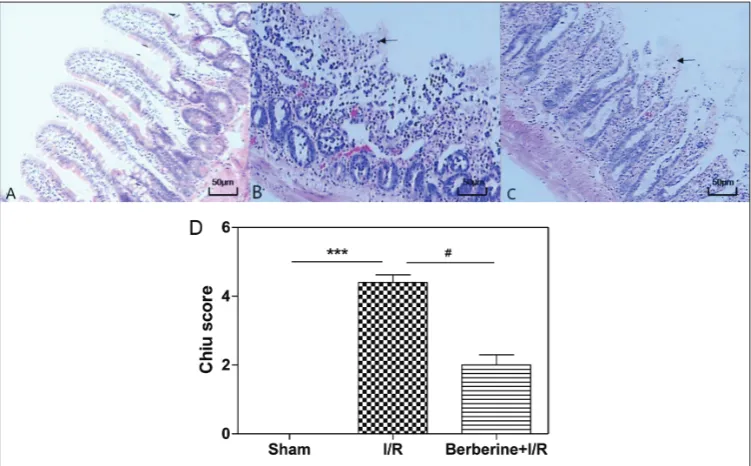
Intestinal injury was evaluated by Chiu’s scoring after microscopy (×100). In the sham group (Fig. 1A), the intestinal mucosa was normal. The gap of epithelial cells was in the normal range and we failed to identify any enlarged gap between epithelial cells. There were no significant pathological changes in the intestinal tissue of the sham and berberine groups. In the IR injury group (Fig.1B), severe edema of mucosal villi and infiltration of necrotic epithelial and
inflamma-tory cells were observed, and intestinal glands showed evidence of severe injury. In addition, a large number of intestinal villi were severed, the gap of epithelial cells increased significantly and blood and lymph vessels expanded markedly, indicating severe mucosal dam-age. Berberine pretreatment significantly reduced the histological damage compared to the IR group. We also observed edema of mucosal villi, infiltration of ne-crotic epithelial and inflammatory cells (Fig. 1C). The score of intestinal mucosa pathology is given in Fig. 1D. Compared with the IR group, rats preconditioned with berberine (200 mg/kg) prior to intestinal IR had significantly reduced intestinal IR injury and signifi-cant reduction in Chiu’s scores (p <0.05; Fig. 1D).
Intestinal tissue levels of MPO, MDA and SOD
The MPO activity was used to reflect neutrophil mi-gration into the small intestine. As shown in Fig. 2A, the activity of MPO in the berberine+IR group was lower than in the IR group (p<0.05). IR was associated with a significant increase in the lipid peroxidation product MDA, which was reduced by berberine pre-treatment (Fig. 2B). SOD is an important free radial
scavenging enzyme, and by measuring its activity we can quantify the body’s ability to eliminate the oxygen radical that derived from the inflammation reaction. SOD activity in the IR group was significantly lower than that in the sham group; berberine markedly re-stored SOD activity (p<0.001; Fig. 2C).
Serum levels of DAO
DAO is an enzyme, also named as histaminase, that ca-talyses the convertion of histidine to histamine, which is related to the metabolism, oxidation and inactiva-tion of histamine. The highest content of the enzyme is observed in the digestive tract and placenta. An in-creased DAO level indicates intestinal epithelial injury. In the present study, the serum levels of DAO in the IR injury group were higher than those in the sham group (p<0.001). DAO was reduced in the berberine+IR group compared to the IR group (p<0.01; Fig. 3).
Intestinal mucosa levels of SIgA
SIgA acts as an important immunologic barrier that could protect gut mucosa from luminal pathogens. The SIgA level in the intestinal mucous was decreased in the IR group compared to the sham group. The secretion of SIgA in the berberine+IR group was increased significantly compared to the IR group (p<0.001; Fig.4).
DISCUSSION
IR injury is a major problem associated with high morbidity and mortality after trauma, hemorrhagic shock, abdominal aortic aneurysm surgery and small bowel transplantation because tissue hypoxia, inflam-mation and cell infiltration result in the loss of the mucosal barrier [4, 23-26]. Of the internal organs, the intestine is probably the most sensitive to IR in-jury [4]. Intestinal IR inin-jury can lead to, or further enhance, oxidative stress during IR, resulting in com-plications in postoperative myocardial functional re-covery. Therefore, effective approaches to prevent IR
Fig.2. Intestinal tissue levels of MPO (A), MDA (B) and SOD (C). Ten rats were present in each group. ***p<0.001 compared with the sham group. #p<0.05 compared with the IR group. ###p<0.001 compared with the IR group.
Fig.3. Serum levels of DAO. Ten rats were present in each group. ***p<0.001 compared with the sham group. ##p<0.01 compared with the IR group.
injury are important. Owing to the relative failure of the clinical treatment of reperfusion injury and in light of previous studies, the importance of target-ing reperfusion injury in cases of intestinal ischemic injury has been brought into question. Recent studies demonstrated that ukrain, an alkaloid thiophosphoric acid derivative of greater celandine (Chelidonium ma-jus, a member of the Papaveraceae family), helps to prevent intestinal tissue breakdown during intestinal IR injury and that this effect can be achieved by an-tioxidant activities [27]. Gu et al. [28] suggested that IR-induced intestinal tight junction dysfunction can be improved by berberine, thereby demonstrating the therapeutic potential of berberine for intestinal IR.
Berberine has a 3000-year-long history of use as a Chinese medicine due to its potent antimicrobial, antiprotozoal, antidiarrheal and antitrichomal actions [29]. Clinical investigations of berberine have demon-strated a wide spectrum of pharmacological effects. Several reports highlighting significant antihyperten-sive, antiarrhythmic, antihyperglycemic, anticancer, antidepressant, anxiolytic, neuroprotective, antioxi-dant, anti-inflammatory, analgesic and hypolipidemic activities of berberine are available [30-32]. Moreover, laboratory studies have shown several molecules and signaling pathways that account for its therapeutic ef-fects [33-36]. In the present study, we investigated the potential protective effect of berberine on IR injury in rat intestine.
We identified MPO activity levels and quantified the tissue neutrophil contents. While extracellular MPO activity is thought to correspond to MPO-induced tissue damage, intracellular activity should correlate with tissue neutrophil numbers. According to our study, berberine can attenuate neutrophil ac-cumulation in IR injury and further reduce excessive tissue damage by the reduction of intestinal MPO activity. There is increasing evidence showing the in-volvement of oxidative stress in IR injury [4,37]. In this study, we investigated the antioxidant activities of berberine. MDA has been recognized as an im-portant lipid peroxidation indicator, since subjects affected by several diseases have increased MDA levels [38]. SOD plays an important role in defense against oxidant injury, and its activity can reflect the scaveng-ing capacity of endogenous free radicals. There is a
sharp rise in oxygen free radical generation during ischemia-reperfusion, resulting in SOD consumption during the elimination of oxygen free radicals [4]. It has been reported that oxidative stress occurs in intes-tinal IR injury in mice, and that it is manifested by a significant increase in serum MDA and a decrease in serum SOD [39]. In our study, reperfusion of the isch-emic intestinal circulation led to a profound increase in MDA levels in intestinal tissues, which is inhibited by pretreatment with berberine. On the other hand, SOD levels in intestinal tissues were significantly in-creased in the berberine+IR group when compared to the IR group.
Diamine oxidase (DAO), which catalyzes oxidative deamination of histamine-like diamines, increases in the plasma during intestinal ischemia, inflammation and other stress [40]. Intestinal barrier functioning was directly assessed by examining DAO activity. DAO is a type of structural enzyme in the intestine, present in high amount; after injury of the intestinal epithelium, DAO is released into the blood [41]. In this work, the serum levels of DAO in the berberine+IR group was significantly decreased compared to the IR group. These results suggested that berberine had a beneficial effect on intestinal barrier function.
It has been reported that the immune function of the intestinal mucosa declines after ischemia-reper-fusion, and that it is closely related to intestinal bacte-rial replacement [42]. SIgA is the principal immune defense against luminal pathogens at gut mucosal sur-faces. It also has anti-inflammatory activities that may be important for the maintenance of mucosal surface integrity. In this experiment, the secretion of SIgA in the berberine+IR group was significantly increased compared to the IR group; berberine may serve to maintain intestinal barrier function and thereby de-crease the systemic inflammatory response by increas-ing the secretion of SIgA.
Acknowledgments: We would like to thank Jiayin Zhu and Qiong zhang from the Laboratory Animal Center of Wenzhou Medical University.
Authors’ contribution: BL conceived and designed the study. TZC performed the experiments and wrote the paper.
Conflict of interest disclosure: The authors declare that they have no competing interests.
REFERENCES
1. Nankervis CA, Giannone PJ, Reber KM. The neonatal intes-tinal vasculature: contributing factors to necrotizing entero-colitis. Semin Perinatol. 2008;32(2):83-91.
2. Olivier C, Alexandre N. Gastro-intestinal vascular emergen-cies. Best Pract Res Clin Gastroenterol. 2013;27(5):709-25. 3. Fishman JE, Sheth SU, Levy G, Alli V, Lu Q, Xu D, Qin Y, Qin
X, Deitch EA. Intraluminal nonbacterial intestinal compo-nents control gut and lung injury after trauma hemorrhagic shock. Ann Surg. 2014;260(6):1112-20.
4. Röth E, Hejjel L, Jaberansari M, Jancso G. The role of free radicals in endogenous adaptation and intracellular signals. Exp Clin Cardiol. 2004;9(1):13-6.
5. Riaz AA, Wan MX, Schäfer T, Dawson P, Menger MD, Jeppsson B, Thorlacius H. Allopurinol and superoxide dis-mutase protect against leucocyte-endothelium interactions in a novel model of colonic ischaemia-reperfusion. Br J Surg. 2002;89(12):1572-80.
6. Amani Nabil S. Febuxostat Improves the Local and Remote Organ Changes Induced by Intestinal Ischemia/Reperfusion in Rats. Digest Dis Sci. 2012;58(3):650-9.
7. Lenka G, Jirí D, Radek M. Quaternary protoberberine alka-loids. J Cheminform. 2007;38(19):150-75.
8. Komal S, Ranjan B, Neelam C, Birendra S. Berberis aristata: A Review. Int J Res Ayurveda and Pharmacy, 2011;2(2):383-8. 9. Kumar A, Ekavali, Chopra K, Mukherjee M, Pottabathini R,
Dhull DK. Current knowledge and pharmacological profile of berberine: An update. Eur J Pharmacol. 2015;761:288-97 10. Battu SK, Repka MA, Maddineni S, Chittiboyina AG, Avery
MA, Majumdar S. Physicochemical characterization of ber-berine chloride: a perspective in the development of a solu-tion dosage form for oral delivery. AAPS PharmSciTech. 2010;11(3):1466-75.
11. Abd El-Wahab AE, Ghareeb DA, Sarhan EE, Abu-Serie MM, El Demellawy MA. In vitro biological assessment of Berberis vulgaris and its active constituent, berberine: antioxidants, anti-acetylcholinesterase, anti-diabetic and anticancer effects. Bmc Complem Altern M. 2013;13(1):218-23.
12. Balestrini L, Isolani ME, Pietra D, Borghini A, Bianucci AM, Deri P, Batistoni R. Berberine exposure triggers developmental effects on planarian regeneration. Sci Rep. 2014;4(6184):649-52.
13. Wang L, Liu L, Shi Y, Cao H, Chaturvedi R, Calcutt MW, Hu T, Ren X, Wilson KT, Polk DB, Yan F. Berberine induces caspase-independent cell death in colon tumor cells through activation of apoptosis-inducing factor. Plos One. 2012;7(5):e36418.
14. Zhang J, Cao H, Zhang B, Cao H, Xu X, Ruan H, Yi T, Tan L, Qu R, Song G, Wang B, Hu T. Berberine potently attenu-ates intestinal polyps’ growth in ApcMin mice and familial adenomatous polyposis patients through inhibition of Wnt signalling. J Cell Mol Med. 2013;17(11):1484-93.
15. Wang L, Cao H, Lu N, Liu L, Wang B, Hu T, Israel DA, Peek RM Jr, Polk DB, Yan F. Berberine Inhibits Proliferation and Down-Regulates Epidermal Growth Factor Receptor through Activation of Cbl in Colon Tumor Cells. Plos One. 2013;8(2): e56666.
16. Wang J, Peng Y, Liu Y, Yang J, Ding N, Tan W. Berberine, a natural compound, suppresses Hedgehog signaling pathway activity and cancer growth. Bmc Cancer. 2015;15(1):1-9. 17. Negar P, Massoud M, Pejman M, Goudarz Sadeghi H. Effects
of berberine on β-secretase activity in a rabbit model of Alzheimer’s disease. Arch Med Sci. 2013;9(1):146-50. 18. Yu L, Li F, Zhao G, Yang Y, Jin Z, Zhai M, Yu W, Zhao L, Chen
W, Duan W, Yu S. Protective effect of berberine against myo-cardial ischemia reperfusion injury: Role of Notch1/Hes1-PTEN/Akt signaling. Apoptosis. 2015;20(6):796-810. 19. Zhang X, Zhao Y, Zhang M, Pang X, Xu J, Kang C, Li M,
Zhang C, Zhang Z, Zhang Y, Li X, Ning G, Zhao L. Structural changes of gut microbiota during berberine-mediated preven-tion of obesity and insulin resistance in high-fat diet-fed rats. Plos One. 2012;7(8): e42529.
20. Sun SF, Zhao TT, Zhang HJ, Huang XR, Zhang WK, Zhang L, Yan MH, Dong X, Wang H, Wen YM, Pan XP, Lan HY, Li P. Renoprotective effect of berberine on type 2 diabetic nephrop-athy in rats. Clin Exp Pharmacol P. 2015;42(6):662–70. 21. Dong H, Wang N, Zhao L, Lu F. Berberine in the Treatment
of Type 2 Diabetes Mellitus: A Systemic Review and Meta-Analysis. Evid-Based Compl Alt. 2012;2012(6):591654. 22. Chiu CJ, Mcardle AH, Brown R, Scott HJ, Gurd FN.
Intes-tinal mucosal lesion in low-flow states. I. A morphologi-cal, hemodynamic, and metabolic reappraisal. Arch Surg. 1970;101(4):478-83.
23. Blikslager AT. Treatment of gastrointestinal ischemic injury. Vet Clin North Am Equine Pract. 2003;19(3):715-27. 24. Gregory KE, Deforge CE, Natale KM, Michele P, Marter LJ,
Van. Necrotizing enterocolitis in the premature infant: neo-natal nursing assessment, disease pathogenesis, and clinical presentation. Adv Neonatal Care. 2011;11(3):e783-e9. 25. Trompeter M, Brazda T, Remy CT, Vestring T, Reimer P.
Non-occlusive mesenteric ischemia: etiology, diagnosis, and inter-ventional therapy. Eur Radiol. 2002;12(5):1179-87.
26. Yasuhara H. Acute mesenteric ischemia: the challenge of gas-troenterology. Surg Today. 2005;35(3):185-95.
27. Akcılar R, Akcılar A, Koçak C, Koçak FE, Bayat Z, Şimşek H, Şahin S, Savran B. Effects of ukrain in rats with intestinal ischemia and reperfusion. J Surg Res. 2015;195(1):67-73. 28. Gu L, Li N, Yu W, Gong J, Li Q, Zhu W, Li J. Berberine reduces
rat intestinal tight junction injury induced by ischemia-reper-fusion associated with the suppression of inducible nitric oxide synthesis. Am J Chin Med. 2013;41(6):1297-312. 29. Han J, Lin H, Huang W. Modulating gut microbiota as an
anti-diabetic mechanism of berberine. Med Sci Monit. 2011;17(7):RA164-RA167.
anti-oxidant system contributes to the effect of berberine amelio-rating memory dysfunction in rat model of streptozotocin-induced diabetes. Behav Brain Res. 2011;220(1):30-41. 31. Habtemariam S. The therapeutic potential of Berberis
dar-winii stem-bark: quantification of berberine and in vitro evi-dence for Alzheimer’s disease therapy. Nat Prod Commun. 2011;6(8):1089-90.
32. Wang Y, Liu J, Ma A, Chen Y. Cardioprotective effect of ber-berine against myocardial ischemia/reperfusion injury via attenuating mitochondrial dysfunction and apoptosis. Int J Clin Exp Med. 2015;8(8):14513-9.
33. Lan T, Shen X, Liu P, Liu W, Xu S, Xie X, Jiang Q, Li W, Huang H. Berberine ameliorates renal injury in diabetic C57BL/6 mice: Involvement of suppression of SphK-S1P signaling pathway. Arch Biochem Biophys. 2010;502(2):112-20. 34. Hai L, Bin D, Sahng Wook P, Hyun-Sook L, Wei C, Jingwen
L. Hepatocyte nuclear factor 1alpha plays a critical role in PCSK9 gene transcription and regulation by the natural hypocholesterolemic compound berberine. J Biol Chem. 2009;284(42):28885-95.
35. Kim WS, Lee YS, Cha SH, Jeong HW, Choe SS, Lee MR, Oh GT, Park HS, Lee KU, Lane MD, Kim JB. Berberine improves lipid dysregulation in obesity by controlling cen-tral and peripheral AMPK activity. Am J Physiol-Endoc M. 2009;296(4):E812-9.
36. Jing Y, Kong WJ, Jiang JD. Learning from berberine: Treating chronic diseases through multiple targets. Sci China Life Sci. 2013;57(09):1-6.
37. Irani K. Oxidant signaling in vascular cell growth, death, and survival: a review of the roles of reactive oxygen species in smooth muscle and endothelial cell mitogenic and apoptotic signaling. Circ Res. 2000;87(3):179-83.
38. Yildiz Y, Serter M, Ek RO, Ergin K, Cecen S, Demir EM, Yenisey C. Protective effects of caffeic acid phenethyl ester on intestinal ischemia-reperfusion injury. Digest Dis Sci. 2009;54(4):738-44.
39. Zhang Z, Pan C, Wang HZ, Li YX. Protective effects of osthole on intestinal ischemia-reperfusion injury in mice. Experimental & Clinical Transplantation Official Jour-nal of the Middle East Society for Organ Transplantation. 2014;12(3):246-52.
40. Changmao C, Wei L, Juntian C, Xinxin L, Shenglin C. Diamine oxidase as a marker for diagnosis of superior mesenteric arterial occlusion. Hepato-gastroenterol. 2012;59(59):155-8.
41. Cakmaz R, Büyükaşık O, Kahramansoy N, Erkol H, Cöl C, Boran C, Buğdaycı G. A combination of plasma DAO and citrulline levels as a potential marker for acute mesenteric ischemia. Libyan J Med. 2013;8(5): 1-6.